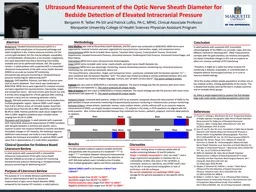
/
Bedside  sonographic  measurement of optic nerve Bedside  sonographic  measurement of optic nerve

Bedside sonographic measurement of optic nerve - PowerPoint Presentation

brianna
brianna . @brianna
Follow
66 views
Uploaded On 2023-07-23

Bedside sonographic measurement of optic nerve - PPT Presentation

Saadatnia M Isfahan University of medical sciences optic nerve sheath diameter Increased OSON ratio gt25 Optic nerve sheath diameter measurement via bedside ocular ultrasonography in patients ID: 1010765

greater optic sheath nerve optic greater nerve sheath diameter ultrasonography specific sensitive disc elevation papilledema intracranial bedside measurement

Share:

Link:

Embed:

Download Presentation from below link

Download Presentation The PPT/PDF document "Bedside sonographic measurement of opt..." is the property of its rightful owner. Permission is granted to download and print the materials on this web site for personal, non-commercial use only, and to display it on your personal computer provided you do not modify the materials and that you retain all copyright notices contained in the materials. By downloading content from our website, you accept the terms of this agreement.


Presentation Transcript

1. Bedside sonographic measurement of optic nerveSaadatnia MIsfahan University of medical sciences

2. optic nerve sheath diameter

3.

4.

5.

6.

7. Increased OS-ON ratio >2.5

8. Optic nerve sheath diameter measurement via bedside ocular ultrasonography in patients with suspected intracranial event in the emergency department is a useful method to determine ICP increase and its severity.

9. Optic disk elevation

10.

11. A minimum disc height of 0.6 mm, obtained via ultrasound can be 82% sensitive and 76% specific for papilledemaOptic nerve sheath diameter of greater than five mm, measured three mm posterior to the orbit, can be a sensitive (88%) and specific (93%) marker for elevated intracranial pressure of greater than 20 cm H2O

12. Point of care ultrasonography Optic disc elevation of greater than 0.6 mm, in addition to optic nerve sheath diameter of greater than five mm, have been shown to be correlated with papilledema. Visualization of papilledema by POCUS in the ED may serve as an effective adjunct or alternative to traditional funduscopic examinations in the diagnosis and management of IIH.

13.

14.

15.

16.

17.

18.

19. Terson syndrome

20.

21. Thank you for your attention