/
Cell structure –  AnswerIT Cell structure –  AnswerIT

Cell structure – AnswerIT - PowerPoint Presentation

attentionallianz
attentionallianz . @attentionallianz
Follow
342 views
Uploaded On 2020-06-16

Cell structure – AnswerIT - PPT Presentation

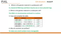
Where is the genetic material in a prokaryotic cell In a bacterial DNA loop and there may be one or more plasmid rings 2 Where is the genetic material in a eukaryotic cell The DNA is in chromosomes enclosed in a nucleus ID: 779481

cells cell area structure cell cells structure area surface dna size microscope light concentration part function questionit solution water

Share:

Link:

Embed:

Download Presentation from below link

Download The PPT/PDF document "Cell structure – AnswerIT" is the property of its rightful owner. Permission is granted to download and print the materials on this web site for personal, non-commercial use only, and to display it on your personal computer provided you do not modify the materials and that you retain all copyright notices contained in the materials. By downloading content from our website, you accept the terms of this agreement.


Presentation Transcript

Slide1

Cell structure – AnswerIT

Where is the genetic material in a prokaryotic cell?

In a bacterial DNA loop and there may be one or more plasmid rings.2. Where is the genetic material in a eukaryotic cell?The DNA is in chromosomes enclosed in a nucleus. 3. Copy and complete the table.4. Why do scientists use prefixes?To make very small numbers more manageable

Prefix

Multiple

Standard form

centi

(cm)

1 cm = 0.01 m

x

10

-2

milli (mm)

1 mm = 0.001 m

x

10

-3

micro (𝛍m)

1

𝛍m

=

0.000 001 m

x

10

-6

nano (nm)

1

nm = 0.000 000 001 m

x

10

-9

Slide2

Cell structure – AnswerIT

5.

Name the structures A to L on the diagrams below and label cells 1, 2 and 3.

Slide3

Cell structure – AnswerIT

6. Copy and complete the table and tick the correct column for each one.

Cell part

Function

Animal

Plant

Bacteria

Nucleus

Contains genetic material, which controls the activities of the cell

Cytoplasm

Most chemical processes take place here, controlled by enzymes

Cell membrane

Controls the movement of substances into and out of the cell

Mitochondria

Most energy is released by respiration here

Ribosomes

Protein synthesis happens here

Cell wall

Strengthens the cell – made of cellulose

Chloroplasts

Contain chlorophyll, absorbs light energy for photosynthesis

Permanent vacuole

Filled with cell sap to help keep the cell turgid

Bacterial DNA

Loop

of DNA NOT found in a nucleus

Plasmid

(DNA)

Small ring of DNA often used as a vector

in genetic modification

Slide4

Cell structure part 2 - Animal cell specialisation

The

structure of different cells helps them to carry out a particular function within the organism. These cells are called specialised cells.Name of animal cellDiagramStructure and function

Sperm

Function is to fertilise an egg.

Streamlined with a long tail to

swim to the egg.

Acrosome in the head containing enzymes to digest the egg cell membrane.

Large number of mitochondria in the mid section to release energy for movement.

Nerve

Function is to carry electrical signals.

Long

to carry signals long distances.

Branched connections to connect to other nerve cells and form a network around the body.

Insulating sheath to enhance transmission of electrical signals.

MuscleFunction is .Contain a large number of mitochondria to release energy from respiration for movement.Long so that there is enough space to contract.

Slide5

Cell structure part 2 - Plant cell specialisation

Name

of plant cellDiagramStructure and functionRoot hairFunction is to absorb water and minerals from the soil.Hair like projections to increase the surface area.(Note that root hair cells have no chloroplasts this is because they do not need them as they are in the soil) Xylem

Function is to carry water and minerals in

plants.

Form hollow xylem tubes made of dead tissue.

Long cells with walls toughened by lignin.

Water and minerals flow from the roots towards the leaves only in one direction in a process called

TRANSPIRATION

.

Phloem

Function is to carry glucose around the plant.

Form phloem tubes made of living tissue.

Cells have end plates with holes in them.

Glucose in solution moves from the leaves to growth and storage tissues in a process called

TRANSLOCATION.

Slide6

Cell structure part 3 – QuestionIT

Define the term resolution.

The shortest distance between two objects that can be seen clearly.2. Copy and complete the table below.3. What are the advantages of the electron microscope? Electron microscopes have a higher magnification and resolution than light microscopes; scientists can see more sub-cellular structures.4. Name the smallest cell structures that can be seen by the light microscope?Nuclei and mitochondria5. What are the smallest cell structures that can be seen by the electron microscope? Internal structures of mitochondria and chloroplasts.

Feature

Light (optical) microscope

Electron microscope

Radiation used

Light rays

Electron beams

Max

magnification

~

1500 times

~ 2 000 000 times

Resolution

200nm

0.2nm

Size of microscope

Small

and portable

Very large and not portable

Cost

~

£100

for a school one

Several £100,000 to £1 million plus

Slide7

Cell structure part 3 – AnswerIT

6. Write down the magnification equation.

7. Rearrange the equation to change the subject for the two other factors.8. A magnified cell structure has a diameter of 375𝛍m.The actual diameter of the structure is 2.5𝛍m. Calculate how many times the structure has been magnified.M = I/A M = 375/2.5 = 150 M = 150 times9. The actual length of a cell structure is 3𝛍m.It is magnified 1,500 times. Calculate the length of the magnified cell structure in mm. I = M x A I = 1500 x 3 I = 4500 𝛍m 4500 / 1000 = 4.5mmreal size of the object (A)

magnification (M) = size of image (

I)

size of image (

I) =

magnification (M) x real size of the object (A)

magnification (M)

real size of the object (A) = size of image (

I)

Slide8

Cell structure part 3 – QuestionIT

10. Name the parts of the light microscope in the diagram below.

Slide9

Cell structure part 3 – QuestionIT

11. Describe how you would make an onion cell slide.

Place thin section of onion epidermis onto slide.Place a drop of iodine in the middle of the slide to stain the onion. Gently lower cover slip onto the onion without trapping air bubbles.Soak up any excess liquid with a paper towel.12. Describe how you would use the light microscope to view onion cells. Switch on the light source and place your slide on the stage.Use the lowest objective lens and turn the focusing wheel to move the lens close to the slide.Slowly adjust the focusing wheel until you can see a clear image.

Increase the magnification by changing the objective lens and re-focus.

Slide10

Cell structure part 3 – QuestionIT

(Biology ONLY)

13. What is ‘binary fission’? Cell division where two identical cells to the parent cell are formed.14. Why do you need to sterilise Petri dished and culture mediums before use? To kill any unwanted microorganisms. 15. What would you use an inoculating loop for? To transfer bacteria onto the agar. 16. How do you sterilise an inoculating loop? By heating in a Bunsen flame. 17. How would you secure the lid of the Petri dish? With tape but not sealed all the way around. 18. What temperature would you incubate the samples at in a school and why should you use this temperature? 25oC, to prevent the growth of pathogens harmful to humans.19. How can you test the effectiveness of antibiotics and disinfectants on bacteria? Inoculate agar with bacteria, place discs soaked in the solutions (water as a control) and place the discs on the agar containing bacteria. Incubate at 25

o

C.

20. What is the zone of inhibition?

An area where bacteria don’t grow.

Slide11

Cell Division– AnswerIT

1. What are chromosomes?

Coiled strands of DNA molecules.2. What is a gene? A section of DNA that codes for a protein.3. What is DNA? A genetic material found in the nucleus that codes for proteins. 4. Where in a cell do you find chromosomes? In the nucleus of cells. 5. How many chromosome pairs do you find in a human body cell?23 pairs6. What are the three stages of the cell cycle? Growth, DNA synthesis and mitosis. 7. What is mitosis and what is it used for in animals and plants? Growth, repair and asexual reproduction. 8. What is a stem cell? An undifferentiated cell. 9. Which type of human stem cell can differentiate into any human cell? Embryo stem cells.

Slide12

Cell Division– AnswerIT

10. What is therapeutic cloning?

Where patients are given stem cells containing the same genes as theirs. 11. What are the risks of therapeutic cloning? Viral infections.12. State two reasons why people may object to the use of stem cells in therapeutic cloning.Ethical and religious reasons. 13. What are meristems? Structures in plants that contain stem cells. 14. State two reasons that plants are cloned? To clone rare species of plants and to clone crop plants with useful characteristics.

Slide13

Transport in cells – QuestionIT

Define ‘diffusion’?

The spreading of the particles from a region where they are of a higher concentration to an area of lower concentration. 2. State three places where diffusion occurs in the body. Small intestines, lungs, kidneys3. What is a concentration gradient? The difference in concentration between two areas next to each other. 4. What three factors affect rate of diffusion? Concentration gradient, temperature, surface area.5. What is surface area to volume ratio? The size of a surface compared to its volume. 6. Which has the largest surface area to volume ratio an elephant or a meerkat? The meerkat.

Slide14

Transport in cells – QuestionIT

7. State how the following are adapted for diffusion:

a. The small intestines. Have villi to increase surface area, good blood supply, thin membranes. b. The lungs. Have alveoli to increase surface area, good blood supply, thin membranes, they are ventilated. c. Gills in fish. Have gill filaments and lamella to increase surface area, good blood supply, thin membranes. d. Roots. Have root hair cells to increase surface area.e. Leaves. Large surface area, thin and stomata.8. Define osmosis? Osmosis is the diffusion of water from a dilute solution to a concentrated solution through a partially permeable membrane.9. What is a partially permeable membrane? A membrane that lets some but not all substances through.

Slide15

Transport in cells – QuestionIT

10. What happens to an animal cell in a hypertonic solution?

It will lose water by osmosis and shrivel. 11. What happens to an animal cell in a hypotonic solution? It will absorb water by osmosis and get bigger/burst. 12. Define active transport. Active transport moves substances from a more dilute solution to a more concentrated solution (against a concentration gradient) using energy from respiration.13. Why does active transport need to occur in root hair cells? Because the minerals are at a higher concentration in the roots than in the soil.14. Why does active transport need to occur in the gut? Because the glucose in the blood is at a higher concentration than in the gut.