/
Endoscopic Transnasal Repair of Bilateral Choanal Atresia in Endoscopic Transnasal Repair of Bilateral Choanal Atresia in

Endoscopic Transnasal Repair of Bilateral Choanal Atresia in - PDF document

audrey
audrey . @audrey
Follow
342 views
Uploaded On 2022-08-22

Endoscopic Transnasal Repair of Bilateral Choanal Atresia in - PPT Presentation

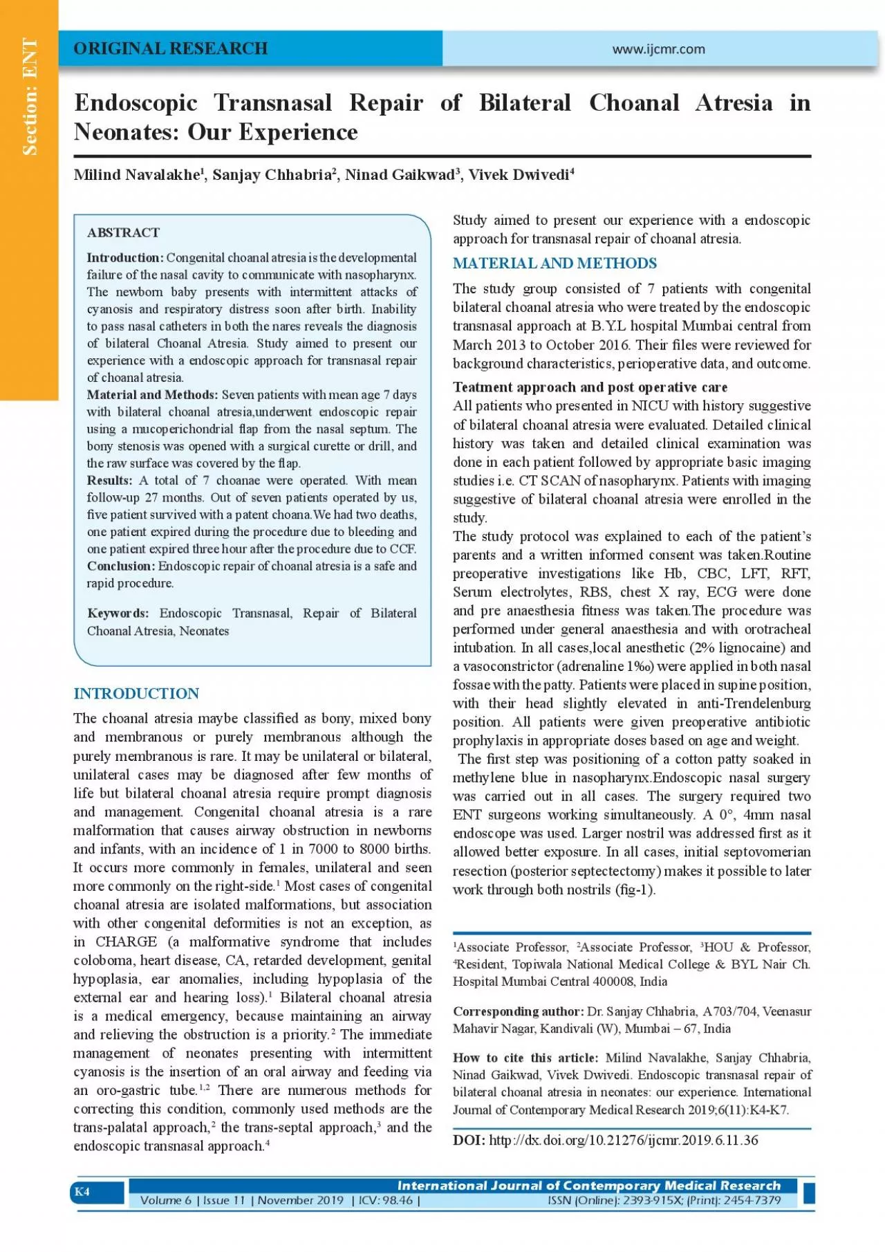
K4 Neonates Our Experience Milind Navalakhe 1 Sanjay Chhabria 2 Ninad Gaikwad 3 Vivek Dwivedi 4 ORIGINAL RESEARCH Introduction Congenital choanal atresia is the developmental failure of the na ID: 940140

atresia choanal x00660069 nasal choanal atresia nasal x00660069 endoscopic bilateral patients x0066006c cases surgery transnasal patient repair approach congenital

Share:

Link:

Embed:

Download Presentation from below link

Download Pdf The PPT/PDF document "Endoscopic Transnasal Repair of Bilatera..." is the property of its rightful owner. Permission is granted to download and print the materials on this web site for personal, non-commercial use only, and to display it on your personal computer provided you do not modify the materials and that you retain all copyright notices contained in the materials. By downloading content from our website, you accept the terms of this agreement.


Presentation Transcript

K4 Endoscopic Transnasal Repair of Bilateral Choanal Atresia in Neonates: Our Experience Milind Navalakhe 1 , Sanjay Chhabria 2 , Ninad Gaikwad 3 , Vivek Dwivedi 4 ORIGINAL RESEARCH Introduction: Congenital choanal atresia is the developmental failure of the nasal cavity to communicate with nasopharynx. The newborn baby presents with intermittent attacks of to pass nasal catheters in both the nares reveals the diagnosis of bilateral Choanal Atresia. Study aimed to present our experience with a endoscopic approach for transnasal repair Material and Methods: INTRODUCTION and membranous or purely membranous although the purely membranous is rare. It may be unilateral or bilateral, unilateral cases may be diagnosed after few months of life but bilateral choanal atresia require prompt diagnosis and management. Congenital choanal atresia is a rare malformation that causes airway obstruction in newborns and infants, with an incidence of 1 in 7000 to 8000 births. It occurs more commonly in females, unilateral and seen more commonly on the right-side. 1 Most cases of congenital choanal atresia are isolated malformations, but association with other congenital deformities is not an exception, as in CHARGE (a malformative syndrome that includes coloboma, heart disease, CA, retarded development, genital hypoplasia, ear anomalies, including hypoplasia of the external ear and hearing loss). 1 Bilateral choanal atresia is a medical emergency, because maintaining an airway and relieving the obstruction is a priority. 2 The immediate management of neonates presenting with intermittent cyanosis is the insertion of an oral airway and feeding via an oro-gastric tube. 1,2 There are numerous methods for correcting this condition, commonly used methods are the trans-palatal approach, 2 the trans-septal approach, 3 and the endoscopic transnasal approach. 4 approach for transnasal repair of choanal atresia. MATERIAL AND METHODS The study group consisted of 7 patients with congenital bilateral choanal atresia who were treated by the endoscopic transnasal approach at B.Y.L hospital Mumbai central from March 2013 to October 2016. Their �les were reviewed for background characteristics, perioperative data, and outcome. Teatment approach and post operative care All patients who presented in NICU with history suggestive of bilateral choanal atresia were evaluated. Detailed clinical history was taken and detailed clinical examination was done in each patient followed by appropriate basic imaging suggestive of bilateral choanal atresia were enrolled in the study. The study protocol was explained to each of the patient’s parents and a written informed consent was taken.Routine preoperative investigations like Hb, CBC, LFT, RFT, Serum electrolytes, RBS, chest X ray, ECG were done and pre anaesthesia �tness was taken.The procedure was performed under general anaesthesia and with orotracheal intubation. In all cases,local anesthetic (2% lignocaine) and a vasoconstrictor (adrenaline 1‰) were applied in both nasal fossae with the patty. Patients were placed in supine position, with their head slightly elevated in anti-Trendelenburg position. All patients were given preoperative antibiotic prophylaxis in appropriate doses based on age and weight. The �rst step was positioning of a cotton patty soaked in methylene blue in nasopharynx.Endoscopic nasal surgery was carried out in all cases. The surgery required two ENT surgeons working simultaneously. A 0°, 4mm nasal endoscope was used. Larger nostril was addressed �rst as it allowed better exposure. In all cases, initial septovomerian resection (posterior septectectomy) makes it possible to later 1 Associate Professor, 2 Associate Professor, www.ijcmr.com Section: ENT International Journal of Contemporary Medical Research Volume 6 | Issue 11 | November 2019 | ICV: 98.46 |ISSN (Online): 2393-915X; (Print): 2454-7379 3 HOU & Professor, 4 Resident, Topiwala National Medical College & BYL Nair Ch. Hospital Mumbai Central 400008, India Corresponding author: Dr. Sanjay Chhabria, A703/704, Veenasur Mahavir Nagar, Kandivali (W), Mumbai – 67, India How to cite this article: Milind Navalakhe, Sanjay Chhabria, Ninad Gaikwad, Vivek Dwivedi. Endoscopic transnasal repair of bilateral choanal atresia in neonates: our experience. International Journal of Contemporary Medical Research 2019;6(11):K4-K7. DOI: http://dx.doi.org/10.21276/ijcmr.2019.6.11.36 K5 Initial

ly, on the roomy side, a vertical, hemitrans�xation incision was taken on the septum, approximately 1cm anterior to the atretic plate, this incision continues along the �oor of the fossa obliquely in the direction up to the posterior choana. A mucosal �ap is then raised to expose the osteocartilaginous septum and the atretic plate. This �ap should be preserved during the entire surgery (�g-2). A vertical incision was made on the septum at the level of the bonycartilagenous junction. This incision was extended superiorly, at a right angle, at the level of the free edge of the middle concha to the choanal border. Analogous to the previous side, a mucosal �ap was raised to expose the septum and the atretic plate of opposite side. Once both �aps were raised, a posterior septectomy was performed and both the atretic plates were removed using different instruments, depending on the type of atresia. The atretic plate can be removed with Kerrison punch, curettes, microdebrider and sometimes a drill was required. After baring the atretic plate, it is punctured at the weakest point preferably infero-medially in order to avoid inadvertently penetrating in to the intracranial area or the eye socket. The puncture was generally made with the tip of the suction, even in purely osseous cases and when it was not possible to puncture with suction tip a burr or chisel was used. After opening the atresia (which was con�rmed by visualization of methylene blue patty in nasopharynx), it was extended circumferentially with forceps or microdebrider for the soft tissues and with the burr for the bone in the pterygoid and sphenopalatine region. It was important to separate the pharyngeal mucosa from the nasal mucosa properly on the lateral edge of the neochoana in order to expose the medial plate of the pterygoid processes. The septectomy must include part of the perpendicular plate of the ethmoid bone and the posterior part of the vomer and the rostrum sphenoidale. This step is fundamental to avoid restenosis. Once a broad communication between the nasal fossae and nasopharynx is established, the �aps must be carefully replaced to cover the bone exposed on the �oor and roof of the neochoana. The �aps are placed in the most convenient way possible; however, we usually rotate the ipsilateral �ap superiorly to cover the choanal arch and the contralateral �ap is rotated inferiorly to cover the �oor of the nasal cavity. At times, depending on the amount of mucosa preserved, it may be advisable to resect the excess mucosa; the adenoids if enlarged were debrided. Drilled after microdebrided In all cases, after achieving haemostasis, topical mitomycin-c Postoperative care The patient was shifted to NICU and was monitored for 48 hrs and taken again in OT and pack was removed under anaesthesia and diagnostic nasal endoscopy was done simultaneosly to asess neochoana. The patient was kept under observation and vitals were monitored. Nasal lavage with saline was done and Vaseline ointment was applied in each nostril. If patient’s post operative recovery was satisfactory, he was discharged with strict instruction to come back if child developed stridor or had cynotic spells. The �rst check scopy was done 1 month post-op.All patients were advised nasal doucing with wáter and topical application of vaseline.The next check scopy was done after 6 months to verify choanal patency.If the air blast was good and choana was patent on nasal endoscopy the patient was asked to follow up annualy. The mean follow-up time for our patients was 27 months. RESULT In our series of 7 patients 2 were males and 5 were females. The mean age at the time of surgery was 7 days (range, 3 days–17 days). 3 patients had associated malformation, one patient had low anorectal malformation,one patient had PDA Figure-1: CT scan showing Choanal Atresia Figure-2: Mucosal Flap being raised Figure-3: Widely Opened Choanal Atresia K6 and low set ear and one patient was a case of frontonasal Patent cavity after 6 month of operation CT scan was done in all cases which revealed presence of mixed variety of choanal atresia in 3 patients,bony variety of choanal atresia in 3 cases and 1 patient had membranous choanal atresia.In all the cases the surgery was scheduled on emergency basis. Mitomycin-c soaked patty was applied post su

rgery for 5 minutes before anterior nasal packing in all cases. After pack removal nose breathing was observed in all patients both awake and while sleeping. In all patients oral feeds was initiated on the same day the nasal pack was removed.If there was no respiratory dif�culty or aspiration,patients were discharged on the fourth day following surgery. Out of seven patients of bilateral choanal atresia operated by us, �ve patient survived with a very good postoperative result and are under regular follow up.We had two deaths, one patient expired during the procedure due to bleeding and one patient expired three hour after the procedure due to CCF. DISCUSSION The analysis of the outcomes achieved in 5 patients in our series of 7 cases was good. We have not observed a single case of restenosis. Bilateral choanal atresia is not a highly prevalent pathology and that most of the studies present a heterogeneous case series consisting of fewer than 30 patients undergoing surgery 5,6,7,8,9,10 , so our outcomes are worthy of being taken into consideration The mean follow-up time for our patients was 27 months. All were examined on the basis of their history, symptoms and signs that would be grounds for suspecting possible restenosis like dif�culty in breathing while eating or mouth breathing while sleeping, as well as by diagnostic nasal endoscopy. As has been shown in literature restenosis takes place, within the �rst year after surgery 11 and our mean follow up is 27 months, we can be fairly con�dent in accepting the High resolution CT is the key radiological study to con�rming diagnosis. Performing a diagnostic CT scan, particularly in early infancy, has the disadvantage that it may be necessary to do so under sedation or general anaesthesia. Nonetheless, we believe that it is an indispensable study to identify the type of atresia and possible anatomical variations particularly, bearing in mind that these patients may have more than one malformation. Choanal atresia having a large bony component display a higher rate of restenosis as they exhibit neo-osteogenesis phenomena. 12 It is therefore important for the choanal framework to be suf�ciently expanded and lined with mucosa. The need for revision surgery is greater in choanal atresia associated with other malformations. 13 The aim of treatment for choanal atresia is to restore the nasal air �ow without injuring structures that produce craniofacial development by means of a safe and ef�cacious technique. Bilateral choanal atresia diagnosed in the newborn are usually a neonatal paediatric emergency. Oral feeding in these children can lead to pulmonary aspiration. Symptoms can vary from mild respiratory distress with feeding to severe obstruction of the airway. Surgery should be performed early, (orotracheal intubation if there are signs of respiratory distress). In our series, the primary cases of bilateral choanal atresia underwent surgery in the �rst few days of life. As seen in our series, the endoscopic approach has no technical or space limitations. Different surgical approaches have been used to treat choanal atresia like transmaxillary, transseptal, transpalatine, transnasal, sublabial, paralateronasal and by nasal endoscopy. 14 Depending on the time and school of thought, one or another has been used. The ideal method should be the one which achieves the highest rate of permanent success in restoring communication between the nasal fossae and the nasopharynx, while causing as little morbidity as possible. We have tried to show that the endoscopic nasal approach for choanal atresia has a great advantage and should be considered over others. It is important to point out two fundamental steps in this kind of surgery: increasing the surface of the choanal frame by eliminating the posterior part of the vomer until a “single” neochoana is formed and the creation of the mucosal �aps that will cover the exposed bony surfaces on the edge of the choana, thus decreasing the formation of granulation tissue and scarring phenomena. Both these surgical gestures lower the incidence of restenosis. It has been shown that the all but total elimination of the vomer does not affect facial growth 15 After obtaining adequate opening of the choanae, some authors apply topical mitomycin-c or place a temporary stent to prevent choanal closure. In our series, w

e have used mitomycin-c and the outcomes achieved have been good.The use of the antimitotic mitomycin-c is supported by several authors 16,17 to prevent the growth of scar tissue. CONCLUSION Bilateral choanal atresia is an emergency which requires a team approach involving pediatrics, ent, radiology and anaesthesia services. Cyclical cynosis with respiratory distress and absence of air bubbles in mucoid nasal discharge Figure-4: Patent Choana 6 months Post-op K7 in a new borne should lead to suspicion of this condition and attempt must be made to pass infant feeding tubes through both the nostrils.If the attempts fail urgent CT scan and other investigation should be done and once the clinical suspicion is con�rmed by imaging patient should be taken up for endoscopic management and creation of neochoana. We believe this method is safe and effective in restoring communication between the nasal fossae and the nasopharynx, while causing as little morbidity as posible. Use of mitomycin-c makes it possible to achieve stable, long-term choanal patency. REFERENCES 1. Leclerc JE, Fearon B.Choanal atresia and associated anomalies. Int J Pediatr Otorhinolaryngol 1987; 13: 265-72. 2. Azar RH, Younis RT. Transnasal repair of choanal atresia using telescopes. Arch Otolaryngol Head Neck Surg 1995; 121: 517-20. 3. Samadi DS, Shah UK, Handler SD. Choanal atresia: a twenty year review or medical comorbidities and surgical outcomes. Laryngoscope 2003; 113: 254-8. 4. Heneger AS, Strom M: Choanal atresia: A new embryonic theory and its in�uence on surgical management. Laryngoscope 2009; 92: 913-2 Dr Sanjay Chhabria (Assist Professor) 5. N. Friedman,R. Mitchell,C. Bailey,D. Albert,S. Leighton Management and outcome of choanal atresia correction Int J Pediatr Otorhinolaryngol, 52 (2000), pp. 45-51 6. N. Teissier,F. Kaguelidou,V. Kouloigner,M. Francois,T. van den Abeele Predictive factors for success after transnasal endoscopic treatment of choanal atresia Arch Otolaryngol Head Neck Surg, 2008;134: 57-61. 7. T.Van den Abbeele,M. Francois,P. Narcy Transnasal endoscopic treatment of choanal atresia without prolonged stenting Arch Otolaryngol Head Neck Surg 2002;128:936-940. 8. J. Gosepath,V.E. Santamaria,B.M. Lippert,W.J. Mann Forty-one cases of congenital choanal atresia over 26 years—retrospective analysis of outcome and technique. Rhinology 2007;45:158-163. 9. H.E. Romeh,O.A. Albirmawy A 13-year experience and predictors for success in transnasal endoscopic repair of congenital choanal obliteration Int J Pediatr Otorhinolaryngol. 2010;74:737-742. 10. A.S. Hengerer, T.M. Brickman, A. Jeyakumar. Choanal atresia: embryologic analysis and evolution of treatment, a 30-year experience. Laryngoscope 2008;118:862-866. 11. M.A. Richardson,J.D. Osguthorpe Surgical management of choanal atresia Laryngoscope 1998; 98:915-918. 12. S. Ayari,D. Abedipour,D. Bossard,P. Froehlich CT- assisted surgery in choanal atresia Acta Otolaryngol 2004;124:502-504. 13. N. Friedman,R. Mitchell,C. Bailey,D. Albert,S. Leighton Management and outcome of choanal atresia correction Int J Pediatr Otorhinolaryngol 2000;52:45-51 14. A.C. Stamm,S.S. Pignatari Nasal septal cross-over �ap technique: a choanal atresia micro-endoscopic surgical repair Am J Rhinol, 2001;15:138-143. 15. M.A. El-Ahl,M.W. El-Anwar Stentless endoscopic transnasal repair of bilateral choanal atresia starting with resection of vómer Int J Pediatr Otorhinolaryngol. 2012;76:1002-1006. 16. J. Gosepath, V.E. Santamaria, B.M. Lippert, W.J. Mann Forty-one cases of congenital choanal atresia over 26 years—retrospective analysis of outcome and technique. Rhinology. 2007;45:23-28. 17. M.K. Bozkurt,B. Keles,A. Azimov,K. Ozturk,H. Arbag The use of adjunctive topical mitomycin in endoscopic congenital choanal atresia repair Int J Pediatr Otorhinolaryngol. 2010;23:74733-74736. Source of Support: Nil; Con�ict of Interest: None Submitted: 25-05-2016; Accepted: 21-07-2016; Published: 28-11-2019 Section: ENT Section: ENT Endoscopic Transnasal Repair of Bilateral Choanal Atresia in Neonates International Journal of Contemporary Medical Research ISSN (Online): 2393-915X; (Print): 2454-7379 | ICV: 98.46 |Volume 6 | Issue 11 | November 2019 Section: ENT Endoscopic Transnasal Repair of Bilateral Choanal Atresia in Neonates International Journal of Contemporary Medical Research Volume 6 | Issue 11 | November 2019 | ICV: 98.46 |ISSN (Online): 2393-915X; (Print): 2454-7