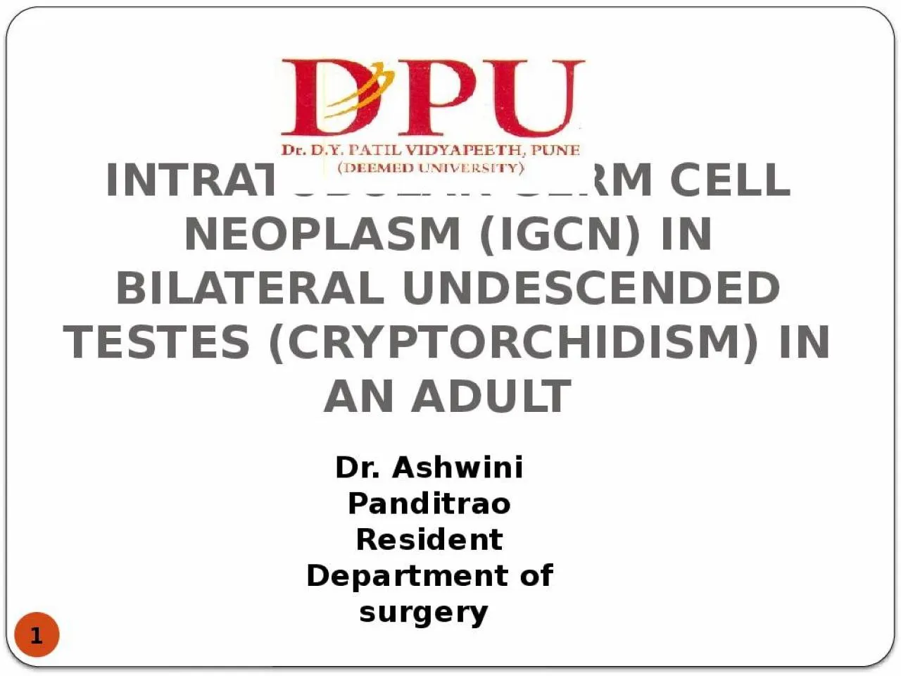
/
INTRATUBULAR GERM CELL NEOPLASM (IGCN) IN BILATERAL UNDESCENDED TESTES (CRYPTORCHIDISM) INTRATUBULAR GERM CELL NEOPLASM (IGCN) IN BILATERAL UNDESCENDED TESTES (CRYPTORCHIDISM)

INTRATUBULAR GERM CELL NEOPLASM (IGCN) IN BILATERAL UNDESCENDED TESTES (CRYPTORCHIDISM) - PowerPoint Presentation

susan
susan . @susan
Follow
342 views
Uploaded On 2022-05-18

INTRATUBULAR GERM CELL NEOPLASM (IGCN) IN BILATERAL UNDESCENDED TESTES (CRYPTORCHIDISM) - PPT Presentation

1 Dr Ashwini Panditrao Resident Department of surgery INTRODUCTION 2 Inguinal masses are a clinical enigma and always pose a dilemma for the surgeon Preoperative diagnosis is always difficult due to varied presentation of the tumours ID: 911915

patients bilateral cell germ bilateral patients germ cell testis inguinal testicular tumours tumour cryptorchidism normal testes undescended incidence cancer

Share:

Link:

Embed:

Download Presentation from below link

Download Presentation The PPT/PDF document "INTRATUBULAR GERM CELL NEOPLASM (IGCN) I..." is the property of its rightful owner. Permission is granted to download and print the materials on this web site for personal, non-commercial use only, and to display it on your personal computer provided you do not modify the materials and that you retain all copyright notices contained in the materials. By downloading content from our website, you accept the terms of this agreement.


Presentation Transcript

Slide1

INTRATUBULAR GERM CELL NEOPLASM (IGCN) IN BILATERAL UNDESCENDED TESTES (CRYPTORCHIDISM) IN AN ADULT

1

Dr. Ashwini PanditraoResidentDepartment of surgery

Slide2

INTRODUCTION

2

Inguinal masses are a clinical enigma and always pose a dilemma for the surgeon.Pre-operative diagnosis is always difficult due to varied presentation of the tumours.The incidence of bilateral testicular germ cell tumour (BGCT) ranges between 1 and 5%.Synchronous bilateral germ cell tumour of the testis is rare and its association with bilateral cryptorchidism is even rarer.Risk factors for germ cell tumour are-

infertility,

cryptorchidism,

micro calcifications, and

genetic predisposition

Slide3

3

CASE REPORT

A 35yr old male patientComplaints of swelling in bilateral inguinal region since 6 monthsPain in abdomen since 6 monthsNo history of pain radiating to back

No history of nausea and/or vomiting

No history of altered bowel habits

No history of weight loss

No history of exposure

General Examination-

Pulse- 82/min

Blood pressure- 130/80 mm of Hg

No evidence of pallor, icterus, clubbing, oedema, lymphadenopathy, no supraclavicular lymphadenpathy

Slide4

4

Local Examination-

Inspection-Oval swelling of approximately 3×2 cms in bilateral inguinal regionCough impulse was absentSkin over the swelling is normalNo visible pulsations

Bilateral scrotum empty

Palpation-

Temperature –normal

Non tender

3×2 cms swelling in bilateral inguinal region

Firm in consistency

Smooth surface

Skin over the swelling normal

Slide5

5

Systemic Examination-

Per Abdomen-No lumpProvisional Diagnosis- Bilateral Undescended Testes

Slide6

6

INVESTIGATIONS

HaemoglobinTotal leukocyte countLiver function testsBlood ureaBlood glucose level

Renal function test

Serum electrolytes

Urine – routine and microscopy

Sperm count- azoospermia

Beta HCG- 2.09 mIU/ml (normal range- 0-4mIU/ml)

Alpha feto protein- 1.26 IU/ml (normal range- 0-8.5 ng/ml)

LDH- 383 IU/L

(normal range- 140-280 U/L)

Within normal

limit

Slide7

7

Chest X-Ray- normalUltrasonography abdomen – normal study

Ultrasonography inguinoscrotal region- Hyperdense lesion in right and left inguinal region with probability of both testes at deep inguinal ring.

Slide8

8

CT scan pelvis-

1. 2 well defined hyperdense lesions noted in right and left inguinal region, right measuring 2×1.5 cm and left measuring 4×1.3 cm 2. Both lesions are located in the course of inguinal canal

3. No enhancement on contrast study, likely suggestive of undescended testes

Slide9

9

MRI scan- 2 well defined lesions in bilateral inguinal regions adjacent to urinary bladder, measuring 2×1.5 cms and 2.4×1.3 cms, both located in the course of inguinal canal

Impression- Bilateral Undescended Testes

Slide10

10

OPERATIVE FINDINGS

Patient was posted for high inguinal orchidectomy on both sides.

Slide11

11

HPE of both sides revealed

INTRATUBULAR GERM CELL NEOPLASM

(pTis IGCN,

Stage 0)

Slide12

12

Post operative period uneventfulDuring the last three monthly follow up, patient underwent

Clinical examinationUltrasonography of abdomen Within normal limitEstimation of tumour markers

CT scan abdomen and pelvis- no retroperitoneal, pre- and para-aortic lymphadenopathy

Slide13

13

DESCENT OF TESTES

(During 7

th

week of

fetal development)

(At 3 months of gestation

at internal inguinal ring)

(Begins between 7 and

9 months of gestation and

completes by birth)

Slide14

14

ANOMALIES IN DESCENT OF TESTIS

Slide15

15

DISCUSSION

Terms such as undescended testis, retentio testis, cryptorchidism, and maldescended testis describe a testis that is not normally located at the bottom of the scrotum. Undescended testis may be situated along its normal route of descent or in an ectopic position.

Cryptorchid/ undescended testis neither resides nor can be manipulated into the scrotum.

Slide16

16

Ectopic testis is the one that has an aberrant course of descent, usually after leaving the inguinal canal, femoral, pubopenile, perineal or crossed scrotal.

Retractile testis can be manipulated into the scrotum where it remains without tension.Gliding testis can be manipulated into the upper scrotum but retracts when released.Acquired is the one in which testis previously descended or after orchiopexy or other inguinal surgery (hernia), then “

ascends

” spontaneously

.

Slide17

17

ETIOLOGY OF UNDESCENDED TESTES

The aetiology of cryptorchidism remains largely unknown.Several risk factors like-Intrauterine growth restriction (IUGR)

Prematurity- incidence in premature infants 30%

First- or second- born boys

Perinatal asphyxia

Toxaemia of pregnancy

Slide18

18

The incidence of bilateral germ cell tumour (BGCT) is much greater in younger age group than compared to that of unilateral seminoma which is common in patients older than 30yrs of age.

Testicular intratubular germ cell neoplasia as seen in our case is considered a premalignant lesion for development of germ cell tumours.Incidence of intratubular germ cell neoplasia (IGCN) is high in patients with one or more risk factors (35-85%) and its detection allows curative tumour eradication with minimal morbidity or mortality.

Primary bilateral testicular tumour is rare ,its prevalence ranges from 2.5% to 5%, most tumours being metachronous.

Slide19

19

In a study by Tekin et al.of 552 patients with GCTT,11 patients developed bilateral disease. Out of 11 patients, 7 developed a second tumour metachronously and 4 had synchronous bilateral GCTT.

The incidence of bilateral germ cell tumours was 1.8% (14 of 776 patients) in patients with seminoma and 0.6% (10 of 1655 patients) in patients with non seminomatous germ cell tumours.Cryptorchidism, infertility, or atrophic testis was associated with development of bilateral GCTT in 7 of the 11 patients. All synchronous tumours and most of the sequential tumours had identical histology on both sides.

Slide20

20

Post orchidectomy management of these patients has been dictated by the stage of the tumour in either of the testes and the pathology with the higher malignant potential (NSGCT as compared to pure seminoma).

Bilateral seminomas have a higher tumour burden, therefore, these patients should not be kept on surveillance only; rather should be treated with prophylactic para-aortic lymph node irradiation or one to two cycles of adjuvant chemotherapy. Patients in stage II or higher should be treated with chemotherapy.

Slide21

21

CONCLUSION

Clinical examination and a simple imaging modality like USG are gold standard in earlier diagnosis of the condition.Thus a thorough clinical and radiological evaluation is of paramount importance in patients being investigated for infertility.Bilateral testicular tumours with cryptorchidism are extremely rare and amongst them, synchronous bilateral testicular tumours are even rarer.

Adjuvant therapy in the form of cisplatin-based chemotherapy has markedly increased the survival rate.

Prognosis depends upon the clinical staging and not whether it is unilateral or bilateral involvement.

Our patient will require life long

f

ollow up and androgen replacement.

Slide22

WHAT MAKES OUR CASE UNIQUE

22

Incidence of bilateral testicular tumours with cryptorchidism is extremely rare and among them, synchronous tumours are even rarer which was in our case, less than 0.8%.Option of testes sparing surgery can be given to patients who are aware and accept the risk of subsequent local relapse and realize the importance of compliance and follow up

.

Slide23

23

REFERENCES

Sushma Agrawal, Ranjeet Bajpai, R. K. Agrawal and T. C. Gupta. Bilateral synchronous seminoma with bilateral cryptorchidism of the testis. Indian J Urol.2010; 26(4):587–589 Walid K. Adham, Bharat K. Raval, Maria C. Uzquiano and Luciano B. Lemos., Bilateral Testicular Tumors: Seminoma and Mixed Germ Cell Tumor. Jrnl of RadioGraphics. 2005; 25:835-839.

Coogan CL, Foster RS, Simmons GR, Tognoni PG, Roth BJ, Donohue JP. Bilateral testicular tumors: management and outcome in 21 patients. Cancer. 1998; 83(3):547-52.

Che M, Tamboli P, Ro JY, Park DS, Ro JS, Amato RJ, Ayala AG. Bilateral testicular germ cell tumors: Twenty-year experience at M. D. Anderson Cancer Center. Br J Cancer. 2002; 95(6):1228-33.

Slide24

24

5. García Morúa A, Gutiérrez García JD, Ortiz Lara Gerardo E, Martínez Montelongo R, Gómez Guerra Lauro S. Synchronous bilateral testicular seminoma in an adult patient with bilateral cryptorchidism: A case report and literature review.Actas Urol Esp.2010;34(2):210-1.

6. Tekin A, Aygun YC, Aki FT, Ozen H. Bilateral germ cell cancer of the testis: A report of 11 patients with a long-term follow-up. BJU Int. 2000; 85(7):864-8.7. Géczi L, Gomez F, Bak M, Bodrogi I. The incidence, prognosis, clinical and histological characteristics, treatment, and outcome of patients with bilateral germ cell testicular cancer in Hungary. J Cancer Res Clin Oncol. 2003; 129(5):309-15.

8. HolzbeierleinJM, Sogani PC, Sheinfeld J. Histology and clinical outcomes in patients with bilateral testicular germ cell tumors: the Memorial Sloan Kettering Cancer Center experience 1950 to 2001. J Urol.2003; 169(6):2122-5.

Slide25

THANK YOU

25