/
A Rare Case Report Bilateral Choanal A Rare Case Report Bilateral Choanal

A Rare Case Report Bilateral Choanal - PDF document

isabella
isabella . @isabella
Follow
342 views
Uploaded On 2022-08-22

A Rare Case Report Bilateral Choanal - PPT Presentation

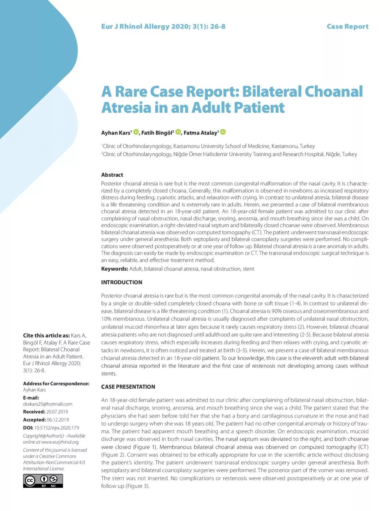
Atresia in an Adult Patient Ayhan Kars 1 Fatih Bingöl 2 Fatma Atalay 1 Address for Correspondence Ayhan Kars Email drakars25hotmailcom 20072019 Accepted 06122019 DOI 105152ejra20 ID: 940141

atresia choanal nasal bilateral choanal atresia bilateral nasal patient case adult rare crossref endoscopic lateral year atres observed transnasal

Share:

Link:

Embed:

Download Presentation from below link

Download Pdf The PPT/PDF document "A Rare Case Report Bilateral Choanal" is the property of its rightful owner. Permission is granted to download and print the materials on this web site for personal, non-commercial use only, and to display it on your personal computer provided you do not modify the materials and that you retain all copyright notices contained in the materials. By downloading content from our website, you accept the terms of this agreement.


Presentation Transcript

A Rare Case Report: Bilateral Choanal Atresia in an Adult Patient Ayhan Kars 1 , Fatih Bingöl 2 , Fatma Atalay 1 Address for Correspondence: Ayhan Kars E-mail: drakars25@hotmail.com 20.07.2019 Accepted: 06.12.2019 DOI: 10.5152/ejra.2020.179 Abstract Posteror choanal atresa s rare but s the most common congental malformaton of the nasal cavty. It s characte - rzed by a completely closed choana. Generally, ths malformaton s observed n newborns as ncreased respratory dstress durng feedng, cyanotc attacks, and relaxaton wth cryng. In contrast to unlateral atresa, blateral dsease s a lfe threatenng condton and s extremely rare n adults. Heren, we presented a case of blateral membranous choanal atresa detected n an 18-year-old patent. An 18-year-old female patent was admtted to our clnc after complanng of nasal obstructon, nasal dscharge, snorng, anosma, and mouth breathng snce she was a chld. On blateral choanal atresa was observed on computed tomography (CT). The patent underwent transnasal endoscopc surgery under general anesthesa. Both septoplasty and blateral coanoplasty surgeres were performed. No compl - catons were observed postoperatvely or at one year of follow up. Blateral choanal atresa s a rare anomaly n adults. The dagnoss can easly be made by endoscopc examnaton or CT. The transnasal endoscopc surgcal technque s an easy, relable, and eectve treatment method. Keywords: Adult, blateral choanal atresa, nasal obstructon, stent I NTRODUCTION Posterior choanal atresia is rare but is the most common congenital anomaly of the nasal cavity. It is characterized by a single or double-sided completely closed choana with bone or soft tissue (1-4). In contrast to unilateral dis ease, bilateral disease is a life threatening condition (1). Choanal atresia is 90% osseous and osseomembranous and 10% membranous. Unilateral choanal atresia is usually diagnosed after complaints of unilateral nasal obstruction, unilateral mucoid rhinorrhea at later ages because it rarely causes respiratory stress (2). However, bilateral choanal atresia patients who are not diagnosed until adulthood are quite rare and interesting (2-5). Because bilateral atresia causes respiratory stress, which especially increases during feeding and then relaxes with crying, and cyanotic at - tacks in newborns, it is often noticed and treated at birth (3-5). Herein, we present a case of bilateral membranous choanal atresia detected in an 18-year-old patient. To our knowledge, this case is the eleventh adult with bilateral stents. C ASE PRESENTATION An 18-year-old female patient was admitted to our clinic after complaining of bilateral nasal obstruction, bilat - eral nasal discharge, snoring, anosmia, and mouth breathing since she was a child. The patient stated that the physicians she had seen before told her that she had a bony and cartilaginous curvature in the nose and had to undergo surgery when she was 18 years old. The patient had no other congenital anomaly or history of trau - ma. The patient had apparent mouth breathing and a speech disorder. On endoscopic examination, mucoid discharge was observed in both nasal cavities. The nasal septum was deviated to the right, and both choanae (Figure 2). Consent was obtained to be ethically appropriate for use in the scientific article without disclosing the patient’s identity. The patient underwent transnasal endoscopic surgery und

er general anesthesia. Both septoplasty and bilateral coanoplasty surgeries were performed. The posterior part of the vomer was removed. The stent was not inserted. No complications or restenosis were observed postoperatively or at one year of follow up (Figure 3). Cite this article as: Kars A, Bingöl F, Atalay F. A Rare Case Report: Bilateral Choanal Atresia in an Adult Patient. Eur J Rhinol Allergy 2020; 3(1): 26-8. Eur J Rhinol Allergy 2020; 3(1): 26-8 Case Report Clinic of Otorhinolaryngology, Kastamonu University School of Medicine, Kastamonu, Turkey 2 Clinic of Otorhinolaryngology, Nide Ömer Halisdemir University Training and Research Hospital, Nide, Turkey Copyright@Author(s) - Available online at www.eurjrhinol.org Content of this journal is licensed under a Creative Commons Attribution-NonCommercial 4.0 International License. D ISCUSSION Choanal atresia was rst described by Johann Roderer in 1755 (4, 6). Pos - terior choanal atresia is rare but is the most common congenital anomaly of the nasal cavity; it is characterized by a single or double-sided complete closure of the choanae with bone or soft tissue (1-4). Choanal atresia is 90% osseous and osseomembranous and 10% membranous (2). Membra - nous atresia was detected in our patient. This anomaly is observed in 1 in 5000-7000 live births and is ass ociated with other congenital anomalies in 50% of patients (1). Diseases such as microcephaly, micrognathia, Down syndrome, humeroradial synostosis, palatal defects, Treacher Collins syn - drome, Crouzon syndrome, Apert syndrome, Pfeier syndrome, nasopha - ryngeal anomalies, branchial anomalies, mandibulofacial synostosis, and nasal ethmoidal encephalocele may accompany choanal atresia, but the most common accompanying anomaly is CHARGE syndrome (2, 4). The re - fore, it is necessary to evaluate the cases detected in detail (3). The etiology and pathogenesis of choanal atresia remains incompletely understood. In one study, a mutation in retinol dehydrogenase 10 related to A vitamin metabolism was shown to play a role in its etiology (7). Ac - cording to one theory, choanal atresia occurs as a result of an abnormality in the migration and development of neural crest cells (3, 4). According to another theory, it is caused by the remnants of the nasobuccal and bucco - pharyngeal mucosa (4). Atresic plaque is located in front of the posterior vomer and the palate, and the plaque thickness is 1-12 mm (8). Nasal endoscopic examination is important in the diagnosis of choanal atresia but does not provide information on the type of a tresia. Therefore, the most used and most ideal method for diagnosis of choanal atresia and identifying the type of choanal atresia is CT (2). Membranous atresia was demonstrated with CT in our patient. Bilateral choanal atresia is a condition that must be urgently treated in newborns (1). Because newborns learn mouth breathing at 4-6 weeks of age, they must initially breath through their nose (2, 3). In bilateral atresia, this situation presents respiratory stress, which especially increases during feeding and then relaxes with crying, and cyanotic attacks immediately after birth (2-4). Death is frequent in cases in which early diagnosis is not possible. Therefore, bilateral choanal atresia is rarely encountered in older patients (2). Only ten cases of bilater al choanal atresia were reported in the literature before our case. In the treatment of choanal atresia, the open transpalatal technique has been replaced by a minimally invasive transnasal endoscopic technique (9). The transnasal endoscopic method is a simple, safe, and eective ap - proach to treat this disease (2). The most common postoperative com - plication is restenosis. A stent can be used to prevent this complicatio

n, but the use of a stent is controversial (1). Some researchers advocate the superiority of stent use (10). We used the transnasal endoscopic method similar to that used in the cases in the literature in our patient. Due to the risk of a foreign body reaction, the need to use long-term antibiotics, and the risk of necrosis in the columella region, we decided not to use a stent. We aimed to prevent re-narrowing by removing the posterior portion of the vomer. No restenosis was observed in our patient at one year. Only 1 of 10 adult patients with bilateral atresia reported in the literature required revision surgery due to restenosis (5). CONCLUSION Bilateral choanal atresia is an extremely rare condition in adults. It can be easily diagnosed by nasal endoscopic examination or CT. Transnasal en - doscopic choanaplasty is the current treatment approac h. This treatment should be carefully considered in patients with complaints of continuous bilateral nasal obstruction, bilateral nasal discharge, snoring, anosmia , and mouth breathing. Figure 3. Endoscopic view of the right and left choana opening at one year postoperatively Figure 2. Axial section computed tomography (CT) view of bilateral choanal atresia Figure 1. Endoscopic appearance of preoperative left and right choanal atresia Kars et al. Blateral Choanal Atresa n an Adult Eur J Rhnol Allergy 2020; 3(1): 26-8 27 Informed Consent: Written informed consent was obtained from the patient who participated in this study. Peer-review: Externally peer-reviewed. Author Contributions: Concept - A.K.; Design - F.B. Supervision - F.A.; Fundings - F.A.; Materials - F.B.; Data Collection and/or Processing - A.K.; Analysis and/or Inter - pretation - F.A.; Literature Search - A.K.; Writing Manuscript - A.K.; Critical Review - F.B. Conict of Interest: The authors have no conicts of interest to declare. Financial Disclosure: The authors declared that this study has received no nan - cial support. REFERENCES Anajar S, Hassnaoui J, Rouadi S, Abada R, Roubal M, Mahtar M. A rare case re - port of bilateral choanal atresia in an adult. Int J Surg Case Rep 2017; 37: 127-9. [ CrossRef ] Bakr S, Ozbay M, Kn V, Gün R, Yorganclar E. Bilateral choanal atresia in adults. Kulak Burun Bogaz Ihtis Derg 2014; 24: 114-7. [ CrossRef ] Aksoy F, Demirhan H, Yildirim YS, Ozturan O. Bilateral choanal atresia in an adult - management with mitomycin C and without stents: A case report. Cases J 2009; 2: 9307. [ CrossRef ] Tatar EÇ, Ozdek A, Akcan F, Korkmaz H. Bilateral congenital choanal atresia encountered in late adulthood. J Laryngol Otol 2012; 126: 949-51. [ CrossRef ] Verma RK, Lokesh P, Panda NK. Congenital bilateral adult choanal atresia undi - agnosed until the second decade: How we did it. Allergy Rhinol 2016; 7: 82-3. [ CrossRef ] Ferguson CF, Flake CG. Subglottic hemangioma as a cause of respiratory ob - struction in infants. Ann Otol Rhino Laryngol 1961; 70: 1095-112. [ CrossRef ] Kurosaka H, Wang Q, Sandell L, Yamashiro T, Trainor PA. Rdh10 loss-of-function and perturbed retinoid signaling underlies the etiology of choanal atresia. Hum Mol Genet 2017; 26: 1268-79. [ CrossRef ] Ramsden JD, Campisi P, Forte V. Choanal atresia and choanal stenosis. Otolar - yngol Clin North Am 2009; 42: 339-52. [ CrossRef ] Bangiyev JN, Govil N, Sheyn A, Haupert M, Thottam PJ. Novel application of steroid eluting stents in choanal atresia repair: A case series. Ann Otol. Rhinol Laryngol 2017; 126: 79-82. [ CrossRef ] Panda NK, Simhadri S, Ghosh S. Bilateral choanal atresia in an adult: Is it com - patible with life? J Laryngol Otol. 2004; 118: 244-5. [ CrossRef ] Kars et al. Blateral Choanal Atresa n an Adult Eur J Rhnol Allergy 2020; 3(1): 26-8 2