/
10  μ m 10  μ m 10  μ 10  μ m 10  μ m 10  μ

10 μ m 10 μ m 10 μ - PowerPoint Presentation

ava
ava . @ava
Follow
342 views
Uploaded On 2022-06-18

10 μ m 10 μ m 10 μ - PPT Presentation

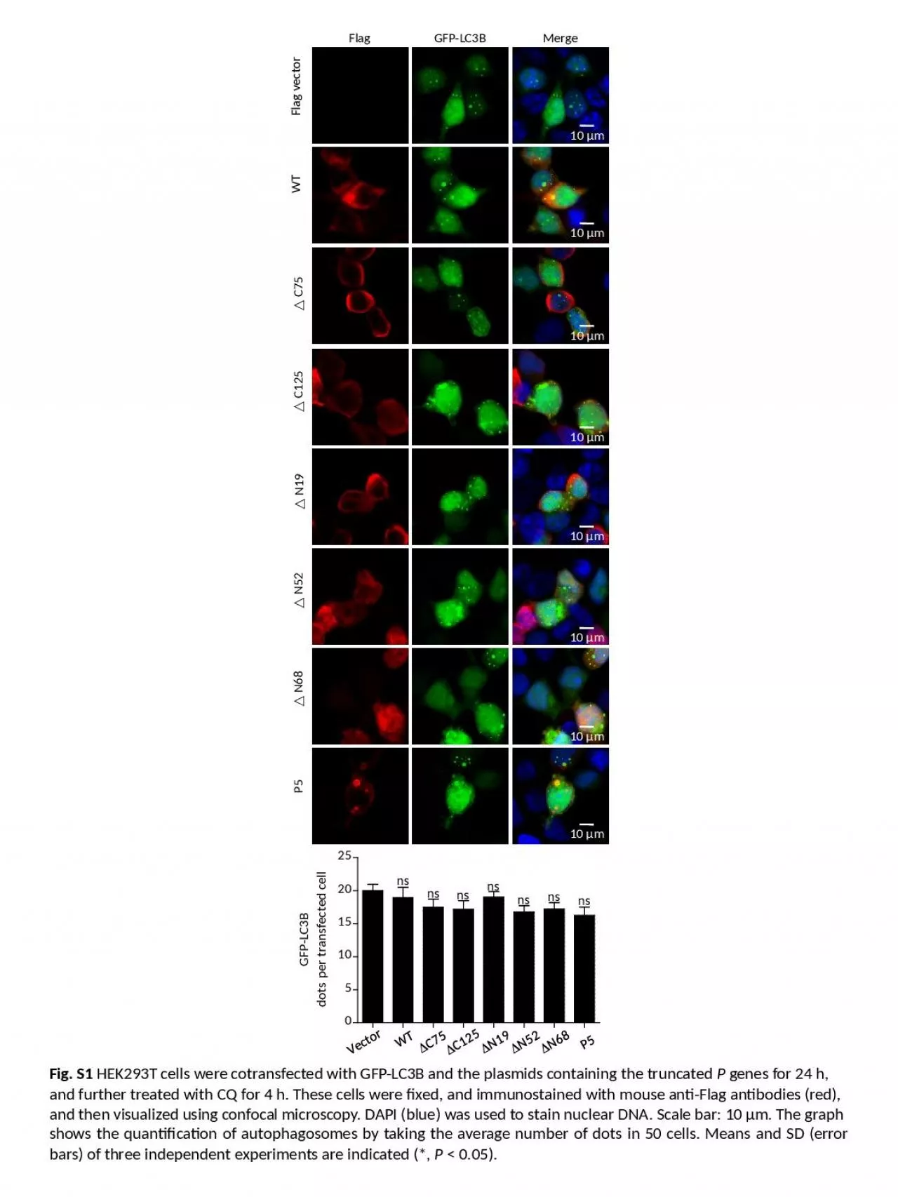
m 10 μ m 10 μ m 10 μ m 10 μ m 10 μ m Flag GFPLC3B Merge Flag vector WT C75 C125 N19 N52 N68 P5 Fig S1 HEK293T cells were ID: 920302

merge flag cells myc flag merge myc cells n68 n19 n52 truncated anti confocal blue cotransfected scale red fig

Share:

Link:

Embed:

Download Presentation from below link

Download Presentation The PPT/PDF document "10 μ m 10 μ m 10 μ" is the property of its rightful owner. Permission is granted to download and print the materials on this web site for personal, non-commercial use only, and to display it on your personal computer provided you do not modify the materials and that you retain all copyright notices contained in the materials. By downloading content from our website, you accept the terms of this agreement.


Presentation Transcript

Slide1

10

μ

m

10

μ

m

10

μ

m

10

μ

m

10

μ

m

10

μ

m

10

μ

m

10

μ

m

Flag

GFP-LC3B

Merge

Flag vector

WT

△ C75

C125

N19

N52

N68

P5

Fig. S1

HEK293T cells were

cotransfected

with GFP-LC3B and the plasmids containing the truncated

P

genes for 24 h,

and further treated with CQ for 4 h. These cells were fixed, and

immunostained

with mouse anti-Flag antibodies (red),

and then visualized using

confocal

microscopy. DAPI (blue) was used to stain nuclear DNA. Scale bar: 10

μm

. The graph

shows the quantification of

autophagosomes

by taking the average number of dots in 50 cells. Means and SD (error bars) of three independent experiments are indicated (*,

P

< 0.05).

Slide2

Flag-P

Myc

-

Δ

N52

Merge

Flag-

Δ

N19

Myc

-

Δ

N68

Merge

Flag-

Δ

N19

Myc

-

Δ

N52

Merge

Flag-P

Myc

-

Δ

N19

Merge

Flag-P

Myc

-

Δ

N68

Merge

Flag-

Δ

N19

Myc-

P5

Merge

Flag-

P

Myc-

P5

Merge

10

μ

m

Flag-

Δ

N68

Myc-P5

Merge

Flag-

ΔN52

Myc-P5

Merge

Flag-

ΔN52

Myc

-

Δ

N68

Merge

10

μ

m

10

μ

m

10

μ

m

10

μ

m

10

μ

m

10

μ

m

10

μ

m

10

μ

m

10

μ

m

Merge

Fig. S2

The truncated protein PΔN82 is required for a ring-like structure. N2a cells were

cotransfected

with Flag and

Myc

tagged plasmids encoding the truncated P genes for 24 h, fixed, and

immunostained

with mouse anti-Flag antibody (green) and rabbit anti-

Myc

(red), and then visualized by

confocal

microscopy. DAPI (blue) stained nuclear DNA. Scale bar: 10

μm

.

Slide3

GFP-LC3B

LAMP1

Merge

Flag-P5

+EBSS

Flag

Flag+EBSS

Flag-P5

Flag+CQ

Flag-P5+CQ

P5

10

μ

m

10

μ

m

10

μ

m

10

μ

m

10

μ

m

10

μ

m

Fig. S3

Autophagosomes

fail to fuse with

lysosomes

in Flag-P5-transfected cells. N2a cells were

cotransfected

with Flag-P5 and GFP-LC3B for 24 h, and were treated with EBSS or CQ for 4 h. Cells were

fixed

, and

immunostained

with rabbit anti-LAMP1

mAb

(red), and mouse anti-Flag

mAb

(blue), and observed using

confocal

microscopy to analyze fusion of

autophagosomes

with

lysosomes

. Scale bar: 10

μm

. The graph shows the quantification of

autolysosomes

by taking the average number of dots in 50 cells. Means and SD (error bars) of three independent experiments are indicated (*,

P

< 0.05; **,

P

< 0.01; ***,

P

< 0.001).

Slide4

10

μ

m

10

μ

m

10

μ

m

Fig. S4

The truncated P proteins

colocalize

with BECN1. N2a cells were

cotransfected

with the plasmids encoding the truncated P genes and Myc-BECN1 for 24 h, and Flag (green), BECN1 (red) and DAPI (blue) were detected by using the indicated antibodies in

confocal

microscopy. Scale bar: 10 μm.

Myc-BECN1

Merge

P△ C75

P

P△ C125

P△ N19

△ N52

△ N68

10

μ

m

10

μ

m

10

μ

m

10

μ

m

10

μ

m

10

μ

m

Flag

Related Contents


Next Show more