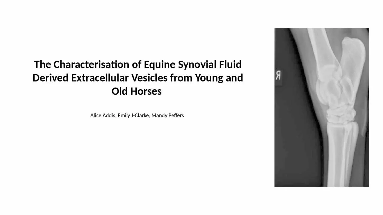
/
The Characterisation of Equine Synovial Fluid Derived Extracellular Vesicles from Young The Characterisation of Equine Synovial Fluid Derived Extracellular Vesicles from Young

The Characterisation of Equine Synovial Fluid Derived Extracellular Vesicles from Young - PowerPoint Presentation

badra
badra . @badra
Follow
64 views
Uploaded On 2024-01-29

The Characterisation of Equine Synovial Fluid Derived Extracellular Vesicles from Young - PPT Presentation

Alice Addis Emily JClarke Mandy Peffers Equine Osteoarthritis Lameness due to joint injury and disease is the most prevalent cause of diminished athletic function and wastage Osteoarthritic changes ID: 1041786

equine cartilage joint osteoarthritis cartilage equine osteoarthritis joint mir extracellular vesicles amp expression journal research veterinary lameness degradation age

Share:

Link:

Embed:

Download Presentation from below link

Download Presentation The PPT/PDF document "The Characterisation of Equine Synovial ..." is the property of its rightful owner. Permission is granted to download and print the materials on this web site for personal, non-commercial use only, and to display it on your personal computer provided you do not modify the materials and that you retain all copyright notices contained in the materials. By downloading content from our website, you accept the terms of this agreement.


Presentation Transcript

1. The Characterisation of Equine Synovial Fluid Derived Extracellular Vesicles from Young and Old Horses Alice Addis, Emily J-Clarke, Mandy Peffers

2. Equine OsteoarthritisLameness due to joint injury and disease is the most prevalent cause of diminished athletic function and wastageOsteoarthritic changes:Degeneration of the articular cartilageChanges within the subchondral bone, the synovial membrane and the joint capsuleSecondary effects to to adjoining connective tissue structuresAlter the joints ability to provide a smooth and nearly frictionless motion as the hose movesNo options for clinical resolution are available

3. Extracellular Vesicles (EVs)Nanoparticles enveloped in a phospholipid bilayer membraneParticipate in intracellular communications and are protective carriers for biologically active signalling molecules.Secreted by most mammalian cellsAct as biomarkers for the progression of disease How do their characteristics and their cargo change with age?

4. AIMS of the projectDo EV characteristics change with age?Mean EV sizeTotal Evs/ml of sampleD10:D50:D90 ratioDoes EV cargo change with age?Eca-mir199a-3pEca-mir-148aEca-mir-146a

5. Methods

6. Nanoparticle Tracking Analysis (NTA)Visualize EVs and provide data on their size and concentrationProperties of light scattering and Brownian motion provide the nanoparticle size distribution of the samples in liquid suspensionGaussian distribution using a Shapiro-wilk normality testIdentified outliers and removedUnpaired T-test using GraphPad PRISM (version 9.2.0)

7. Real time qPCRRNA extractioncDNA synthesisqPCR

8. Selection of miRNA primersMir148a-3pOverexpression promotes cartilage production and inhibits cartilage degradation Mir199a-3pEvidence of involvement in equine osteoarthritis physiopathologyAnabolic and catabolic regulation of key gene expression: MMP13, Adamt-5 or Col2A1 Mir146a-5pRole in reducing inflammationChondrogenic differentiation potential

9. 2^-(dCT) to calculate relative gene expression

10. Mir148a-3pAgeing and chondrocyte pathway involving the proliferation of cells and tumor lines Over expression protects cartilage from degradation and promotes its productionDisease modifying compound OA cartilage shows a decreased expression, as well as changes in cartilage matrix degradation and production

11. Mir199a-3p

12. Mir146a-5pTumour necrosis, osteoclastogenesis and inflammatory responses Classed as an inflammRs for its ability to mediate inflammatory pathwaysA biomarker of inflammation related to ageing Arthritic mouse model: IV administration resulted in suppression of bone and cartilage destruction

13. ConclusionThe size and concentration of EVs were the same when young and old samples were compared Older horses showed a trend of decreased expression of eca-mir199a-3p, eca-mir-148a, eca-mir-146aWhat is the therapeutic potential of these miRNAs?Could they be used as a biomarker for equine osteoarthritis?

14. References Balaskas, P. et al. (2020) 'Small Non-Coding RNAome of Ageing Chondrocytes', International Journal of Molecular Sciences, 21(16), pp. 0-22.Boere, J. (2017) 'EXTRACELLULAR VESICLES IN SYNOVIAL FLUID: Dynamics during joint inflammation and articular development and promise for joint regeneration and restoration of joint homeostasis', PhD thesis. Utrecht University. Available at: http://dspace.library.uu.nl/handle/1874/355071 (Accessed at 6 January 2022).Broeckx, SY. et al. (2019) 'The use of equine chondrogenic-induced mesenchymal stem cells as a treatment for osteoarthritis: A randomised, double-blinded, placebo-controlled proof-of-concept study', Equine Vet J, 51(6), pp. 787-794.Théry, C. et al (2018) 'Minimal information for studies of extracellular vesicles 2018 (MISEV2018): a position statement of the International Society for Extracellular Vesicles and update of the MISEV2014 guidelines', Journal of Extracellular Vesicles, 7(1), pp. 2-47.Desjardin, C. et al. (2014) 'Next-generation sequencing identifies equine cartilage and subchondral bone miRNAs and suggests their involvement in osteochondrosis physiopathology', BMC Genomics, 15(798), pp. 2-11.Gibson, G. & Asahara, H. (2013) 'microRNAs and Cartilage', Journal of Orthopaedic Research, 31(9), pp. 1333-1344.Goldring, S. R. & Goldring, M. B. (2006) 'Clinical aspects, pathology and pathophysiology of osteoarthritis.', Journal of Musculoskeletal and Neuronal Interactions, 6(4), pp. 376-378.Goodrich, L. R. & Nixon, J. A. (2006) 'Medical treatment of osteoarthritis in the horse – A review'. The Veterinary Journal, 171(3), pp. 51-69.Malda, J. et al. (2016) 'Extracellular vesicles — new tool for joint repair and regeneration', Nature Reviews Rheumatology, 12(4), pp. 243-249.McCoy, A. (2016) 'Recent Advances in Equine Osteoarthritis', University of Illinois College of Veterinary Medicine. Unpublished.Mirza, M. H. et al. (2016) 'Gait changes vary among horses with naturally occurring osteoarthritis following intra-articular administration of autologous platelet-rich plasma', Frontiers in Veterinary Science, 3(29), pp. 1-9.Neundorf, R. H. et al. (2010) 'Determination of the prevalence and severity of metacarpophalangeal joint osteoarthritis in Thoroughbred racehorses via quantitative macroscopic evaluation', American Journal of Veterinary Research, 71(11), pp. 1284-1293.Niemelä, T. M., Tulamo, R.-M. & Hielm-Björkman, A. K. (2016) 'A randomised, double-blinded, placebo- controlled clinical study on intra-articular hyaluronan treatment in equine lameness originating from the metacarpophalangeal joint', BMC Veterinary Research, 12(40), pp. 1-8.'Lameness' (2019) Available at: https://dictionary.cambridge.org/dictionary/english/lameness (Accessed 6 January 2021).Olivieri, F. et al. (2021) 'miR-21 and miR-146a: The microRNAs of inflammaging and age-related diseases', Ageing Research Reviews, 70(2), pp. 1-19.Pauley, K. M. & Cha, S. (2011) 'MicroRNA-146a in rheumatoid arthritis: a new therapeutic strategy', Immunotherapy, 3(7), p. 829–831.Pauley, K. M. et al. (2008) 'Upregulated miR-146a expression in peripheral blood mononuclear cells from rheumatoid arthritis patients', Arthritis Research and Therapy, 10(4), pp. 1-10.Robbins, P. D. (2017) 'Extracellular vesicles and aging', Stem Cell Investigation, 4(98), pp. 1-7.United States Department of Agriculture (2000) Lameness and laminitis in U.S. horses. Available at: http://www.aphis.usda.gov/animal_health/nahms/equine/downloads/equine98/Equine98_dr_Lameness.pdf (Accessed: 10 December 2021).Vonk, L. et al. (2014) 'Overexpression of hsa-miR-148a promotes cartilage production and inhibits cartilage degradation by osteoarthritic chondrocytes', Osteoarthritis and Cartilage, 22(5), pp. 145-153.Weeren, P. R. v. & Brama, P. A. J. (2001) 'Physiology and pathology of the equine joint', Pferdeheilkunde, 17(4), pp. 307-318.