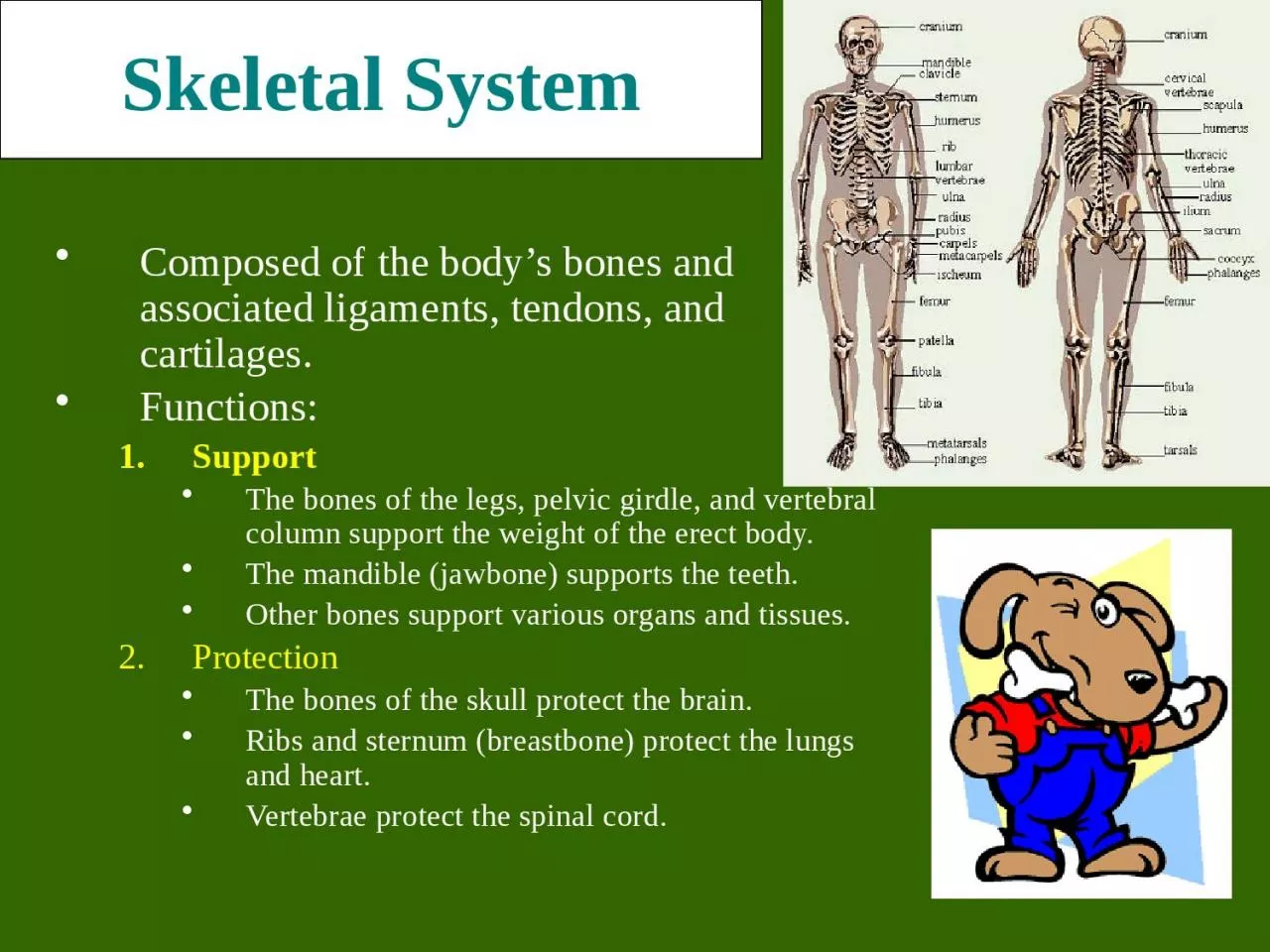
/
Skeletal System Composed of the body’s bones and associated ligaments, tendons, and Skeletal System Composed of the body’s bones and associated ligaments, tendons, and

Skeletal System Composed of the body’s bones and associated ligaments, tendons, and - PowerPoint Presentation

barbara
barbara . @barbara
Follow
27 views
Uploaded On 2024-02-09

Skeletal System Composed of the body’s bones and associated ligaments, tendons, and - PPT Presentation

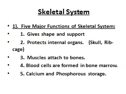
Functions Support The bones of the legs pelvic girdle and vertebral column support the weight of the erect body The mandible jawbone supports the teeth Other bones support various organs and tissues ID: 1044971

bones bone blood tissue bone bones tissue blood matrix osteoblasts cartilage spongy marrow ossification cells compact growth activity shaft

Share:

Link:

Embed:

Download Presentation from below link

Download Presentation The PPT/PDF document "Skeletal System Composed of the body’s..." is the property of its rightful owner. Permission is granted to download and print the materials on this web site for personal, non-commercial use only, and to display it on your personal computer provided you do not modify the materials and that you retain all copyright notices contained in the materials. By downloading content from our website, you accept the terms of this agreement.


Presentation Transcript

1. Skeletal SystemComposed of the body’s bones and associated ligaments, tendons, and cartilages.Functions:SupportThe bones of the legs, pelvic girdle, and vertebral column support the weight of the erect body.The mandible (jawbone) supports the teeth.Other bones support various organs and tissues.Protection The bones of the skull protect the brain.Ribs and sternum (breastbone) protect the lungs and heart.Vertebrae protect the spinal cord.

2. Skeletal SystemFunctions:Movement Skeletal muscles use the bones as levers to move the body.Reservoir for minerals and adipose tissue99% of the body’s calcium is stored in bone.85% of the body’s phosphorous is stored in bone.Adipose tissue is found in the marrow of certain bones.Hematopoiesis A.k.a. blood cell formation.All blood cells are made in the marrow of certain bones.

3. Interesting Facts· The human hand has 27 bones; your face has 14!· The longest bone in your body? Your thigh bone, the femur – it' s about 1/4 of your height. The smallest is the stirrup bone in the ear which canmeasure 1/10 of an inch.· Did you know that humans and giraffes have the same number of bones in their necks? Giraffe neck vertebrae are just much, much longer!· You have over 230 moveable and semi-moveable joints in your body. When you were born you had over 300 bones. As you grew, some of these bones began to fuse together. The result? An adult has only 206 bones!

4. Bone ClassificationThere are 206 named bones in the human body.Each belongs to one of 2 large groups:Axial skeletonForms long axis of the body.Includes the bones of the skull, vertebral column, and rib cage.These bones are involved in protection, support, and carrying other body parts.Appendicular skeletonBones of upper & lower limbs and the girdles (shoulder bones and hip bones) that attach them to the axial skeleton.Involved in locomotion and manipulation of the environment.

5. Clinical ConditionsOsteomalaciaLiterally “soft bones.”Includes many disorders in which osteoid is produced but inadequately mineralized.Causes can include insufficient dietary calciumInsufficient vitamin D fortification or insufficient exposure to sun light.RicketsChildren's form of osteomalaciaMore detrimental due to the fact that their bones are still growing.Signs include bowed legs, and deformities of the pelvis, ribs, and skull.What about the above x-ray is indicative of rickets?

6. Clinical ConditionsOsteomyelitisOsteo=bone + myelo=marrow + itis=inflammation.Inflammation of bone and bone marrow caused by pus-forming bacteria that enter the body via a wound (e.g., compound fracture) or migrate from a nearby infection.Fatal before the advent of antibiotics.

7. Clinical ConditionsOsteoporosisGroup of diseases in which bone resorption occurs at a faster rate than bone deposition.Bone mass drops and bones become increasingly porous.Compression fractures of the vertebrae and fractures of the femur are common.Often seen in postmenopausal women because they experience a rapid decline in estrogen secretion; estrogen stimulates osteoblast and inhibits osteoclast activity.Based on the above, what preventative measures might you suggest?

8. Clinical ConditionsGigantismChildhood hypersecretion of growth hormone by the pituitary gland causes excessive growth.Acromegaly Adulthood hypersecretion of GH causes overgrowth of bony areas still responsive to GH such as the bones of the face, feet, and hands.Pituitary dwarfismGH deficiency in children resulting in extremely short long bones and maximum stature of 4 feet.

9. Bone Classification4 types of bones:Long Bones Much longer than they are wide.All bones of the limbs except for the patella (kneecap), and the bones of the wrist and ankle.Consists of a shaft plus 2 expanded ends.Your finger bones are long bones even though they’re very short – how can this be?Short BonesRoughly cube shaped.Bones of the wrist and the ankle.Femur Carpal Bones

10. Bone ClassificationTypes of bones:Flat BonesThin, flattened, and usually a bit curved.Scapulae, sternum, (shoulder blades), ribs and most bones of the skull.Irregular BonesHave weird shapes that fit none of the 3 previous classes.Vertebrae, hip bones, 2 skull bones ( sphenoid and the ethmoid bones).SternumSphenoid Bone

11. Bone StructureBones are organs. Thus, they’re composed of multiple tissue types. Bones are composed of:Bone tissue (a.k.a. osseous tissue).Fibrous connective tissue.Cartilage.Vascular tissue.Lymphatic tissue.Adipose tissue.Nervous tissue.

12. All bones consist of a dense, solid outer layer known as compact bone and an inner layer of spongy bone – a honeycomb of flat, needle-like projections called trabeculae.Bone is an extremely dynamic tissue!!!!Above: Note the relationship btwn the compact and spongy bone.Below: Close up of spongy bone.

13. Note the gross differences between the spongy bone and the compact bone in the above photo.Do you see the trabeculae?

14. Compare compact and spongy bone as viewed with the light microscope

15. Bone StructureBone tissue is a type of connective tissue, so it must consist of cells plus a significant amount of extracellular matrix.Bone cells:OsteoblastsBone-building cells.Synthesize and secrete collagen fibers and other organic components of bone matrix. Initiate the process of calcification.Found in both the periosteum and the endosteumThe blue arrows indicate the osteoblasts. The yellow arrows indicate the bone matrix they’ve just secreted.

16. Bone Structure2. OsteocytesMature bone cells.Osteoblasts that have become trapped by the secretion of matrix.No longer secrete matrix.Responsible for maintaining the bone tissue.Yellow arrows indicate osteocytes – notice how they are surrounded by the pinkish bone matrix.Blue arrow shows an osteoblast in the process of becoming an osteocyte.On the right, notice how the osteocyte is “trapped” within the pink matrix

17. 3. OsteoclastsHuge cells derived from the fusion of as many as 50 monocytes (a type of white blood cell).Cells that digest bone matrix – this process is called bone resorption and is part of normal bone growth, development, maintenance, and repair.Concentrated in the endosteum.On the side of the cell that faces the bone surface, the PM is deeply folded into a ruffled border. Here, the osteoclast secretes digestive enzymes (how might this occur?) to digest the bone matrix. It also pumps out hydrogen ions (how might this occur?) to create an acid environment that eats away at the matrix. What advantage might a ruffled border confer?Why do we want a cell that eats away at bone? (Hint: bone is a very dynamic tissue.)

18. Here, we see a cartoon showing all 3 cell types. Osteoblasts and osteoclasts are indicated. Note the size of the osteoclast (compare it to the osteoblast), and note the ruffled border. Why is there a depression underneath the osteoclast? What is the name of the third cell type shown here? What do you think the tan material represents?

19. Bone StructureBone Matrix:Consists of organic and inorganic components.1/3 organic and 2/3 inorganic by weight.Organic component consists of several materials that are secreted by the osteoblasts:Collagen fibers and other organic materialsThese (particularly the collagen) provide the bone with resilience and the ability to resist stretching and twisting.

20. Inorganic component of bone matrixConsists mainly of 2 salts: calcium phosphate and calcium hydroxide. These 2 salts interact to form a compound called hydroxyapatite.Bone also contains smaller amounts of magnesium, fluoride, and sodium.These minerals give bone its characteristic hardness and the ability to resist compression.Three-dimensional array of collagen molecules. The rod-shaped molecules lie in a staggered arrangement which acts as a template for bone mineralization. Bone mineral is laid down in the gaps. Note collagen fibers in longitudinal & cross section and how they occupy space btwn the black bone cells.

21. This bone: a. Has been demineralized b. Has had its organic component removed

22. Long Bone StructureShaft plus 2 expanded ends.Shaft is known as the diaphysis.Consists of a thick collar of compact bone surrounding a central marrow cavityIn adults, the marrow cavity contains fat - yellow bone marrow.Expanded ends are epiphysesThin layer of compact bone covering an interior of spongy bone.Joint surface of each epiphysis is covered w/ a type of hyaline cartilage known as articular cartilage. It cushions the bone ends and reduces friction during movement.

23. Long Bone StructureThe external surface of the entire bone except for the joint surfaces of the epiphyses is covered by a double-layered membrane known as the periosteum.Outer fibrous layer is dense irregular connective tissue.Inner cellular layer contains osteoprogenitor cells and osteoblasts.Periosteum is richly supplied with nerve fibers, lymphatic vessels and blood vessels.These enter the bone of the shaft via a nutrient foramen.Periosteum is connected to the bone matrix via strong strands of collagen.

24. Long Bone StructureInternal bone surfaces are covered with a delicate connective tissue membrane known as the endosteum.Covers the trabeculae of spongy bone in the marrow cavities and lines the canals that pass through compact bone.Contains both osteoblasts and osteoclasts.

25. Structure of Short, Irregular, and Flat BonesThin plates of periosteum-covered compact bone on the outside and endosteum-covered spongy bone within.Have no diaphysis or epiphysis because they are not cylindrical.Contain bone marrow between their trabeculae, but no marrow cavity.In flat bones, the internal spongy bone layer is known as the diploë, and the whole arrangement resembles a stiffened sandwich.

26. Bone MarrowBone marrow is a general term for the soft tissue occupying the medullary cavity of a long bone, the spaces amid the trabeculae of spongy bone, and the larger haversian canals.There are 2 main types: red & yellow.Red bone marrow = blood cell forming tissue = hematopoietic tissueRed bone marrow looks like blood but with a thicker consistency. It consists of a delicate mesh of reticular tissue saturated with immature red blood cells and scattered adipocytes.Notice the red marrow and the compact bone

27. Distribution of MarrowIn a child, the medullary cavity of nearly every bone is filled with red bone marrow.In young to middle-aged adults, the shafts of the long bones are filled with fatty yellow bone marrow.Yellow marrow no longer produces blood, although in the event of severe or chronic anemia, it can transform back into red marrowIn adults, red marrow is limited to the axial skeleton, pectoral girdle, pelvic girdle, and proximal heads of the humerus and the femur.Note the compact bone on the bottom and marrow on the bottom.

28. Microscopic Structure of Compact BoneConsists of multiple cylindrical structural units known as osteons or haversian systems.Imagine these osteons as weight-bearing pillars that are arranged parallel to one another along the long axis of a compact bone.The diagram below represents a long bone shaft in cross-section. Each yellow circle represents an osteon. The blue represents additional matrix filling in the space btwn osteons. The white in the middle is the marrow cavity.

29. OsteonsEach osteon consists of a single central canal, known as a haversian canal, surrounded by concentric layers of calcified bone matrix.Haversian canals allow the passage of blood vessels, lymphatic vessels, and nerve fibers.Each of the concentric matrix “tubes” that surrounds a haversian canal is known as a lamella.All the collagen fibers in a particular lamella run in a single direction, while collagen fibers in adjacent lamellae will run in the opposite direction. This allows bone to better withstand twisting forces.

30. Running perpendicular to the haversian canals are Volkmann’s canals. They connect the blood and nerve supply in the periosteum to those in the haversian canals and the medullary cavity.

31. OsteonsLying in between intact osteons are incomplete lamellae called interstitial lamellae. These fill the gaps between osteons or are remnants of bone remodeling.There are also circumferential lamellae that extend around the circumference of the shaft. There are inner circumferential lamellae surrounding the endosteum and outer circumferential lamellae just inside the periosteum.

32. Spider-shaped osteocytes occupy small cavities known as lacunae at the junctions of the lamellae. Hairlike canals called canaliculi connect the lacunae to each other and to the central canal.Canaliculi allow the osteocytes to exchange nutrients, wastes, and chemical signals to each other via intercellular connections known as gap junctions.

33.

34. Here, we have a close up and a far away view of compact bone. You should be able to identify haversian canals, concentric lamellae, interstitial lamellae, lacunae, and canaliculi.

35. Microscopic Structure of Spongy BoneAppears poorly organized compared to compact bone.Lacks osteons.Trabeculae align along positions of stress and exhibit extensive cross-bracing.Trabeculae are a few cell layers thick and contain irregularly arranged lamellae and osteocytes interconnected by canaliculi.No haversian or Volkmann’s canals are necessary. Why?

36. Bone DevelopmentOsteogenesis (a.k.a. ossification) is the process of bone tissue formation.In embryos this leads to the formation of the bony skeleton.In children and young adults, ossification occurs as part of bone growth.In adults, it occurs as part of bone remodeling and bone repair.

37. Formation of the Bony SkeletonBefore week 8, the human embryonic skeleton is made of fibrous membranes and hyaline cartilage.After week 8, bone tissue begins to replace the fibrous membranes and hyaline cartilage.The development of bone from a fibrous membrane is called intramembranous ossification. Why?The replacement of hyaline cartilage with bone is known as endochondral ossification. Why?

38. Intramembranous OssificationSome bones of the skull (frontal, parietal, temporal, and occipital bones), the facial bones, the clavicles, the pelvis, the scapulae, and part of the mandible are formed by intramembranous ossification Prior to ossification, these structures exist as fibrous membranes made of embryonic connective tissue known as mesenchyme.

39. Mesenchymal cells first cluster together and start to secrete the organic components of bone matrix which then becomes mineralized through the crystallization of calcium salts. As calcification occurs, the mesenchymal cells differentiate into osteoblasts.The location in the tissue where ossification begins is known as an ossification center.Some osteoblasts are trapped w/i bony pockets. These cells differentiate into osteocytes.

40. The developing bone grows outward from the ossification center in small struts called spicules.Mesenchymal cell divisions provide additional osteoblasts.The osteoblasts require a reliable source of oxygen and nutrients. Blood vessels trapped among the spicules meet these demands and additional vessels branch into the area. These vessels will eventually become entrapped within the growing bone.

41. Initially, the intramembranous bone consists only of spongy bone. Subsequent remodeling around trapped blood vessels can produce osteons typical of compact bone.As the rate of growth slows, the connective tissue around the bone becomes organized into the fibrous layer of the periosteum. Osteoblasts close to the bone surface become the inner cellular layer of the periosteum.

42. Endochondral OssificationBegins with the formation of a hyaline cartilage model which will later be replaced by bone.Most bones in the body develop via this model.More complicated than intramembranous because the hyaline cartilage must be broken down as ossification proceeds.We’ll follow limb bone development as an example.

43. Endochondral Ossification – Step 1Chondrocytes near the center of the shaft of the hyaline cartilage model increase greatly in size. As these cells enlarge, their lacunae expand, and the matrix is reduced to a series of thin struts. These struts soon begin to calcify.The enlarged chondrocytes are now deprived of nutrients (diffusion cannot occur through calcified cartilage) and they soon die and disintegrate.

44. Endochondral Ossification – Step 2Blood vessels grow into the perichondrium surrounding the shaft of the cartilage. The cells of the inner layer of the perichondrium in this region then differentiate into osteoblasts.The perichondrium is now a periosteum and the inner osteogenic layer soon produces a thin layer of bone around the shaft of the cartilage. This bony collar provides support.

45. Endochondral Ossification – Step 3Blood supply to the periosteum, and capillaries and fibroblasts migrate into the heart of the cartilage, invading the spaces left by the disintegrating chondrocytes. The calcified cartilaginous matrix breaks down; the fibroblasts differentiate into osteoblasts that replace it with spongy bone.Bone development begins at this primary center of ossification and spreads toward both ends of the cartilaginous model.While the diameter is small, the entire diaphysis is filled with spongy bone.Notice the primary ossification centers in the thigh and forearm bones of the above fetus.

46. Endochondral Ossification – Step 4The primary ossification center enlarges proximally and distally, while osteoclasts break down the newly formed spongy bone and open up a medullary cavity in the center of the shaft.As the osteoblasts move towards the epiphyses, the epiphyseal cartilage is growing as well. Thus, even though the shaft is getting longer, the epiphyses have yet to be transformed into bone.

47. Endochondral Ossification – Step 5Around birth, most long bones have a bony diaphysis surrounding remnants of spongy bone, a widening medullary cavity, and 2 cartilaginous epiphyses.At this time, capillaries and osteoblasts will migrate into the epiphyses and create secondary ossification centers. The epiphysis will be transformed into spongy bone. However, a small cartilaginous plate, known as the epiphyseal plate, will remain at the juncture between the epiphysis and the diaphysis.Articular cartilageEpiphyseal plate

48.

49. Growth in Bone LengthEpiphyseal cartilage (close to the epiphysis) of the epiphyseal plate divides to create more cartilage, while the diaphyseal cartilage (close to the diaphysis) of the epiphyseal plate is transformed into bone. This increases the length of the shaft.

50. As a result osteoblasts begin producing bone faster than the rate of epiphyseal cartilage expansion. Thus the bone grows while the epiphyseal plate gets narrower and narrower and ultimately disappears. A remnant (epiphyseal line) is visible on X-rays (do you see them in the adjacent femur, tibia, and fibula?)At puberty, growth in bone length is increased dramatically by the combined activities of growth hormone, thyroid hormone, and the sex hormones.

51. Growth in Bone ThicknessOsteoblasts beneath the periosteum secrete bone matrix on the external surface of the bone. This obviously makes the bone thicker.At the same time, osteoclasts on the endosteum break down bone and thus widen the medullary cavity.This results in an increase in shaft diameter even though the actual amount of bone in the shaft is relatively unchanged.

52. FracturesDespite its mineral strength, bone may crack or even break if subjected to extreme loads, sudden impacts, or stresses from unusual directions.The damage produced constitutes a fracture.The proper healing of a fracture depends on whether or not, the blood supply and cellular components of the periosteum and endosteum survive.

53. Fracture RepairStep 1:Immediately after the fracture, extensive bleeding occurs. Over a period of several hours, a large blood clot, or fracture hematoma, develops.Bone cells at the site become deprived of nutrients and die. The site becomes swollen, painful, and inflamed.Step 2:Granulation tissue is formed as the hematoma is infiltrated by capillaries and macrophages, which begin to clean up the debris. Some fibroblasts produce collagen fibers that span the break , while others differentiate into chondroblasts and begin secreting cartilage matrix.C. Osteoblasts begin forming spongy bone.D. This entire structure is known as a fibrocartilaginous callus and it splints the broken bone.

54. Step 3:Bone trabeculae increase in number and convert the fibrocartilaginous callus into a bony callus of spongy bone. Typically takes about 6-8 weeks for this to occur.Fracture RepairStep 4:During the next several months, the bony callus is continually remodeled. Osteoclasts work to remove the temporary supportive structures while osteoblasts rebuild the compact bone and reconstruct the bone so it returns to its original shape/structure.

55. Fracture TypesFractures are often classified according to the position of the bone ends after the break:Open (compound)  bone ends penetrate the skin.Closed (simple)  bone ends don’t penetrate the skin.Comminuted  bone fragments into 3 or more pieces. Common in the elderly (brittle bones).Greenstick  bone breaks incompletely. One side bent, one side broken. Common in children whose bone contains more collagen and are less mineralized.Spiral  ragged break caused by excessive twisting forces. Sports injury/Injury of abuse.Impacted  one bone fragment is driven into the medullary space or spongy bone of another.

56.

57. What kind of fracture is this?It’s kind of tough to tell, but this is a _ _ _ _ _ _ fracture.

58. Bone RemodelingBone is a dynamic tissue. What does that mean?Wolff’s law holds that bone will grow or remodel in response to the forces or demands placed on it. Examine this with the bone on the left.

59. Check out the mechanism of remodeling on the right!Why might you suspect someone whose been a powerlifter for 15 years to have heavy, massive bones, especially at the point of muscle insertion?Astronauts tend to experience bone atrophy after they’re in space for an extended period of time. Why?

60. Nutritional Effects on BoneNormal bone growth/maintenance cannot occur w/o sufficient dietary intake of calcium and phosphate salts.Calcium and phosphate are not absorbed in the intestine unless the hormone calcitriol is present. Calcitriol synthesis is dependent on the availability of the steroid cholecalciferol (a.k.a. Vitamin D) which may be synthesized in the skin or obtained from the diet.Vitamins C, A, K, and B12 are all necessary for bone growth as well.

61. Hormonal Effects on BoneGrowth hormone, produced by the pituitary gland, and thyroxine, produced by the thyroid gland, stimulate bone growth.GH stimulates protein synthesis and cell growth throughout the body.Thyroxine stimulates cell metabolism and increases the rate of osteoblast activity. In proper balance, these hormones maintain normal activity of the epiphyseal plate (what would you consider normal activity?) until roughly the time of puberty.

62. Hormonal Effects on BoneAt puberty, the rising levels of sex hormones (estrogens in females and androgens in males) cause osteoblasts to produce bone faster than the epiphyseal cartilage can divide. This causes the characteristic growth spurt as well as the ultimate closure of the epiphyseal plate.Estrogens cause faster closure of the epiphyseal growth plate than do androgens. Estrogen also acts to stimulate osteoblast activity.

63. Hormonal Effects on BoneOther hormones that affect bone growth include insulin and the glucocorticoids.Insulin stimulates bone formationGlucocorticoids inhibit osteoclast activity.Parathyroid hormone and calcitonin are 2 hormones that antagonistically maintain blood [Ca2+] at homeostatic levels. Since the skeleton is the body’s major calcium reservoir, the activity of these 2 hormones affects bone resorption and deposition.

64. CalcitoninReleased by the C cells of the thyroid gland in response to high blood [Ca2+].Calcitonin acts to “tone down” blood calcium levels.Calcitonin causes decreased osteoclast activity which results in decreased break down of bone matrix and decreased calcium being released into the blood.Calcitonin also stimulates osteoblast activity which means calcium will be taken from the blood and deposited as bone matrix.Notice the thyroid follicles on the right. The arrow indicates a C cell

65. Calcitonin Negative Feedback LoopIncreased Blood [Ca2+]Increased calcitonin release from thyroid C cells.Increased osteoblast activityDecreased osteoclast activity

66. Parathyroid HormonePTH increases calcitriol synthesis which increases Ca2+ absorption in the small intestine.PTH decreases urinary Ca2+ excretion and increases urinary phosphate excretion. Released by the cells of the parathyroid gland in response to low blood [Ca2+].Causes blood [Ca2+] to increase.PTH will bind to osteoblasts and this will cause 2 things to occur:The osteoblasts will decrease their activity and they will release a chemical known as osteoclast-stimulating factor. Osteoclast-stimulating factor will increase osteoclast activity.

67. Increased PTH release by parathyroid glandBinds to osteoblast causing decreased osteoblast activity and release of osteoclast-stimulating factorOSF causes increased osteoclast activityDecreased bone deposition and increased bone resorptionIncreased calcitriol synthesisIncreased intestinal Ca2+ absorptionDecreased Ca2+ excretionIncreased Blood [Ca2+]Decreased Blood [Ca2+]