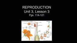
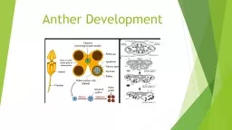
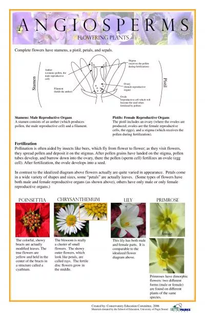
/
Structure  of mature anther, pollen Structure  of mature anther, pollen

Structure of mature anther, pollen - PowerPoint Presentation

barbara
barbara . @barbara
Follow
67 views
Uploaded On 2023-10-30

Structure of mature anther, pollen - PPT Presentation

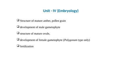
grain development of male gametophyte structure of mature ovule development of female gametophyte Polygonam type only fertilization Unit IV Embryology a The Stamen Stamen in a flower consists of two parts the long narrow stalk like filament and upper broader knobl ID: 1027142

cells pollen ovule cell pollen cells cell ovule anther wall layer sac type called embryo nucleus mother form structure

Share:

Link:

Embed:

Download Presentation from below link

Download Presentation The PPT/PDF document "Structure of mature anther, pollen" is the property of its rightful owner. Permission is granted to download and print the materials on this web site for personal, non-commercial use only, and to display it on your personal computer provided you do not modify the materials and that you retain all copyright notices contained in the materials. By downloading content from our website, you accept the terms of this agreement.


Presentation Transcript

1. Structure of mature anther, pollen graindevelopment of male gametophytestructure of mature ovule,development of female gametophyte (Polygonam type only)fertilizationUnit - IV (Embryology)

2. a) The Stamen:Stamen in a flower consists of two parts, the long narrow stalk like filament and upper broader knob-like bi-lobed antherThe proximal end of the filament is attached to the thalamus or petal of the flower. The number and length of stamens vary in different species.b) Structure of anther:A normal bithecous or dithecous anther is made up of two anther lobes, which are connected by a strip of sterile part called connective. Two anther lobes contain four elongated cavities or pollen sacs (microsporangia)Structure of mature anther

3. c) Structure of microsporangium (pollen sac):Young anther while it is still in flower bud in T.S. reveals the presence of outermost epidermis. The outermost wall layer lying just below the epidermis is called endothecium or fibrous layer (Fig. 2.5 C), because wall (two radial and inner) develop fibrous thickenings on them except at the junctions of two pollen sacs. Below the endothecium, there are 1-3 middle layers of parenchyma cells.The cells of innermost wall layer are radially elongated and rich in protoplasmic contents. This layer is called tapetum. The tapetum forms the nutritive tissue nourishing the developing microspores. The cells of tapetum may be multinucleate or may have large polyploid nucleus. The tapetal cells provide nourishment to young microspore mother cells either by forming a plasmodium (amoeboid or invasive type) or through diffusion (parietal or secretory type).The pollen sac wall encloses a number of archesporial cells that further forms microspore mother cells (microsporocytes). In the beginning microspore mother cells are polygonal and closely packed, but as the anther enlarges, the pollen sac becomes spacious and gets loosely arranged. A few microspore mother cells become non- functional and are finally absorbed by developing microspores.

4.

5. d) Structure of microspore (Pollen grain):Pollen grains develop from the diploid microspore mother cells in pollen sacs of anthers. Typically, pollen grain is a haploid, unicellular body with a single nucleus. Pollen grains are generally spherical measuring about 25-30 micrometeres in diameter. The outer surface of microspores may have spines, ridges or furrows which may vary in other ways in different species.There may be oval, ellipsoidal, triangular, lobed or even crescent-shaped pollen grains. The cytoplasm is surrounded by a two layered wall. The outer layer exine is thick and sculptured or smooth. It is cuticularised and the cutin is of special type called sporopollenin which is resistant to chemical and biological decomposition. In insect pollinated pollen grains, the exine is covered by a yellowish, viscous and sticky substance called pollen kit.Pollen grains are well preserved as fossils because of the presence of sporopollenin. At certain places the exine remains tirin. The thin areas are known as germ pores, when they are circular in outline and germ furrows when they are elongated. The cytoplasm is rich in starch and unsaturated oils.

6. Development of anther (microsporangium):(a) Development of micro-sporangia is eusporangiate type (i.e, from a group of initial cells)(b) Few cells in the hypodermal region become differentiated as archesporial cells. In Boerhavia and Dionaea, there is only one archesporial cell.(c) The archesporial cell divides periclinally (along the periphery) to form outer – primary parietal layer and inner – sporogenous layer.(d) The primary parietal layer lies just beneath the epidermis and divides again periclinally to form 3-5 concentric layers. These layers give raise the wall of the sporangium, along with epidermis.(e) The innermost layer of the wall is called tapetum, which serves to provide nourishment to the developing pollen grains.(f) The layer just below the epidermis is called endothecium.The sporogenous layer may function directly as pollen mother cell or it may divide to form many pollen mother cells.

7.

8. Anther wall: a) The mature anther wall comprises of an epidermis, followed by an endothecium, 2-3 middle layers and innermost tapteum.(b) Endothecium consists of radially elongated cells, which possess fibrous bands and these are hygroscopic (moisture absorbing) in nature. These help in dehiscence of anther (splitting of anther to release spores).Tapetum:It is the innermost nourishing layer of the anther wall present below the middle layer. It is usually single layered and is rich in reserve food material.

9. The ovule is part of the makeup of the female reproductive organ in seed plants. It’s the place where female reproductive cells are made and contained, and it is what eventually develops into a seed after fertilization, only for the seed to then ripen and produce a complete adult plant. Ovules are contained in ovaries at the bottom of a vase-like structure, the carpel, which has a neck called a style and an opening at the top, called a stigma.Components of OvulesThe ovule is made up of the nucellus, the integuments that form the outermost layer, and the female gametophyte (called an embryo sac in flowering plants), which are found at the very center.The NucellusThe nucellus is the largest part of the ovule. It houses the embryo sac as well as nutritive tissue and actually remains present in some flowering plants after fertilization as a source of nutrients for the embyo.Structure of mature ovule

10. Types of OvulesOvules have been separated into six categories based on their shapes:Orthotropous (Atropous): This is where the body of these ovules is straight so that the chalaza, where the nucellus and integuments merge, the funicle, which attaches the ovule to the placenta, and the micropyle are all aligned.Anatropous: In this case, the ovules become completely inverted during development so that the micropyle lies close to the hilum. The hilum is a scar that marks the point where the seed was attached to the fruit wall by the funicle.Hemi-anatropous: The body of these ovules becomes at a right angle in relation to the funicle, so it looks like the ovule is lying on its side.Campylotropous: The body of this type is bent and the alignment between the chalaza and micropyle is lost. The embryo sac is only slightly curved.Amphitropous: The body of the ovule is very much curved that the embryo sac and the ovule itself take the shape of a horseshoe.Circinotropous: The funicle in this case is especially long that it creates a nearly full circle around the ovule whose micropyle is ultimately pointing upwards.

11. Development of female gametophyte The ovule or the megasporangium develops as a small protuberance of the placental tissue. In the very young ovule a single hypodermal cell is differentiated as the archesporium.This archesporium cell may or may not cut off some parietal cells and then becomes the megaspore mother cell. The megaspore mother cell now undergoes meiosis or reduction division, and, usually, a linear row of four haploid megaspore cells (‘linear tetrad’) is formed.Meanwhile, two integuments develop from the base of the ovule. Of the linear tetrad of megaspores, usually the lower­ most one enlarges and becomes the functional megaspore while the three on top disinte­grate.The functional megaspore now develops the female gametophyte or the embryo sac.In Angiosperms, the development of the female gametophyte is completely endosporous, i.e., within the megaspore. In a typical case, the nucleus of the embryo sac, which is the same as the functional megaspore, divides into two, then four and finally, eight daughter nuclei four of which are located at each pole.

12.

13. Then, one nucleus from each pole moves to the centre of the embryo sac and fuses there forming the fusion or secondary nucleus.Finally, the embryo sac or the female gametophyte becomes organized.Three nuclei at the base form the antipodal cells. The secondary nucleus remains at the centre.On the top, three cells from the egg apparatus which consists of two flask-shaped synergids and a round egg cell (or ovum or oosphere) hanging between and below them.The synergids usually are somewhat notched by an indentation and they show striations at the tip (‘filiform apparatus’).There is prominent vacuolation below the synergid nuclei and above the egg nucleus, showing accumulation of cytoplasm at different regions.While the above method of gametophyte formation is usually described as the typical or normal type it has been found that many Angiosperms do not conform to this rule but show variations.

14.

15. FertilizationFertilization, the pollen tube has to enter the ovule and eventually the embryo sac (female gametophyte) before delivering the male gametes for fertilization.The egg apparatus, located at the micropylar end, is made up of the central egg and the two surrounding synergids.The three antipodal cells are located at the opposite end. The two sets are separated by a large central cell with two nuclei; the nuclei fuse to form the polar nucleus before fertilization.Pollen tube enters one of the synergids and discharges the two male gametes. One of them fuses with the egg to give rise to the zygote and the other fuses with the polar nucleus of the central cell to give rise to the primary endosperm cell.These two fusion events, termed double fertilization are unique to flowering plants. The zygote develops into the embryo and the primary endosperm cell develops into the endosperm which nourishes the embryo.The ovule develops into the seed and the ovary into the fruit. This completes sexual reproduction in flowering plants.

16.

17. Structure of Pollen Grain and Pistil:It is necessary to give a brief account of the structural details of the pollen grain and the pistil which are relevant to pollen screening.Pollen grains, when shed, are made up of two cells – a vegetative cell and a generative cell or three cells – a vegetative cell and the two male gametes.In two celled pollen, the generative cell divides to form the two male gametes during pollen tube growth in the pistil.Pollen wall is very complex; it is made up of two layers.The inner intine, comparable to the cellulosic wall of any other plant cell and the outer exine made up of highly resistant material, the sporopollenin. Both intine and exine contain extracellular components which are highly heterogeneous; they contain proteins, glycoproteins, carbohydrates, lipids and few other compounds in small quantities.

18. The stigma shows great variation in its morphology. Stigmas of different species can be grouped into two categories; the dry type in which the stigma surface is free from any visible exudate and the wet type in which the surface is covered with exudate of varying quantity.Each of them can be divided into several groups. Irrespective of its morphological variations, stigma surface invariably contains extracellular material, similar to those present in the pollen wall.In the dry type of stigma, they form a thin lining on the stigmatic papillae and in the wet type, they are present as a part of the exudate.The style is also basically of two types, solid and hollow. In the former, a solid strand of transmitting tissue with large intercellular spaces connects the stigma with the ovary. The intercellular spaces contain extracellular components secreted by the cells of the transmitting tissue. In hollow style, a canal bordered by one or a few layers of glandular cells – canal cells, connects the stigma with the ovary.Here also, extracellular components fill up the whole canal or the inner lining of the canal cells. Pollen tubes invariably grow through the intercellular spaces of the transmitting tissue (solid styles) or through the stylar canal on the surface of the canal cells