PDF-Doi 104274vhdViral Hepatitis Journal 2018
Author : bety | Published Date : 2022-08-16
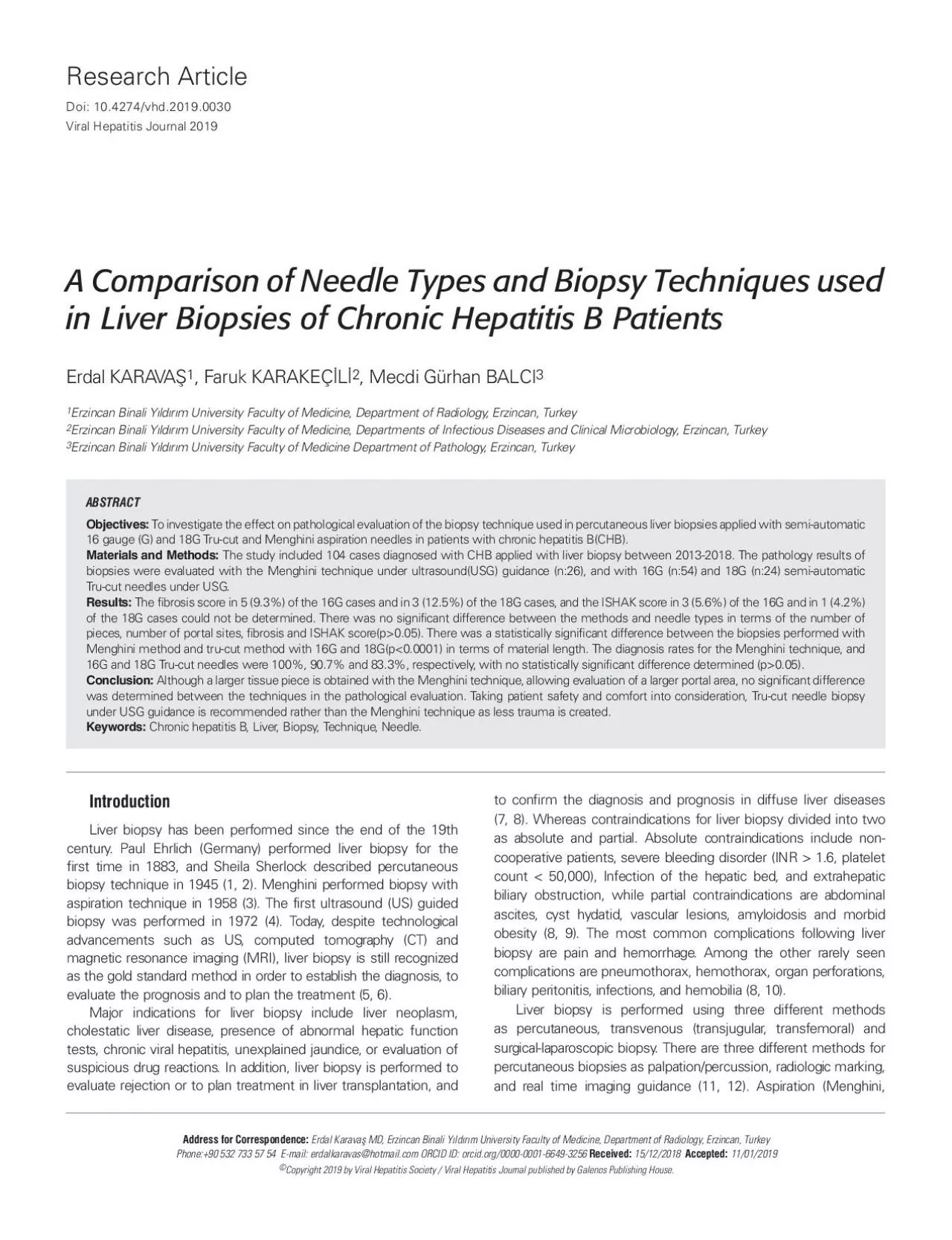
Liver biopsy has been performed since the end of the 19th century Paul Ehrlich Germany performed liver biopsy for the first time in 1883 and Sheila Sherlock described
Presentation Embed Code
Download Presentation
Download Presentation The PPT/PDF document "Doi 104274vhdViral Hepatitis Journal 201..." is the property of its rightful owner. Permission is granted to download and print the materials on this website for personal, non-commercial use only, and to display it on your personal computer provided you do not modify the materials and that you retain all copyright notices contained in the materials. By downloading content from our website, you accept the terms of this agreement.
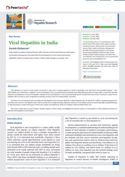
Doi 104274vhdViral Hepatitis Journal 2018: Transcript
Download Rules Of Document
"Doi 104274vhdViral Hepatitis Journal 2018"The content belongs to its owner. You may download and print it for personal use, without modification, and keep all copyright notices. By downloading, you agree to these terms.
Related Documents