/
Ectopic Pregnancy Management Tubal and interstitial Ectopic Pregnancy Management Tubal and interstitial

Ectopic Pregnancy Management Tubal and interstitial - PDF document

bety
bety . @bety
Follow
342 views
Uploaded On 2022-09-07

Ectopic Pregnancy Management Tubal and interstitial - PPT Presentation

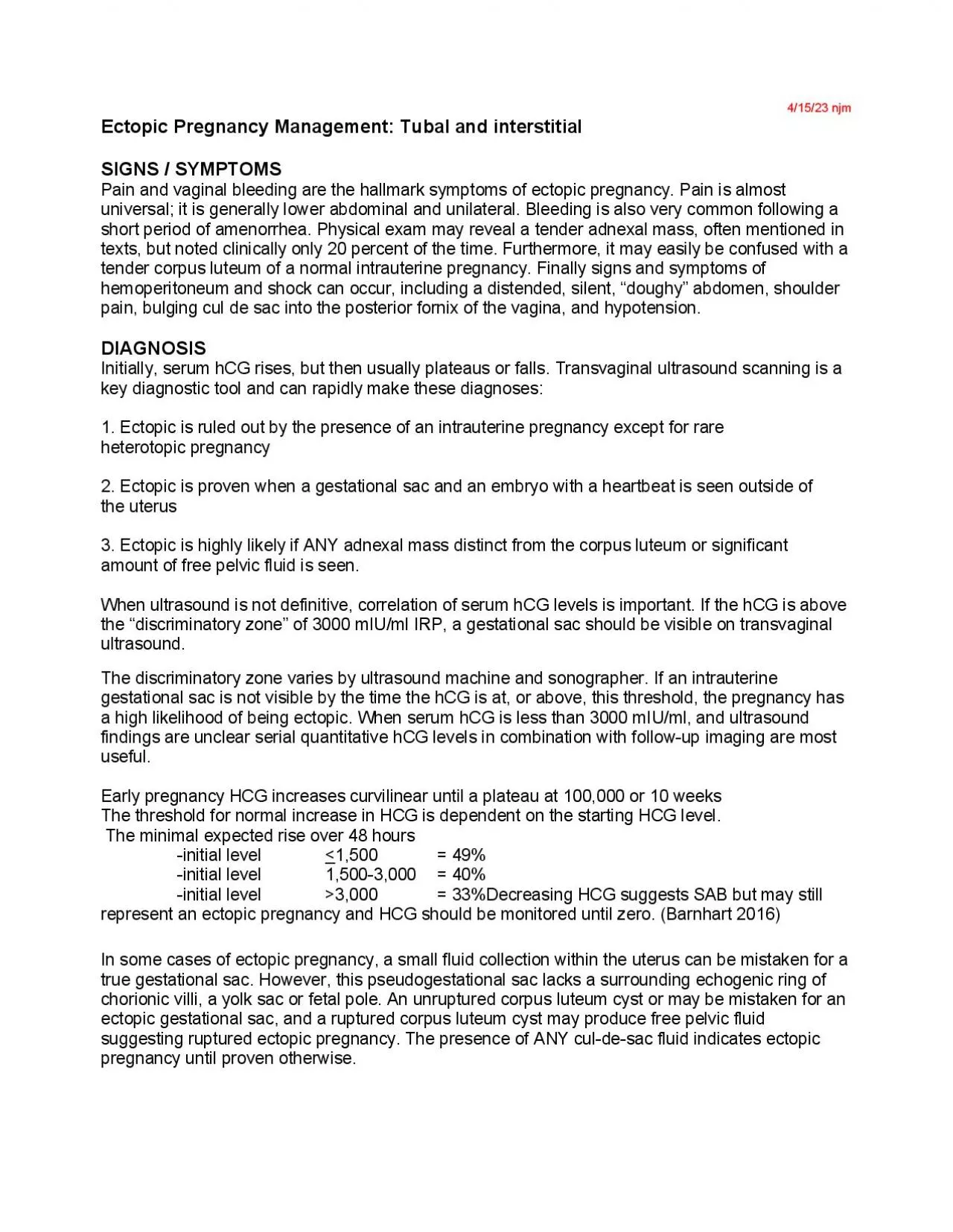
101121 njm SIGNS SYMPTOMS Pain and vaginal bleeding are the hallmark symptoms of ectopic pregnancy Pain is almost universal it is generally lower abdominal and unilateral Bleeding is also v ID: 952669

hcg pregnancy sac ectopic pregnancy hcg ectopic sac management dose mtx day surgical ultrasound therapy methotrexate check tubal serum

Share:

Link:

Embed:

Download Presentation from below link

Download Pdf The PPT/PDF document "Ectopic Pregnancy Management Tubal and i..." is the property of its rightful owner. Permission is granted to download and print the materials on this web site for personal, non-commercial use only, and to display it on your personal computer provided you do not modify the materials and that you retain all copyright notices contained in the materials. By downloading content from our website, you accept the terms of this agreement.


Presentation Transcript

10/11/21 njm Ectopic Pregnancy Management: Tubal and interstitial SIGNS / SYMPTOMS Pain and vaginal bleeding are the hallmark symptoms of ectopic pregnancy. Pain is almost universal; it is generally lower abdominal and unilateral. Bleeding is also very common following a short period of amenorrhea. Physical exam may reveal a tender adnexal mass, often mentioned in texts, but noted clinically only 20 percent of the time. Furthermore, i t may easily be confused with a tender corpus luteum of a normal intrauterine pregnancy. Finally signs and symptoms of hemoperitoneum and shock can occur, including a distended, silent, “doughy” abdomen , shoulder pain, bulging cul de sac into the posterior fornix of the vagina, and hypotension. DIAGNOSIS Initially, serum hCG rises, but then usually plateaus or falls. Transvaginal ultrasound scanning is a key diagnostic tool and can rapidly make these diagnoses: 1. Ectopic is ruled out by the presence of a n intrauterine pregnancy except for rare heterotopic pregnancy 2. Ectopic is proven when a gestational sac and an embryo with a heart beat is seen outside of the uterus 3. Ectopic is highly likely if ANY adnexal mass distinct from the corpus luteum or sig nificant amount of free pelvic fluid is seen. When ultrasound is not definitive, correlation of serum hCG levels is important. If the hCG is above the “disc riminatory zone” of 3000 mIU/ml IRP, a gestational sac should be visible on transvaginal ultrasound. The discriminatory zone varies by ultrasound machine and sonographer. If an intrauterine gestational sac is not visible by the time the hCG is at, or above, this threshold, the pregnancy has a high likelihood of being ectopic. When serum hCG is less tha n 3000 mIU/ml, and ultrasound findings are unclear serial quantitative hCG levels in combination with follow - up imaging are most useful. Early pregnancy HCG increases curvilinear until a plateau at 100,000 or 10 weeks The threshold for normal increase in HCG is dependent on the starting HCG level. The minimal expected rise over 48 hours - initial level 1,500 = 49% - initial level 1,500 - 3,000 = 40% - initial level �3,000 = 33%Decreasing HCG suggests SAB but may still represent an ectopic pregnancy and HCG should be monitored until zero . (Barnhart 2016) In some cases of ectopic pregnancy, a small fluid coll

ection within the uterus can be mistaken for a true gestational sac. However, this pseudogestational sac lacks a surrounding echogenic ring of chorionic villi, a yolk sac or fetal pole. An unruptured co rpus luteum cyst or may be mistaken for an ectopic gestational sac, and a ruptured corpus luteum cyst may produce free pelvic fluid suggesting ruptured ectopic pregnancy. The presence of ANY cul - de - sac fluid indicates ectopic pregnancy until proven otherwi se. If c horionic villi are not confirmed, hCG levels should be monitored. A plateau or increase in hCG postprocedure should prompt further work - up for retained products or treatment for an ectopic pregnancy (ACOG 2018) When hCG levels are decreasing and ultrasound cannot confirm pregnancy location, the pregnancy still needs to be followed closely. 20% of women with ectopic pregnancy will have a decline in hCG similar to the decline seen with a SAB (Silva 2006) When hCG levels are not rising normally an d ultrasound cannot confirm pregnancy location, a dilation and cu ret t age (D&C) or manual vacuum aspiration (MVA) may yield chorionic villi or a gestational sac 38 percent of the time. When this happens - a failed intrauterine pregnancy is diagnosed - and t reatment for an ectopic pregnancy avoided. When suspicion for ectopic pregnancy is high but cannot be confirmed with noninvasive testing, laparoscopy can both confirm the diagnosis and accomplish treatment. Pregnancy of unknown location When a pregnant patient with pain and/or bleeding has an US, particularly a transabdominal ultrasound ( TAUS ) , that has no findings (ie, no IUP, adnexal mass, or echogenic fluid), the differential diagnosis is normal early IUP, nonviable IUP, or ectopic pre gnancy. Approximately 15 to 26 percent of women with ectopic pregnancy will have a "normal" initial US. In a hemodynamically stable patient, an US with no findings should be repeated when the human chorionic gonadotropin (hCG) reaches the discriminatory z one for endometrial findings or in three to four days, since the gestational sac of an IUP grows approximately 1 mm per day and is visible on US when it reaches 3 mm or greater. In 11 to 16 percent of cases with an indeterminate initial scan, ectopic pregn ancy is evident on follow - up US - Pelvic ultrasonography is the most useful imaging modality for women with pregnancy of unknown location. ( Jurkovic 2010) - Approximately 15 to 26 percent of women with ectopi

c pregnancy will have an initial US with no findings. The serum human chorionic gonadotropin above which a singleton gestational sac should be visualized on ultrasound examination is 3000 IU/L with transva ginal. US - Sonographic identification of an intrauterine gestational sac is suggestive, but not diagnostic, of an intrauterine pregnancy. An endometrial pseudosac can be seen in up to 20 percent of women with an ectopic pregnancy. - The presence of a yolk sac or an embryo within the endometrium is diagnostic of an intrauterine pregnancy. - Adnexal findings that are suggestive, but not diagnostic, of a tubal pregnancy are a "tubal ring" or a non - cystic adnexal mass. Sonographic confirmation of a tubal preg nancy is made when an extrauterine sac with a yolk sac and/or embryo is visualized - Echogenic peritoneal free fluid almost always represents hemoperitoneum. The presence or absence of peritoneal free fluid is not a reliable indicator of whether an ectopic pregnancy has ruptured. Terminology ‘Indeterminate’ is a term used in clinical practice that has led to confusion. Some practitioners have used the term to mean ‘pregnancy of indeterminate site’ while others mean ‘pregnancy of indeterminate viability’. The term ‘indeterminate’ should no longer be used and should be replaced with the two separate terms below. Both terms should only be used after assessment by TVS. ‘Pregnancy of unknown location’ : No signs of either intra - o r extrauterine pregnancy or retained products of conception in a woman with a positive pregnancy test . Pregnancy of ‘uncertain viability’ : Intrauterine sac (16 - 24 mm mean diameter) with no o bvious fetus ; crown – rump length mm with no obvious fetal he art activity. In order to confirm or refute viability, a repeat scan at a minimal interval of 11 days is necessary. Finding’s diagnostic of Pregnancy Failure by Transvaginal Ultrasound - CRL � 7mm with no fetal cardiac motion - Mean sac diameter � 25 mm and no embryo - Absence of embryo with fetal cardiac motion � 2 wks after a scan that showed a gestational sac without a yolk sac - Absence of embryo with fetal cardiac motion � 11 days after a scan that showed a gestational sac with a yolk sac Management: 1.) Ectopic Pregnancy With early diagnosis the management of ectopic pregnancy has firmly moved into the outpatient real

m. Current treatment options favor medical and laparoscopic management with expectant management reserved for cases with a d eclining quantitative hCG 1,000 and open surgical management limited to cases of tubal rupture and hemoperitoneum. Surgical management via laparoscopy or open laparotomy can involve complete removal of the fallopian tube (salpingectomy) or efforts to remove the ectopic pregnancy and conserve the tube (salpingostomy). In the presence of a healthy contralateral tube there is no clear evidence that salpingotomy should be used in preference to salpingectomy. ( Silva 1993) Laparoscopic salpingostomy should be considered as the primary treatment when managing tubal pregnancy in the presence of contralateral tubal disease and the desire for future fe rtility. (RCOG 2004) In comparing systemic methotrexate with tube - sparing laparoscopic surgery, randomized trials have shown no difference in overall tubal preservation, tubal patency, repeat ectopic pregnancy, or future pregnancies. (ACOG 2008) Ectopic pregnancies located in the tubal cornua, cesarean scar, interstitial area or uterine cervix are difficult to diagnose and treat , plus radiologic diagnosis can be more challenging. 3 - D ultrasound and or MRI may aid in diagnosis. Table 1 shows indications fo r surgical management. (See Section 2) Table 1. Indications for Surgical Management of Ectopic Pregnancy Unstable vital signs or signs of hemoperitoneum Uncertain diagnosis Advanced ectopic pregnancy (high hCG, large mass, cardiac activity) Unreliable follow - up Any contraindication to observation or methotrexate Expectant or medical treatment are options in hemodynamically stable women who are carefully selected and informed accor ding to the criteria in Tables 2 and 3 . Expectant management is an option for clinically stable asymptomatic women with an ultrasound diagnosis of ectopic pregnancy and a decreasing serum hCG, initially less than serum 1000 I u/l. (RCOG 2004) Human chorionic gonadotropin level is the best predictor of successful treatment with methotrexate. A systematic review of several studies showed that failures with single dose methotrexate occurred 3.7 percent of the time when hCG levels were below 5,000 mIU/ml vs. 14.3 percent when higher than this cut - off. The failure rate for women w ho had an initial concentration between 5000 and 9999 mIU/mL was significantly higher than for those who had in

itial levels between 2000 and 4999 mIU/mL (OR 3.8, 95% CI 1.2 - 12.3). ( Menon 2007) Thus, single methotrexate is used when hCG levels are below 5,000 mIU/ml. ( Menon 2007) If hCG levels are higher than 5,000 mIU/mL, then multiple doses may be appropriate (Alleyassin 2006 ). Table2. Criteria for Expectant Management Include: Minimal pain or bleeding Patient reliable for follow - up No evidence for tu bal rupture Starting hCG level less than 1000 m I U /ml and falling Ectopic or adnexal mass less than three centimeters, or not detected No embryonic heartbeat Table 3. Criteria for Medical Management of Ectopic Pregnancy With Methotrexate Stable vital signs and low level of symptomatology No medical contraindication for methotrexate therapy ( Normal liver enzymes, creatinine , complete blood count and platelet count) Unruptured ectopic pregnancy Ectopic mass four centimeters or less Starting hCG levels l ess than 5000 (If 5 - 10K m I U /ml – Two or Multidose) Absence of embryonic cardiac activity (If present – Two or Multidose) Willing to stay in an area with surgical backup until HCG is negative Expectant management is used most often when the actual location of the pregnancy cannot be determined. If the initial hCG level is less than 200 m I U/mL, 88% of patients experience spontaneous resolution . ( Korhonen 1994) Medical management with methotrexate, a folic acid antagonist, is appropriate for properly selecte d patients and has been shown in randomized trials to be safe and effective; it also can be less costly and result in equal or better subsequent fertility than conservative surgical treatment. As with expectant treatment, patient selection and close follow - up are key to safety and success. Single dosage intramuscular regimens are commonly calculated at one mg/kg or 50 mg/m 2 . The dose is rounded up to the nearest 5 mg. Body surface area (BSA) may be calculated based upon height and weight on the day of trea tment using the formula BSA = square root ((cm X kg)/3600) or a BSA calculator : http://www.uptodate.com/contents/calcul ator - body - surface - area - mosteller - square - root - method?source=see_link&utdPopup=true Guidelines for single , two dose and multiple do se regimens are shown in Table 4 . Single dose regimen Serum hCG testing is performed on the fourth and seventh post - treatment days and then followed weekly until the level reaches

5 mIU/ml, which may take three to four weeks. The hCG initially rises slightly but should fall 15 percent between days four and seven; if not, the dosage should be repeated, or surgical t herapy performed. The methotrexate dosage should be repeated no more than once before surgical consultation is obtained. Because some patients managed expectantly or with methotrexate will eventually require surgery, prompt consultation is essential for th e non - surgeon. During therapy the patient should be advised: - Avoid coitus - Avoid ETOH - No Folic Acid or folic acid containing vitamins, e. g., prenatal vitamins - Pain frequently develops on the 2 nd to 7 th day after therapy (consider non - NSAIDs) - Blood type a Rh should be known in all patients prior to therapy - Avoid pelvic exams during surveillance of MTX therapy due to theoretical risk of tubal rupture - Avoid sun exposure to limit risk of MTX dermatitis - Avoid nonsteroidal anti - inflammatory drugs, as the interaction with MTX may cause bone marrow suppression, aplastic anemia, or gastrointestinal toxicity - A birth control method is recommended for at least 3 months post therapy. - Stay in an area with surgical backup until HCG is negative Consider the possibility of hete rotopic gestation, in women who have utilized assisted reproductive technologies such as in vitro fertilization or gamete intrafallopian transfer . (Dimitry 1990) For women who have conceived naturally, the presence of an intrauterine pregnancy makes the likelihood of ectopic pregnancy extremely rare as the incidence of heterotopic gestation has been reported to be between 1 in 4,000 and 1 in 8,000. 2.) Intersti tial ectopic pregnancy: Management Initially treat interstitial pregnancy (located at the junction of the fallopian tube and uterine cavity) with multidose medical therapy if medically stable (Table 4) resorting to surgical therapy if there is any deterior ation in clinical status. (Stovall 1989) There are no high - quality data comparing single versus multidose MTX therapy for interstitial pregnancy. 3.) Cervical ectopic and cesarean scar ectopic pregnancy: Management These represent challenging cases and of ten require creative surgical management, including uterine artery embolization or multidose MTX therapy. There is no high - quality data which includes all cases. (Glenn 2018, Chia 2018) Table 4. Single , Two and Multiple Dose Methotrexate Regimens for Treatment of Ect

opic Treatment Single Dose Two - dose Multidose Pretreatment, rule out spontaneous abortion and IUP hCG, creatinine, liver function, CBC w/diff blood type and Rh hCG, creatinine, liver function, CBC w/diff blood type and Rh hCG, creatinine, liver function, CBC w/diff blood type and Rh Day #1 Check hCG value, Administer first dose of MTX 50 mg/m 2 IM Check hCG value, Administer first dose of MTX 50 mg/m 2 IM Check hCG value, Administer first dose of MTX 1 mg/kg IM Followed by Leucovorin 0.1 mg/kg IM on Day 2 Check hCG on day 2 Day #4 Check hCG value Check hCG value, Administer second dose of MTX 50 mg/m 2 IM Administer second dose of MTX (day 3) and Leucovorin (day 4) on and respectively. Check hCG on day 4 Day #7 Check hCG value for 15 % decrease between days 4 and 7. If� 15 % fall, check hCG weekly until not Check hCG value for 15 % decrease between days 4 and 7. If� 15 % fall, check Continue administering doses of MTX and Leucovorin (i.e. course 3 day 5 and 6 and course 4 day 7 and 8) until detectable in serum. If 15 % fall, administer second dose MTX, 50 mg/m 2 IM hCG weekly until not detectable in serum. If 15 % fall, administer Third dose MTX, 50 mg/m 2 IM and recheck on day 11 hCG values have declined by 15 %. Do not exceed 4 courses If� 15 % decline, check hCG weekly until not detectable in serum. Weekly surveillance If follow - up hCG value till zero plateau or rise, consider surgical intervention or repeat dose of MTX as additional therapy. If follow - up hCG value till zero plateau or rise, consider surgical intervention or repeat dose of MTX as additional therapy. If follow - up hCG value till zero plateau or rise, consider surgical intervention or repeat dose of MTX as addi tional therapy. Sources: - Supplement to: Barnhart KT. Ectopic Pregnancy. N Engl J Med 2009; 361:379 - 87. - ACOG Practice Bulletin No. 193. (Two dose) References Tubal ectopic pregnancy. ACOG Practice Bulletin No. 193. American College of Obstetricians and Gynecologists. Obstet Gynecol 2018;131: e91 – 103. Reaffirmed 2019 (Accessed 10/11/21) Diagnosis and Management of Ectopic Pregnancy ( RCOG Green - top Guideline No. 21) Royal College of Obstetricians and Gynaecologists . April 2016 https://www.rcog.org.uk/

en/guidelines - research - services/guidelines/gtg21 / (Accessed 10/11/21) Barnhart KT, Sammel MD, Rinaudo PF, Zhou L, Hummel AC, Guo W. Symptomatic patients with an early viable intrauterine pregnancy; hCG curves redefined. Obstet Gynecol 2004; 104:50 – 5. M enon S, Colins J, Barnhart KT. Establishing a human chorionic gonadotrop in cutoff to guide methotrexate treatment of ectopic pregnancy: a systematic review. Fertil Steril 2007; 87:481 - 4. Stovall TG, Ling FW, Buster JE. Outpatient chemotherapy of unruptured ectopic pregnancy. Fertil Steril. 1989;51(3):435. Korhonen J, Stenman UH, Ylotalo P. Serum human chorionic gonadotropin dynamics during spontaneous resolution of ectopic pregnancy. Fertil Steril 1994; 61:632 – 6. Alleyassin A, Khademi A, Aghahos seini M, Safdarian L, Badenoosh B, Hamed EA. Comparison of success rates in the medical management of ectopic pregnancy with single - dose and multiple - dose administration of methotrexate: a prospective, randomized clinical trial. Fertil Steril 2006; 85:1661 – 6. Barnhart KT. Ectopic Pregnancy. N Engl J Med 2009; 361:379 - 87. Silva P, Schaper A, Rooney B. Reproductive outcome after 143 laparoscopic procedures for ectopic pregnancy. Fertil Steril 1993; :710 – 5. Dimitry ES , Oskarsson T , Margara R , Winston RM . Heterotopic pregnancy associated with assisted reproductive technology. Am J Obstet Gynecol. 1990 Jul;163(1 Pt 1):244 - 5. Jurkovic D. hCG as a patient. Ultrasound in Obstetrics & Gynecology. October 2010 36)4):395 - 399 Doubilet PM , et al Diagnostic criteria for nonviable pregnancy early in the first trimester. N Engl J Med. 2013 Oct 10;369(15):1443 - 51 Barnhart KT, et al. Differences in Serum Human Chorionic Gonadotropins Rise in Early Pregnancy by Race and Value at Presentation. Obstet Gynecol 2016 Sept;128(3 ):504 – 11. Silva C, et al. Human Chorionic Gonadotropin Profile for Women with Ectopic Pregnancy. Obstet Gynecol. 2006 March. 107(3);605 - 10 Glenn TL et al. Cesarean Scar Ectopic Pregnancy: Current Management Strategies. Obstet Gynecol Surv. 2018 Mary; Vol . 73 (5): 293 - 302 Chia V et al. Management of Cervical Ectopic Pregnancies: A Systemic Review. Obstet Gynecol. 2018 May. 131 Suppl 1:186S - 186S Reviewed 10/11/21 njm Revised 8/2 9 /19 njm Revised 4/21/19 njm Revised 4/21/19 njm Reviewed 7/6/18 njm Revised 4 /23/16 njm Revised 2/9/14 njm Reviewed 8/16/13 njm Reviewed 5/18 /11 njm Approved 1/23/