/
BLEEDING IN EARLY PREGNANCY, ABORTION, RECURRENT FETAL LOSS BLEEDING IN EARLY PREGNANCY, ABORTION, RECURRENT FETAL LOSS

BLEEDING IN EARLY PREGNANCY, ABORTION, RECURRENT FETAL LOSS - PowerPoint Presentation

madison
madison . @madison
Follow
65 views
Uploaded On 2023-11-16

BLEEDING IN EARLY PREGNANCY, ABORTION, RECURRENT FETAL LOSS - PPT Presentation

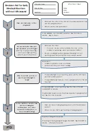
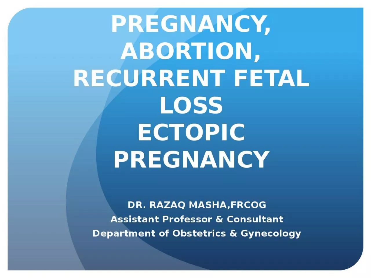
ECTOPIC PREGNANCY DR RAZAQ MASHAFRCOG Assistant Professor amp Consultant Department of Obstetrics amp Gynecology An abortion is pregnancy that ends before the baby can survive outside the uterus because it has not yet reached viability It is therefore defined as a spontaneous loss of a ID: 1032081

abortion pregnancy bleeding ectopic pregnancy abortion ectopic bleeding weeks ultrasound factors early intrauterine cervical vaginal pregnancies evacuation pain inevitable

Share:

Link:

Embed:

Download Presentation from below link

Download Presentation The PPT/PDF document "BLEEDING IN EARLY PREGNANCY, ABORTION, R..." is the property of its rightful owner. Permission is granted to download and print the materials on this web site for personal, non-commercial use only, and to display it on your personal computer provided you do not modify the materials and that you retain all copyright notices contained in the materials. By downloading content from our website, you accept the terms of this agreement.


Presentation Transcript

1. BLEEDING IN EARLY PREGNANCY, ABORTION, RECURRENT FETAL LOSSECTOPIC PREGNANCYDR. RAZAQ MASHA,FRCOGAssistant Professor & ConsultantDepartment of Obstetrics & Gynecology

2. An abortion is pregnancy that ends before the baby can survive outside the uterus because it has not yet reached viability. It is therefore defined as a spontaneous loss of a pregnancy before 24 weeks. An abortion tends to start with bleeding and pain may develop.A threatened abortion is characterized by bleeding early in the pregnancy but the pregnancy continues.An inevitable abortion means that the pregnancy cannot be saved. It may be incomplete, with pregnancy products still in the uterine cavity or complete with nothing remaining.

3. TYPES OF ABORTIONSpontaneous abortion – this is when the abortion occurs naturally as opposed to being induced.Induced abortion – the pregnancy is terminated artificially.Threatened abortion : There is bleeding and sometimes pelvic pain but the cervix is closed and ultrasound indicates an ongoing pregnancy within the uterus.Inevitable abortion – the pregnancy is not continuingComplete abortion – an inevitable abortion and the uterus has completely emptied itselfIncomplete abortion – an inevitable abortion with products of the pregnancy still present in the uterusMissed abortion– there are no reasons to have suspected that the pregnancy is not going to continue but the embryo has died

4. Septic Abortion – The abortion has been complicated by infectionRecurrent or Habitual abortion – most authorities recommend that these terms should be used only for three or more consecutive abortions although there is a tendency towards two.Second Trimester abortion – abortion after thirteen weeks and before 24 weeks

5. Prevalence of AbortionIt is thought that 10-20% of pregnancies miscarry. Most abortions occur in the early weeks of pregnancy.Ultrasound screening of fetal anomaly has shown the incidence of non-viable pregnancy at 10 to 13 weeks to be 2.8%Often the cause of an abortion remains unknown. The most common cause for abortion is a blighted ovum – the trophoblast tissues develop but there is no fetus.Another common cause is a genetic defect. Smoking and obesity may contribute to abortion.

6. Blighted OvumNormally the zygote divides and part becomes the embryo, a part becomes the placental tissue (trophoblast) and the amniotic membranes. When there is blighted ovum, the trophoblast develops alone without the development of the embryo. Blighted ovum has also been referred to as” anembryonic pregnancy”. Nearly half of early abortions are associated with a bighted ovum. It is likely that abnormal chromosomes are more prevalent.

7. ManagementThreatened abortion : Slight bleeding which may persist for weeks, mild pain, internal os is closed. It is then essential to decide whether there is any possibility of continuation of the pregnancy by vaginal ultrasound. Gestation sac can be seen by scan 33 – 35 days after the last menstrual period. * Serial qualitative HCG levels * Increasing titres indicate a live fetus and expectant observation

8. Inevitable AbortionIndicates the pregnancy is doomed to end shortly.Progressive cervical dilatation without passage of tissueBleeding may be moderate in amount and the fetus is deadPain usually moreThe internal os is dilated and products may be felt in the cervical canal.Ultrasound scan will show a non-viable fetusRemove the products from the canal / emergency evacuation.

9. Complete AbortionDiagnosed if patient has passed tissue, has slight pain and slight vaginal bleeding.Transvaginal ultrasound – empty uterusAnti D injection if patient is Rh negative to prevent sensitisation.Incomplete AbortionIf the patient has passed some tissue but the internal cervical os is openEmergency evacuation

10. Missed AbortionIt is defined as retention of dead products of conception in utero for several weeks.Symptoms of early pregnancy disappear.Uterus not only has ceased to enlarge but also has become smaller.Abnormal sonographic findingsIrregular gestation sacTreated with prostaglandins or suction evacuation

11. Septic AbortionUterine infection at any stage of abortion caused by: a. Delay in evacuation of uterus b. Delay in seeking advice c. Incomplete surgical evacuation followed by infection from vaginal organisms after 48 hours, coliform bacillus d. Perforation or cervical tear e. Criminal abortion

12. Treatment should be active to minimize the risk of septic shock:Cervical and HVS, blood cultureBroad spectrum antibioticsEvacuation – caution to avoid perforation

13. Induced Abortion:Therapeutic abortion – termination of pregnancy before the age of fetal viability for the purpose of safe-guarding maternal health e.g. in heart disease, invasive Ca of Cervix or breast. * A certificate of opinion is given by the heart consultant and the obstetrician.Elective (voluntary) abortion is the interruption of pregnancy before viability at the request of the woman but not for reason of impaired maternal health or fetal disease.Illegal abortion is usually performed in unsterile environment by operators with little or no medical trainingIt is often incomplete and complicated by: a. Haemorrhage b. Infection – which may lead to tubal occlusion. Septic shock and death may be the ultimate consequences.

14. Recurrent Miscarriage:Term is used when a woman has had three or more consecutive miscarriages.Genetic Factors Karyotyping of both partners may reveal chromosomal anomaliesAnatomical Factors Uterine anomalies Cervical incompetence Hysteroscopy and HSG. Septum/FibroidEndocrine problems Raised LH in PCO Thyroid disease. Diabetes mellitus

15. Immunological Factors Antiphospholipid antibody syndrome Anti cardiolipid antibodies Lupus anti coagulantMaternal disease SLE, Renal diseaseEnvironmental factors : Smoking / Alcohol

16. Techniques:* Medical : Antiprogesterone (R4 486) – or Mifepristone and prostaglandin OxytocinProstaglandin vaginal suppository applied to the cervix to ripen and soften it leading to dilatation or as an adjunct with Mifepristone.* Surgical : Suction dilatation and curettage

17. Ectopic pregnancyThis is a complication of pregnancy in which the embryo implants outside the uterine cavity. With rate exceptions, ectopic pregnancies are not viable. Furthermore they are dangerous for the mother, since internal haemorrhage is a life-threatening complication. Most ectopic pregnancies occur in the fallopian tubes but can also occur in:The cervixOvariesAbdomenIn a typical ectopic pregnancy, the embryo adheres to the lining of the fallopian tube and burrows into the tubal lining. Most commonly this invades vessels and will cause bleeding.

18. Non tubal ectopic pregnancy2 % of ectopic pregnancies occur in the ovary, cervix or are intra abdominal. Transvaginal ultrasound examination is usually able to detect a cervical pregnancy. An ovarian pregnancy is differentiated from a tubal pregnancy by histology.Heterotopic PregnancyIn rare cases of ectopic pregnancy there may be two fertilized ova, one outside and the other inside. This is called heterotopic pregnancy. Although rare, heterotopic pregnancies are becoming more common, likely due to increased use of IVF. The survival rate of the uterine fetus of an ectopic pregnancy is around 70%.

19. Signs and SymptomsUp to 10% of women with ectopic pregnancy have no symptoms. The symptoms are often non specific and difficult to differentiate from those of : a. Genito urinary disorders b. Gastro intestinal disorders like appendicitis or salpingitis c. Ruptured corpus luteum cyst d. Miscarriage e. Ovarian torsion f. Urinary tract infectionClinical presentation of ectopic pregnancy occurs at a mean of 7 weeks after the last normal menstrual period with a range of 4 – 8 weeks.

20. Early Signs inlcude:Vaginal bleeding : The amount varies although classically there is a complaint of spotting. Heavy bleeding, in the absence of of ultrasound or hCG assessment, may lead to a misdiagnosis of miscarriage.Abdominal painNausea, vomiting and diarrhoeaIn ruptured ectopic pregnancy there may be abdominal distension, abdominal tenderness, peritonism and haemorrhagic shock.

21. Risk Factors for EctopicThere are a numbver of risk factors for ectopic pregnancies. However, in as many as one third to one-half, no risk factors can be identified.Risk factors include:Pelvic inflammatory diseaseInfertilityUse of an intrauterine device (IUCD)Previous exposure to DESTubal surgeryIntra uterine surgery e.g. D & CPrevious ectopic pregnancyTubal ligationSmoking

22. DiagnosisAn ectopic pregnancy should be considered as the cause of abdominal pain or vaginal bleeding in every woman who has a positive pregnancy test.TRANSVAGINAL ULTRASONOGRAPHYAn ultrasound showing a gestational sac with fetal heart in the fallopian tube has a very high specificity of ectopic pregnancy. Transvaginal ultrasonography has a sensitivity of at least 90% for ectopic pregnancy

23. The combination of a positive pregnancy test and the presence of what appears to be a normal intrauterine pregnancy does not exclude an ectopic pregnancy, since there may be either a heterotopic pregnancy or a pseudo sac, which is a collection of fluid within the endometrial cavity that may be seen in up to 20% of woman.The presence of an adnexal mass in the absence of an intrauterine pregnancy on transvaginal sonography increases the likelihood of an ectopic pregnancy 100-fold.Where no intrauterine pregnancy is seen on ultrasound, measuring BHCG levels may aid in the diagnosis.Other diagnostic methods include laparoscopy and laparotomy which can also be performed to visually confirm an ectopic pregnancy. This is generally reserved for women presenting with signs of an acute abdomen and / or hypovolemic shock.

24. (Treatment)Medical : Early treatment of an ectopic pregnancy with methotrexate is a viable alternative to surgical treatment. The methotrexate terminates the growth of the emrbyo and is then resorbed by the woman’s body.Contraindications include liver, kidney or blood disease as well as an ectopic mass > 3.5 cm.Also it may lead to the inadvertent termination of an undetected intrauterine pregnancy, or severe abnormality in any surviving pregnancy. Therefore it is recommended that methotrexate should only be administered when hCG has been serially monitored with a rise less than 35% over 48 hours, which practically excludes a viable intrauterine pregnancy.

25. Surgical:If haemorrhage has already occurred surgical intervention may be necessaryAccess to the pelvis may be gained either by laparoscopy or laparotomy. We either incise the affected fallopian tube and remove only the pregnancy (Salpingostomy) or remove the affected tube with the pregnancy (Salpingectomy)A randomized study in 2013 came to the result that the rates of intrauterine pregnancy 2 years after treatment of ectopic pregnancy are approximately 64% with radical surgery, 67 % with medication and 70% with conservative surgery.