/
The Ultimate Solution for X The Ultimate Solution for X

The Ultimate Solution for X - PDF document

bety
bety . @bety
Follow
360 views
Uploaded On 2021-08-04

The Ultimate Solution for X - PPT Presentation

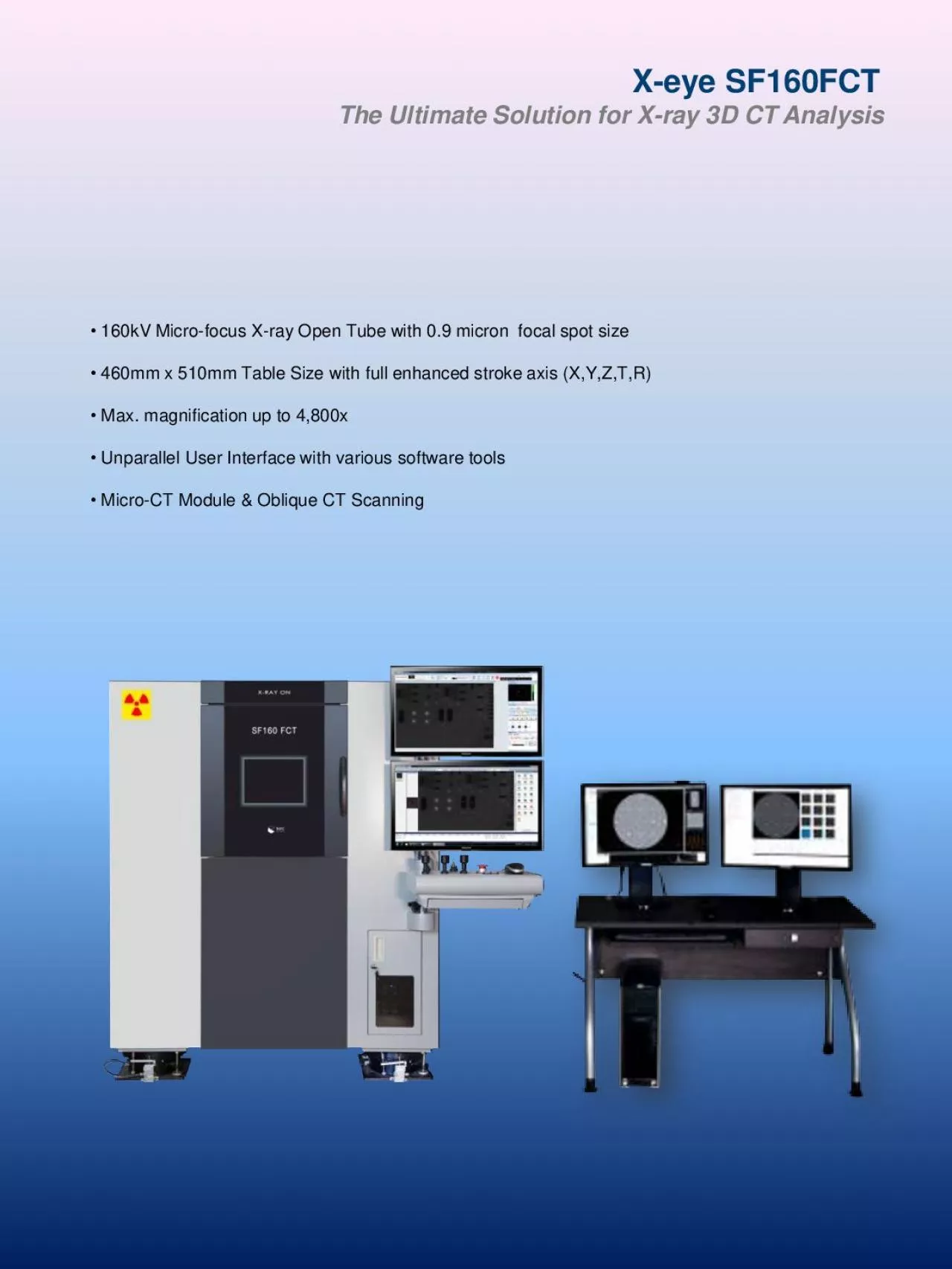
Xeye SF160FCTray 3D CT Analysis160kV Microfocus Xray Open Tube with 09 micron focal spot size460mm x 510mm Table Size with full enhanced stroke axis XYZTR Max magnification up to 4800xUnparallel ID: 856501

oblique ray size object ray oblique object size auto analysis max micro tube inspection table high module open focus

Share:

Link:

Embed:

Download Presentation from below link

Download Pdf The PPT/PDF document "The Ultimate Solution for X" is the property of its rightful owner. Permission is granted to download and print the materials on this web site for personal, non-commercial use only, and to display it on your personal computer provided you do not modify the materials and that you retain all copyright notices contained in the materials. By downloading content from our website, you accept the terms of this agreement.


Presentation Transcript

1 X - eye SF160FCT The Ultimate Solution
X - eye SF160FCT The Ultimate Solution for X - ray 3D CT Analysis • 160kV Micro - focus X - ray Open Tube with 0.9 micron focal spot size • 460mm x 510mm Table Size with full enhanced stroke axis (X,Y,Z,T,R) • Max. magnification up to 4,800x • Unparallel User Interface with various software tools • Micro - CT Module & Oblique CT Scanning The Ultimate Solution for 3D CT Analysis SF160FCT is a high - resolution micro - focus X - ray system for the inspection of semiconductor, PCB assembly, and electronic component. With its superior X - ray imaging, micro - scale hidden defects can be detected in high resolution. SF160FCT equips 160kV micro - focus open tube with 0.8um spot size. The system can magnify the object up to 4,800x and display the X - ray image at any angle using 6 - axis manipulator configuration. 3D CT (Computed Tomography) visualizes all hidden structure and even micro - scale defects inside the object. SEC unique oblique CT technology realizes high - magnified 3D CT visualization of large sample. Generally, it is known that CT scanning is limited by the object size, but oblique CT technology overcomes the size limitation and can be adapted to PCB assemblies, large - size Multilayer boards and even to semiconductor wafers. Auto BGA Inspection Module Auto Teaching (CNC Programming) Jog - sticks & One - click Teaching Buttons 3D CT Rendering Software • Intuitive and Flexible User Interface • No Complicated Jog - stick or Mouse Control • Real - time Image Acquisition • Auto Focus Tracing – Never lose your point of view • Auto Teaching – Maximize Inspection Throughput • GPU Based Ultra - fast 3D CT Reconstruction • Easy Click - in Change Filament Replacement Unparalleled User - friendly Environment Oblique CT Scanning Technology

2
X - eye SF160FCT The Ultimate Solution for X - ray 3D CT Analysis • SMT (Surface Mount Technology) Assembly - BGA, CSP - Open, Crack, Cold Soldering - General Solder Joint – Bridging, Void - Auto Voiding Area Calculation • Semiconductor Packaging, LED - Wire Bonding – Broken Wire, Lifted Wire, Sweeping - Bump, Pattern De - lamination, Void, Crack - 3D Packaging – MCP, TSV, FCB micro defect • Multilayer PCB (Printed Circuit Board) - Multilayer Pattern Open, Short Inspection and Analysis - Via - Hole Alignment, Copper Wall Plate - FPCB (Flexible PCB) – Blind Via Hole (Laser Via) • Electronic Components - Connector – Internal Wire Connection - Camera Module – Component Attachment - General Pattern Open, Short, Hidden Contamination X - ray Images by Application Cone Beam CT is generally used for 3D CT analysis of electronic components. For analysis of flat and large size components such as PCB, Cone Beam CT is restricted to provide high magnified 3D images due to long source - to - object distance (SOD). Oblique CT scans from an oblique direction while the object is rotating horizontally. The unique technology provides high magnified 3D images of flat and large components by horizontal object rotation without geometric interferences, and this realizes short source - to - object distance (SOD). While inspecting the object in 2D, simply click the Oblique - CT scan button without any table switching, and the system will provide you the 3D CT images within minutes. GPU - based CT Reconstruction Engine will improve your throughput tremendously. SOD SDD SDD SOD • SDD : Source to Detector Distance • SOD : Source to Object Distance Cone Beam CT Oblique CT X

3 - ray Tube Tube - type Micro
- ray Tube Tube - type Micro - focus Open Tube Focal Spot Size 0.9um Max. Tube Voltage 160kV Max. Tube Current 200uA X - ray Imaging Detector Flat Panel Detector (3.2M pixel) Active Area 153mm x 204mm (6 x 8 inch) System Magnification Max. 4,800x Manipulator Table Size 460mm x 510mm Max. Sample Loading 5Kg Stroke – X axis 400mm Stroke – Y axis 450mm Stroke – Z axis 200mm Detector Tilt Max. 70 ° Table Rotation Max. 360 ° Workstation Monitor 24 inch LED Dual CPU Intel® Pentium i5 Process Memory 4GB HDD 250GB x 2EA Rack Type General Dimension / Weight 1,350 x 1,450 x 1,660mm, 2,200Kg Power 220 VAC Single Phase 50/60Hz X - ray Safety Leakage Dose less than 1uSv/hr Hardware Specifications Software Features X - eye SF160FCT The Ultimate Solution for X - ray 3D CT Analysis South Korea - Headquarter China - Shanghai Office 415 Factory World, 332 - 2 Woncheon - Dong Room 104,Treasure Island Int°l CBD Centre, Suwon, South Korea 443 - 758 No 258, Rd Chengjiaqiao , Minhang District, Shanghai, China. 200335 Tel: +82 - 31 - 215 - 7341 Fax: +82 - 31 - 215 - 7343 Tel: +86 - 21 - 6083 - 0707 Email: sales@seceng.co.kr Email: studyhan@hotmail.com www.seceng.co.kr/eng www.seceng.co.kr/CHI Specifications in this catalog are subject to change without notice as SEC CO., Ltd. continues to develop the latest technologies and products • User - friend

4 ly Interface - Intuitive, flexi
ly Interface - Intuitive, flexible workspace • Easy - to - use Mouse Manipulation and Navigation - “Click & Center” on X - ray image or Jog - stick control - Navigation Panel shows actual Table Images • Auto BGA Inspection Module - Auto calculation of BGA diameter, Void Percentage - Auto Pass/Fail determination. • Inspection Mode Selection - Database management of X - ray and image adjustment parameters • Image Processing - Averaging, Contrast/Brightness, Binarization, Inverse - Histogram Adjustment - Image Filtering: Sharpening, Enhanced Focus • Various Measurement & Annotation Tool - Measuring: Line, Point to Point, Center to Center - Annotation & Reporting Workstation for 3D CT Monitor 24 inch LCD x 2EA (Dual Monitor) CPU Intel® Pentium i5 Process Memory 8GB HDD 500GB + 250GB(SSD) 3D Reconstruction & Viewer Software Recon. Algorithm Feldkamp Cone - Beam Algorithm Scan Mode Normal Scan (Full, Half), Oblique Scan 3D Recon. Matrix. Max. 2048 3 Recon. Time 10 sec. CT Manipulation Hardware ‘Quick Exchange° CT Manipulator for Cone - beam CT Scanning. High - precision rotation motor mounted perpendicular to X - ray tube & Detector direction. Oblique CT does not require any table switching . 3D CT Module Specifications 3D CT Software Features • VR (Volume Rendering) for 3D • Visualization from any angle • DICOM 3.0 standard compatible • 3D Sync. • Unlimited Oblique Slice / Unlimited level Oblique View • VR with cut plane, MIP, MPR • 3D Measurement with Analysis Features • 3D Zoom • Report Function 3D CT Module is an option for X - eye SF160 Series System