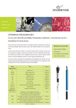
/
Microsensors  for instrumented medical tools for their real time monitoring Microsensors  for instrumented medical tools for their real time monitoring

Microsensors for instrumented medical tools for their real time monitoring - PowerPoint Presentation

briana-ranney
briana-ranney . @briana-ranney
Follow
349 views
Uploaded On 2018-09-21

Microsensors for instrumented medical tools for their real time monitoring - PPT Presentation

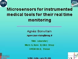
Agnès Bonvilain Agnesbonvilainimagfr TIMA Laboratory Micro amp Nano System Group GRENOBLE France 119 Outline Context Previous works Microfabrication of the microsensors ID: 673948

needle microfabrications medical layer microfabrications needle layer medical connections integration cristallization experimentations conclusion microsensors bonding annealing improvement instrumented system

Share:

Link:

Embed:

Download Presentation from below link

Download Presentation The PPT/PDF document "Microsensors for instrumented medical t..." is the property of its rightful owner. Permission is granted to download and print the materials on this web site for personal, non-commercial use only, and to display it on your personal computer provided you do not modify the materials and that you retain all copyright notices contained in the materials. By downloading content from our website, you accept the terms of this agreement.


Presentation Transcript

Slide1

Microsensors for instrumented medical tools for their real time monitoring

Agnès

BonvilainAgnes.bonvilain@imag.frTIMA LaboratoryMicro & Nano System GroupGRENOBLE, France

1/19Slide2

Outline

Context Previous works Microfabrication of the microsensors

First

experimentations

Improvement of the GF Integration of the connections Conclusion and Perspectives

2/19Slide3

Context

Goal

: give in real time the deformation of the needle during its use

3/19Slide4

y

x

Previous

works

4/19Slide5

Microfabrications

5/19

Diameter = 4’’

Diameter

= 0.6 mm

Length

= 10 cmSlide6

6/19

MicrofabricationsSlide7

7/19

Microfabrications

Sputtering of Si3N4 (200 nm) Evaporation of

Ge + Au (400 + 20 nm

)

Cristallization Annealing (2h at 300°C) Au

Etch

Evaporation of Cr/Au (10 + 200 nm)

Wire

bonding

NeedleSlide8

Microfabrications

8/19

Microsensors on stainless steel

medical

needle

500 µm

500 µmSlide9

Microfabrications

9/19Slide10

10/19

y

x

First

exper

imentationsSlide11

11/19

Experimentations

of the instrumented needleFirst experimentations

GF = 3 (

compared

to the

commercialized gauges which have a GF = 2)Slide12

12/19

Improvement

of the GFWe have worked on the material aspect of the Ge layer to

improve

the GF.

The

cristallization annealing has three important parameters : the temperature

, the

duration

and the

thickness

of the

metal

layer.

In the

litterature

we find

that a 20nm layer of gold allows the MIC

under 300°C.X-ray

diffractometry

allow

us to show

that

the layer must

be

40nm,

because

with

20nm the

begining

of the

cristallization

must

be

325°C.Slide13

13/19

Improvement

of the GFSlide14

14/19

Integration

of the connectionsSlide15

15/19

Integration

of the connectionsWe have adapted the microfabrication to have a better GF,Modification of the cristallization

annealing

parameters,Do not do the Au etching (which do not change the resistivity of the Ge layer and

avoid

damages of the KI-I²

etching

),

No

connecting

pads but

smaller

connecting

tracksSlide16

Integration

of the connections

16/19Slide17

17/19

Conclusion

The experimentations of the new microgauges with their connections at the extremity of the

needle

are in

progress

. We always use the bonding which is

very

fragile.

We

want

now

to put

three

microgauges at

120° with their connections

at the extremity of the

needle

.

We

design a

connector

to

get

free of the

bonding

.

The

next

goal

will

be

to

fabricate

a prototype

that

we

can

integrate

in a

microlocalization

system to test

it

.Slide18

Prototype of an instrumented needle to integrate in a microlocalization

system

Conclusion and Perspectives18/19Slide19

Conclusion and Perspectives

We

have shown in this work that it is possible to instrument a medical tool

to monitor

it

.

We have open a dent in the field of microfabrications applicated to medical

instruments

with

unconventional

shape

and

material

. So

we

can expected

now the development of the instrumentation of

other medical tools

.

19/19Slide20

Acknowledgment

Wenbin

YangMathilde GangneronLéo ZanardelliANR (Agence Nationale de la Recherche)