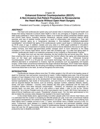
/
 The Electrical System of the Heart  The Electrical System of the Heart

The Electrical System of the Heart - PowerPoint Presentation

briana-ranney
briana-ranney . @briana-ranney
Follow
342 views
Uploaded On 2020-04-05

The Electrical System of the Heart - PPT Presentation

Cardiac Muscle Contraction Depolarization of the heart is rhythmic and spontaneous About 1 of cardiac cells have automaticity are selfexcitable Gap junctions ensure the heart contracts as a unit ID: 775818

node depolarization heart ventricular node depolarization heart ventricular repolarization figure atrial complete wave apex causing impulses fibers waves step

Share:

Link:

Embed:

Download Presentation from below link

Download Presentation The PPT/PDF document " The Electrical System of the Heart" is the property of its rightful owner. Permission is granted to download and print the materials on this web site for personal, non-commercial use only, and to display it on your personal computer provided you do not modify the materials and that you retain all copyright notices contained in the materials. By downloading content from our website, you accept the terms of this agreement.


Presentation Transcript

Slide1

The Electrical System of the Heart

Slide2

Cardiac Muscle Contraction

Depolarization of the heart is rhythmic and spontaneous

About 1% of cardiac cells have automaticity— (are self-excitable)

Gap junctions ensure the heart contracts as a unit

Long absolute refractory period (250 ms)

Slide3

Figure 18.12

Absolute

refractory

period

Tension

development

(contraction)

Plateau

Action

potential

Time (ms)

1

2

3

Depolarization

is

due to Na

+

influx throughfast voltage-gated Na+channels. A positivefeedback cycle rapidlyopens many Na+channels, reversing themembrane potential.Channel inactivation endsthis phase.

Plateau phase isdue to Ca2+ influx throughslow Ca2+ channels. Thiskeeps the cell depolarizedbecause few K+ channelsare open.

Repolarization is due to Ca2+ channels inactivating and K+channels opening. This allows K+ efflux, which brings the membranepotential back to itsresting voltage.

1

2

3

Tension (g)

Membrane potential (mV)

Slide4

Heart Physiology: Electrical Events Don’t copy

Intrinsic cardiac conduction system

A network of noncontractile (autorhythmic) cells that initiate and distribute impulses to coordinate the depolarization and contraction of the heart

Slide5

Heart Physiology: Sequence of Excitation

Sinoatrial (SA) node (pacemaker)

Generates impulses about 75 times/minute (sinus rhythm)

Depolarizes faster than any other part of the myocardium

Slide6

Heart Physiology: Sequence of Excitation

Atrioventricular (AV) node

Smaller diameter fibers; fewer gap junctions

Delays impulses approximately 0.1 second

Depolarizes 50 times per minute in absence of SA node input

Slide7

Heart Physiology: Sequence of Excitation

Atrioventricular (AV) bundle (bundle of His)

Only electrical connection between the atria and ventricles

Slide8

Figure 18.14a

(a) Anatomy of the intrinsic conduction system showing the

sequence of electrical excitation

Internodal pathway

Superior vena cava

Right atrium

Left atrium

Purkinje

fibers

Inter-

ventricular

septum

1

The

sinoatrial (SA)

node

(pacemaker)

generates impulses.

2

The impulses

pause (0.1 s) at the

atrioventricular

(AV) node.

The

atrioventricular

(AV) bundle

connects the atria

to the ventricles.

4

The

bundle branches

conduct the impulses

through the

interventricular septum.

3

The

Purkinje fibers

depolarize the contractile

cells of both ventricles.

5

Slide9

Homeostatic Imbalances

Defects in the intrinsic conduction system may result in

Arrhythmias: irregular heart rhythms

Uncoordinated atrial and ventricular contractions

Fibrillation: rapid, irregular contractions; useless for pumping blood

Slide10

Homeostatic Imbalances

Defective SA node may result in

Ectopic focus: abnormal pacemaker takes over

If AV node takes over, there will be a junctional rhythm (40–60 bpm)

Defective AV node may result in

Partial or total heart block

Few or no impulses from SA node reach the ventricles

Slide11

Extrinsic Innervation of the Heart

Heartbeat is modified by the ANS

Cardiac centers are located in the medulla oblongata

Cardioacceleratory center innervates SA and AV nodes, heart muscle, and coronary arteries through sympathetic neurons

Cardioinhibitory center inhibits SA and AV nodes through parasympathetic fibers in the vagus nerves

Slide12

Figure 18.15

Thoracic spinal cord

The

vagus nerve

(parasympathetic)

decreases heart rate.

Cardioinhibitory center

Cardio-

acceleratory

center

Sympathetic cardiac

nerves

increase heart rate

and force of contraction.

Medulla oblongata

Sympathetic trunk ganglion

Dorsal motor nucleus of vagus

Sympathetic trunk

AV node

SA node

Parasympathetic fibers

Sympathetic fibers

Interneurons

Slide13

Electrocardiography

Electrocardiogram (ECG or EKG): a composite of all the action potentials generated by nodal and contractile cells at a given time

Three waves

P wave: depolarization of SA node

QRS complex: ventricular depolarization

T wave: ventricular repolarization

Slide14

Figure 18.16

Sinoatrial

node

Atrioventricular

node

Atrial

depolarization

QRS complex

Ventricular

depolarization

Ventricularrepolarization

P-Q

Interval

S-TSegment

Q-TInterval

Slide15

Figure 18.17

Atrial depolarization, initiated

by the SA node, causes the

P wave.

P

R

T

Q

S

SA node

AV node

With atrial depolarization

complete, the impulse isdelayed at the AV node.

Ventricular depolarizationbegins at apex, causing theQRS complex. Atrialrepolarization occurs.

P

R

T

Q

S

P

R

T

Q

S

Ventricular depolarizationis complete.

Ventricular repolarizationbegins at apex, causing theT wave.

Ventricular repolarizationis complete.

P

R

T

Q

S

P

R

T

Q

S

P

R

T

Q

S

Depolarization

Repolarization

1

2

3

4

5

6

Slide16

Figure 18.17, step 1

Atrial depolarization, initiated by

the SA node, causes the P wave.

P

R

T

Q

S

SA node

Depolarization

Repolarization

1

Slide17

Figure 18.17, step 2

Atrial depolarization, initiated by

the SA node, causes the P wave.

P

R

T

Q

S

SA node

AV node

With atrial depolarization complete,

the impulse is delayed at the AV node.

P

R

T

Q

S

Depolarization

Repolarization

1

2

Slide18

Figure 18.17, step 3

Atrial depolarization, initiated by

the SA node, causes the P wave.

P

R

T

Q

S

SA node

AV node

With atrial depolarization complete,

the impulse is delayed at the AV node.

Ventricular depolarization begins

at apex, causing the QRS complex.Atrial repolarization occurs.

P

R

T

Q

S

P

R

T

Q

S

Depolarization

Repolarization

1

2

3

Slide19

Figure 18.17, step 4

Ventricular depolarization is

complete.

P

R

T

Q

S

Depolarization

Repolarization

4

Slide20

Figure 18.17, step 5

Ventricular depolarization is

complete.

Ventricular repolarization beginsat apex, causing the T wave.

P

R

T

Q

S

P

R

T

Q

S

Depolarization

Repolarization

4

5

Slide21

Figure 18.17, step 6

Ventricular depolarization is

complete.

Ventricular repolarization beginsat apex, causing the T wave.

Ventricular repolarization iscomplete.

P

R

T

Q

S

P

R

T

Q

S

P

R

T

Q

S

Depolarization

Repolarization

4

5

6

Slide22

Figure 18.17

Atrial depolarization, initiated

by the SA node, causes the

P wave.

P

R

T

Q

S

SA node

AV node

With atrial depolarization

complete, the impulse isdelayed at the AV node.

Ventricular depolarizationbegins at apex, causing theQRS complex. Atrialrepolarization occurs.

P

R

T

Q

S

P

R

T

Q

S

Ventricular depolarizationis complete.

Ventricular repolarizationbegins at apex, causing theT wave.

Ventricular repolarizationis complete.

P

R

T

Q

S

P

R

T

Q

S

P

R

T

Q

S

Depolarization

Repolarization

1

2

3

4

5

6

Slide23

Slide24

Slide25

Figure 18.18

(a) Normal sinus rhythm.

(c) Second-degree heart block.

Some P waves are not conducted through the AV node; hence more P than QRS waves are seen. In this tracing, the ratio of P waves to QRS waves is mostly 2:1.

(d) Ventricular fibrillation. These chaotic, grossly irregular ECG deflections are seen in acute heart attack and electrical shock.

(b) Junctional rhythm.

The SA

node is nonfunctional, P waves

are absent, and heart is paced by

the AV node at 40 - 60 beats/min.

Slide26

Heart Sounds

Two sounds (lub-dup) associated with closing of heart valves

First sound occurs as AV valves close and signifies beginning of systole

Second sound occurs when SL valves close at the beginning of ventricular diastole

Heart murmurs: abnormal heart sounds most often indicative of valve problems

Slide27

In catheter ablation, catheters are threaded through the blood vessels to the inner heart, and electrodes at the catheter tips transmit energy to destroy a small spot of heart tissue.