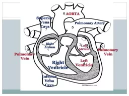
PPT-6.2.U 8 The heart beat is initiated by a group of specialized muscle cells in the right

Author : robaut | Published Date : 2020-06-19

sinoatrial node AND 62U9 The sinoatrial node acts as a pacemaker AND 62U10 The sinoatrial node sends out an electrical signal that stimulates contraction as

Presentation Embed Code

Download Presentation

Download Presentation The PPT/PDF document "6.2.U 8 The heart beat is initiated by..." is the property of its rightful owner. Permission is granted to download and print the materials on this website for personal, non-commercial use only, and to display it on your personal computer provided you do not modify the materials and that you retain all copyright notices contained in the materials. By downloading content from our website, you accept the terms of this agreement.

6.2.U 8 The heart beat is initiated by a group of specialized muscle cells in the right: Transcript


sinoatrial node AND 62U9 The sinoatrial node acts as a pacemaker AND 62U10 The sinoatrial node sends out an electrical signal that stimulates contraction as it is propagated through the walls of the atria and then the walls of the ventricles. . Digital . Laboratory. It’s best to view this in . Slide Show . mode, especially for the quizzes.. This module will take approximately . 45 . minutes to complete. .. After completing this exercise, you should be able to. What is Muscle Tissue?. Composed of cells that are highly specialized to shorten in length by contractility.. Made up of cells that are called . myocytes. .. Elongated in one direction (muscle . fibres. Thorax. Thorax is the superior part of the trunk between the neck and abdomen.. It extends below the neck to the diaphragm.. It contains the primary organs of respiratory and cardiovascular system. A typical human rib cage consists of 24 ribs, the sternum, costal cartilages, and the 12 thoracic vertebrae.. Pamela BL. Introduction . The cardiac muscle forms the muscular wall of the heart that is it forms the myocardium.. Some of the cardiac muscle is also present in the walls of the aorta, pulmonary vein and superior vena cava.. Capillary brings oxygen, nutrients, hormones to cells. Cells give CO. 2. . , hormones, nitrogenous wastes back to capillaries. Nutrients Exchanged. Filtration and diffusion occur due to osmotic and blood pressure. Composed of cells that are highly specialized to shorten in length by contractility.. Made up of cells that are called . myocytes. .. Elongated in one direction (muscle . fibres. ).. Special Terms. Plasma Membrane . Location. Superior surface of diaphragm. Left of the midline. Anterior to the vertebral column, posterior to the sternum. Heart Anatomy. Coverings of the Heart: Anatomy. Pericardium – a double-walled sac around the heart composed of:. Heart Anatomy. Approximately the size of a fist. Location. In the mediastinum between second rib and fifth intercostal space. On the superior surface of diaphragm. Two-thirds to the left of the midsternal line. Understandings. U.D.4.1: structure of cardiac muscle cells allows propagation of stimuli through the heart wall. . U.D.4.2: signals from the sinoatrial node that cause contraction cannot pass directly from atria to ventricles.. It’s best to view this in . Slide Show . mode, especially for the quizzes.. This module will take approximately 45 minutes to complete.. After completing this exercise, you should be able to:.  . Distinguish, at the light microscope level, each of the following::. a closed fist. It is located in the . mediastinum. , which is the area in the center of the chest, under the sternum. The heart acts as a . double-sided pump. , moving deoxygenated blood to the lungs and oxygenated blood to the body. Left Atrium. Left Ventricle. Pulmonary Artery. Superior . Vena . Cava. Inferior. Vena. Cava. Pulmonary . Vein. Pulmonary . Vein. For the following slides. Blue = Deoxygenated. Red = Oxygenated . . 1. . DR.SAEED VOHRA. OBJECTIVES. At the end of the lecture, the student should be able to :. Describe the shape of heart . regarding . apex. , base, . sternocostal. and diaphragmatic surfaces.. Describe the interior of heart . Cultural connection. https://www.youtube.com/watch?v=w-j4ZiMZZTg. Diabetes has become very prevalent in Native American communities. Many people with diabetes experience discomfort in their legs and feet, with symptoms such as cramping, numbness, tingling, and pain. One of the culprits is poor circulation (along with nerve damage)..

Download Document

Here is the link to download the presentation.
"6.2.U 8 The heart beat is initiated by a group of specialized muscle cells in the right"The content belongs to its owner. You may download and print it for personal use, without modification, and keep all copyright notices. By downloading, you agree to these terms.

Related Documents