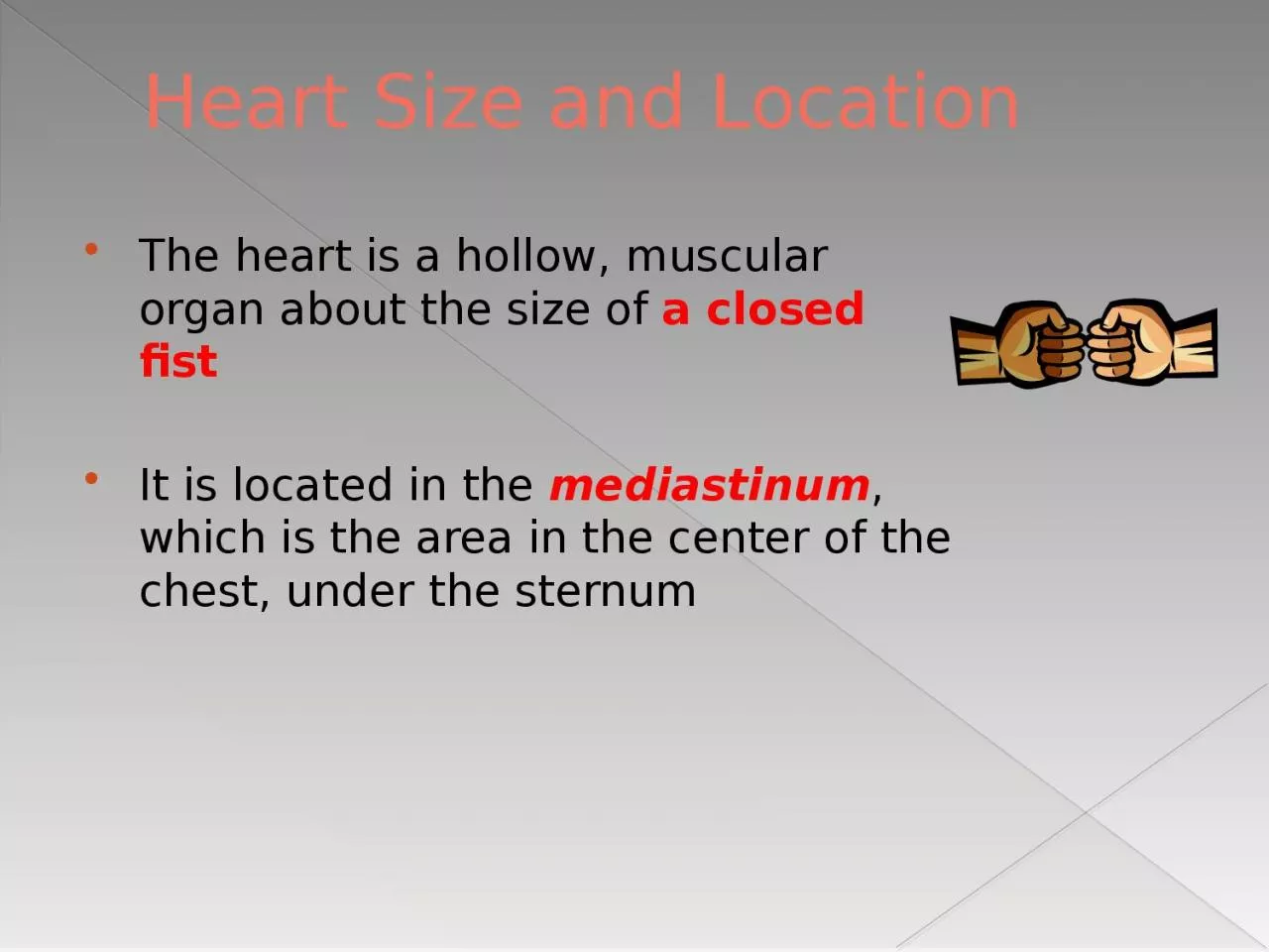
/
Heart Size and Location The heart is a hollow, muscular organ about the size of Heart Size and Location The heart is a hollow, muscular organ about the size of

Heart Size and Location The heart is a hollow, muscular organ about the size of - PowerPoint Presentation

belinda
belinda . @belinda
Follow
345 views
Uploaded On 2022-06-18

Heart Size and Location The heart is a hollow, muscular organ about the size of - PPT Presentation

a closed fist It is located in the mediastinum which is the area in the center of the chest under the sternum The heart acts as a doublesided pump moving deoxygenated blood to the lungs and oxygenated blood to the body ID: 920864

blood heart atrium ventricle heart blood ventricle atrium left cont vessels chambers veins pulmonary body valve valves lungs deoxygenated

Share:

Link:

Embed:

Download Presentation from below link

Download Presentation The PPT/PDF document "Heart Size and Location The heart is a h..." is the property of its rightful owner. Permission is granted to download and print the materials on this web site for personal, non-commercial use only, and to display it on your personal computer provided you do not modify the materials and that you retain all copyright notices contained in the materials. By downloading content from our website, you accept the terms of this agreement.


Presentation Transcript

Slide1

Heart Size and Location

The heart is a hollow, muscular organ about the size of a closed fistIt is located in the mediastinum, which is the area in the center of the chest, under the sternum

Slide2

The heart acts as a

double-sided pump, moving deoxygenated blood to the lungs and oxygenated blood to the body

Slide3

Layers of the Heart

The pericardium is the protective sac that surrounds the heart.The epicardium is the thin, protective outer layer of the heart, composed of

connective tissue.

Slide4

Layers of the Heart Cont.

The myocardium is the thick, middle, muscular layer.The myocardium performs the powerful pumping action (contractions) of the heart.The

endocardium

is the thin, smooth inner lining.

Slide5

Layers of the Heart

Slide6

Chambers of the Heart

The heart is divided into four chambersright atriumleft atriumright ventricle left ventricle

Slide7

Chambers of the Heart Cont.

The atria (plural) are the small top chambers of the heart. An atrium (singular) is one of the top chambers of the heart.There is a right atrium and a left atrium.

Slide8

Chambers of the Heart Cont.

The right atrium receives deoxygenated blood from the superior and inferior vena cava.The left atrium receives oxygenated blood from the lungs via the pulmonary veins.

Slide9

Chambers of the Heart Cont.

The ventricles are the large lower chambers of the heart. There is a right ventricle and a left ventricle.

Slide10

Chambers of the Heart Cont.

The right ventricle receives blood from the right atrium via the tricuspid valve.The left ventricle receives blood from the left atrium via the bicuspid valve.The left ventricle is the largest, strongest chamber and is considered the “

powerhouse

” of the heart.

Slide11

Valves

The heart contains four valves, which keep blood flowing in one direction.

Slide12

Valves Cont.

Semilunar valves: The pulmonary semilunar valve is between the right ventricle and the pulmonary artery.The aortic valve is between the left ventricle and the aorta

Slide13

Valves Cont.

Atrioventricular valves are between an atrium and a ventricle.The tricuspid valve is between the right atrium and the right ventricle.The bicuspid valve is between the left atrium and the left ventricle.

Slide14

Types of Blood Vessels

Veins are blood vessels that carry blood toward the heart.Venules are small veins.Arteries

are blood vessels that carry blood away from the heart.

Arterioles

are small arteries.

Capillaries

are the smallest blood vessels.

Capillaries connect

venules to arterioles

, and they are the exchange site for oxygen, carbon dioxide, nutrients, and other substances that travel through the blood.

Slide15

Major Blood Vessels

The superior vena cava is one of the largest veins in the body. It carries deoxygenated blood from the upper half of the body to the right atrium of the heart.

Slide16

Major Blood Vessels Cont.

The inferior vena cava is also one of the largest veins in the body. It carries deoxygenated blood from the lower half of the body toward the right atrium of the heart.

Slide17

Major Blood Vessels Cont.

Pulmonary is a medical term that refers to the lungs. The pulmonary arteries are arteries that carry deoxygenated blood away from the right ventricle of the heart to the lungs.

There are right and left pulmonary arteries.

Slide18

Major Blood Vessels Cont.

The pulmonary veins are veins that carry newly oxygenated blood from the lungs toward the left atrium of the heart.The aorta is the largest artery in the body. The aorta carries oxygenated blood coming from the left ventricle of the heart to the body.