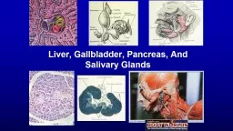
LIVER HISTOLOGY Largest gland of the body Present under diaphragm and occupies the entire right hypochondrium part of epigastrium and left hypochondrium Functions synthesis of urea formation and secretion of bile detoxification of metabolic wastes storage of glycogen metab ID: 914790
Download Presentation The PPT/PDF document "HISTOLOGY OF LIVER DR. AKANKSHA SINGH" is the property of its rightful owner. Permission is granted to download and print the materials on this web site for personal, non-commercial use only, and to display it on your personal computer provided you do not modify the materials and that you retain all copyright notices contained in the materials. By downloading content from our website, you accept the terms of this agreement.
Slide1
HISTOLOGY OF LIVER
DR. AKANKSHA SINGH
Slide2LIVER HISTOLOGY
Largest gland of the body.
Present under diaphragm and occupies the entire right
hypochondrium
, part of
epigastrium
and left
hypochondrium
.
Functions – synthesis of urea , formation and secretion of bile, detoxification of metabolic wastes, storage of glycogen, metabolism of
cholestrol
and fat, synthesis of plasma proteins etc.
Slide3Slide4Slide5Histology of liver
Slide6Slide7C
lassic lobule
It forms
the structural and functional unit of the organ.
Hexagonal in shape.
It has a vein at the centre, the
central vein
Portal tracts in the periphery.
Slide8Slide9Slide10Slide11LIVER ACINUS
It is the
functional unit of the liver.
Diamond shaped.
At the two opposite angles of the diamond shaped
acinus
are
central veins
and at the other two opposite site angles are
portal triads
.
Slide12RAPPAPORT’S CLASSIFICATION OF LIVER (Very important)
The
acinus
is
devided
into 3 zones according to
distance from the blood supply:
Zone1
:
hepatocytes
are closest to blood vessels and they are
best oxygenated
.
Zone2
:
less oxygenated
than zone1.
Zone 3
: which is farthest from blood vessels (near the central vein) has the
poorest oxygen supply
.
Slide13Liver histology
Slide14PORTAL TRIADS-
It consists of
portal vein, bile duct and hepatic artery.
Slide15Slide16Slide17Slide18Slide19Slide20Slide21Slide22Slide23Slide24Slide25Space of Disse
and MALL
The space of Mall is a primary component of the digestive system. This is due to the fact that
bile
produced
by the liver is collected here. Ducts draining the bile manufactured by the liver are surrounded by a mesh of tiny branches of the hepatic artery. Blood flowing from the hepatic artery goes through this space before draining off into the portal veins, while the bile flows out through the bile ducts to the
small intestine
, with some stored in the gallbladder.
The space of
Disse
is a smaller continuation of the space of Mall. This smaller space is located between individual liver cells and tiny
blood vessels
. While the space of Mall enables exchange of arterial and venous blood, along with bile collection and transport out of the liver, the space of
Disse
provides nutrition to individual liver cells. It does this by
perfusing
proteins and other cell nutrition substances from adjacent arterial vessels directly into liver cells, where they are absorbed.
Slide26Slide27Slide28PANCREAS
Retroperitoneal organ.
Extends from the concavity of the duodenum to the
hilum
of the spleen.
Microscopic features: it has 3 components:
stroma
, parenchyma, and duct system.
stroma
:
the gland is covered with
a thin capsule of connective tissue.
From the capsule numerous septa arise which divide the parenchyma of the gland into lobules.
Parenchyma-
having
exocrine and endocrine parts
Exocrine part and duct system
Pancreatic
acini
-
pyramidal shaped cells
with a
small lumen
having
rounded nuclei
at its base.
Cytoplam
is
eosinophilic
at apex
nd
basophilic near the base of the cells
(like parotid gland)
Duct
System-
small duct-
intralobular
duct
lined by
simple
cuboidal
epithelium
and larger duct –
interlobar
duct
lined by
columnar epithelium.
Cenroacinar
cells –
are seen at the centre of the
acini
where duct system begins. Having central nucleus and pale cytoplasm.
Slide30ENDOCRINE PARTS
ISLETS OF LANGERHANS-
Polyhedral cells, arranges in irregular cords. In between the cords there are capillaries.
(
characterstic
feature)
Islet of
langerhans
have 4 types of cells: Alpha, beta,
delta,PP
Cells.
Beta cells are located in the centre of islets, alpha and delta cells are in the periphery and PP CELLS are found throughout the islets.
Slide31Slide32Slide33Gall bladder
(
no
muscularis
mucosa and
submucosa
)
Simple columnar epithelium
Lamina
propria
Muscularis
externa
adventitia
serosa
Slide34Blind sac, present at inferior surface of the liver.
Microscpic
features- from lumen to outside-
Mucosa
:
mucosa has numerous folds.
Lining epithelium
–
simple columnar epithelium with
no goblet cells,
lies over lamina
propria
. Epithelial cells have
microvilli
.
No
musculars
mucosa (imp)
Muscularis
externa
–
smooth muscles are
arreanged
in circular, longitudinal and oblique manner. Numerous collagen and elastic fibers.
Adventitia and
serosa
No
submucosa
in gall bladder.(imp)
Slide35Important questions
What is?
(liver histology very imp for
theroy
exams as well)
1) Classic lobule, portal lobule, liver
acinus
2)
Rappaport’s
classification of liver
3) Space of
disse
and mall
4) Islets
of
langerhans