/
 Histology Histopathology,  Histology Histopathology,

Histology Histopathology, - PowerPoint Presentation

lois-ondreau
lois-ondreau . @lois-ondreau
Follow
342 views
Uploaded On 2020-04-04

Histology Histopathology, - PPT Presentation

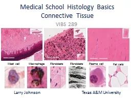
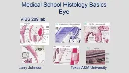
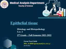
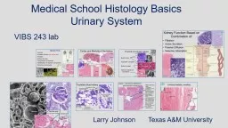
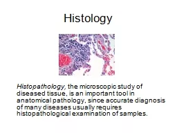
the microscopic study of diseased tissue is an important tool in anatomical pathology since accurate diagnosis of many diseases usually requires histopathological examination of samples Histology Processing ID: 775367

liver blood cells kidney liver blood cells kidney vein cell tissue duct filament gill trunk bile 400x histology hepatic

Share:

Link:

Embed:

Download Presentation from below link

Download Presentation The PPT/PDF document " Histology Histopathology," is the property of its rightful owner. Permission is granted to download and print the materials on this web site for personal, non-commercial use only, and to display it on your personal computer provided you do not modify the materials and that you retain all copyright notices contained in the materials. By downloading content from our website, you accept the terms of this agreement.


Presentation Transcript

Slide1

Histology

Histopathology, the microscopic study of diseased tissue, is an important tool in anatomical pathology, since accurate diagnosis of many diseases usually requires histopathological examination of samples.

Slide2

Histology Processing

Samples fix in 10% NBF.

48hrs -72 hrs -days

Slide3

Paraffin infiltration and embedding into blocks.

Cuts thin sections on slides (3-5um thick).

Slide4

Deparaffinization and Rehydration

Slide5

Stain tissue sections

Hematoxylin and eosin stain (H & E stain)

structures staining blue are called basophilic because of their affinity for the basic dye (hematoxylin) structures staining pink are called acidophilic because of their affinity for the acid dye (eosin),

Slide6

Liver

Hepatocytes

– functional liver cells

Hepatic Sinusoids

– small blood vessel within liver tissue

Hepatic Portal Vein

– brings blood into the liver

Central Vein

– takes blood out of the liver

Bile Duct

– storage and drainage of bile

Arteriole

– delivers blood from artery to capillaries

Glycogen Vacuole

– used for glycogen storage

Slide7

Hepatic sinusoid

Hepatocyte

Glycogen

vacuole

Liver

1000X

Slide8

Erythrocytes visible within the central vein

Hepatic sinusoids

Liver

400x

Slide9

400X

Liver

Arteriole

Slide10

RBC

Arteriole

Central vein

Liver

Slide11

Liver

Bile Duct

Slide12

Liver

Bile Duct

Slide13

Liver

White Blood Cells

Slide14

Head Kidney

Interrenal gland

– Is a cortisol producing tissue..

Chromaffin cell

– neuroendocrine cells that catalyze and secrete epinephrine, norepinephrine, and other hormones

Postcardinal vein

– Blood transport

Hematopoietic tissue

– where new blood cells are formed

Slide15

A

B

400x

C

Interrenal Gland

Chromaffin Cell

Postcardinal vein

Head Kidney

Slide16

Head Kidney

Chromaffin

cells

Cardinal

Vein

Interrenal cells

Slide17

Trunk Kidney

Nephron – a renal tubule that removes waste and helps maintain homeostasisRenal corpuscleGlomerulus – Pressure filters blood plasma into Bowman’s capsuleBasement membrane – glomerulus membrane that does the blood filteringBowman’s capsule – collects filtered blood plasma and connects with proximal tubuleProximal tubule – selectively transports nutrients (amino acids, glucose, salt, ect.) back into the bloodDistal tubule – tubular secretion of molecules to balance pH Melanocytes – pigment cellsCollecting duct and/or Mesonephric duct – collects urea from multiple nephrons and controls futher water reabsorptionConnective tissue – fibres surrounding ducts

Slide18

Proximal

tubules

Distal

tubules

Trunk

Kidney

Renal corpuscle

Slide19

Glomerulus

Basement membrane

Bowmans

capsule

Trunk

Kidney

Slide20

A

B

Mesonephric Duct

Melanocytes

Connective tissue

400x

C

Trunk

Kidney

Slide21

Gills

Filament

– threadlike structure forming respiratory surface

Lamella

– fingerlike projections of the filament

Goblet (or mucous) cell

– secret mucus

Chloride cell

– acid/base regulation by Cl

-

and HCO

3

-

excretion

Cells surface area becomes enhanced during alkalosis and decreases during acidosis

Pillar (or pilaster) cell

– lend support for lamelle

Gill filament cartilage

– center of filament for structural support

Slide22

Gill filaments and lamellae

Lamella

Filament cartilagePillar cellChloride cell

40x

A

B

C

Gill

Slide23

Goblet

cells

Epithelial

cells

Gill

Slide24

Pillar

cells

Erythrocytes

Chloride

cell

Gill

Slide25

Laboratory Steps

Use proper microscope handling procedures (distributed in Lab 2)

Examine histology slides of liver, kidney, and gills

Examine additional histology slides of other organs