/
 Medical School Histology Basics Eye  Medical School Histology Basics Eye

Medical School Histology Basics Eye - PowerPoint Presentation

luanne-stotts
luanne-stotts . @luanne-stotts
Follow
353 views
Uploaded On 2020-04-03

Medical School Histology Basics Eye - PPT Presentation

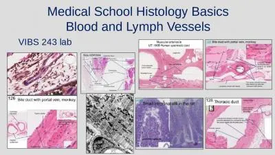
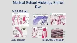
VIBS 243 lab Larry Johnson Texas AampM University OUTLINE OVERVIEW CELLULAR STRUCTURES THROUGH WHICH LIGHT PASSES CORNEA LENS RETINA STRUCTURES WHICH INFLUENCE THE IMAGE ID: 775212

lens iris eye epithelium lens iris eye epithelium muscle cells isbn edition ciliary chamber retina histology 34412 192 anterior

Share:

Link:

Embed:

Download Presentation from below link

Download Presentation The PPT/PDF document " Medical School Histology Basics Eye" is the property of its rightful owner. Permission is granted to download and print the materials on this web site for personal, non-commercial use only, and to display it on your personal computer provided you do not modify the materials and that you retain all copyright notices contained in the materials. By downloading content from our website, you accept the terms of this agreement.


Presentation Transcript

Slide1

Medical School Histology Basics Eye

VIBS 243 lab

Larry Johnson Texas A&M University

Slide2

OUTLINE

OVERVIEWCELLULAR STRUCTURES THROUGH WHICH LIGHT PASSESCORNEA LENSRETINASTRUCTURES WHICH INFLUENCE THE IMAGEIRISCILIARY BODIESTRABECULAR MESHWORK

Aqueous humor

Vitreous humor

Ref code

#

6

Slide3

VITREOUS BODY

(VITREOUS HUMOR)

Aqueous

humor

Ref code

#

6, 10

Slide4

34412

Ref code

# 5

Slide5

Eye – conjuctiva on white of eye

192

sclera

Conjunctiva

Stratified squamous

epithelium withsome goblet cells

Ref code

#

10

Slide6

Eye

192

human and 34412 monkey

Cornea

Pupil

Iris

Anterior chamber

Posterior chamber

Vitreous body

Optic nerve

Sclera

Connective tissue

Chorioid layer

Lens

192

34412

Nerve

34412

Retina

Ciliary body

Aqueous humor

Slide7

Ref code

#

5, 6

Slide8

Eye – corneal layers

192

Bowman's membrane

Corneal stroma

Descemet's Membrane

Corneal

endothelium

Stratified squamous

Epithelium

Corneal fibroblasts

Ref code

#

6

Slide9

corneal layers Iris of Eye (toluidine blue)

292

Bowman's membrane

Corneal stroma

Descemet's

Membrane

Corneal

endothelium

Sclera

Sclera-cornea junction (limbus)

Anterior

chamber

Posterior chamber

Iris

Iris

Ciliary muscle:

smooth muscle

Aqueous

humor

Aqueous

humor

Lens

Slide10

292 Ciliary muscle: smooth muscle

Ciliary muscle

Nerves

Blood vessel

Ciliary processes

lens

192

Slide11

Eye – ciliary muscle

192

Ciliary muscle

Ciliary processes

Anterior

chamber

Sclera-cornea junction (limbus)

Conjunctival

sac

Ciliary muscle (smooth muscle)

Artifactual

space

Posterior chamber

Pupil

Iris

Slide12

192 Eye – secretory cells

192

Ciliary processes

Secretory cells

Secretory cells

become the

pigmented cells

of the iris

Iris

Pigmented cells of

the ciliary processes

become the dilator

muscle of the iris

Pigmented cells

Slide13

Eye (toluidine blue)

292

Trabecular meshwork

Conjunctiva

Air

Limbus

Posterior chamber

Ciliary

processes

Ciliary muscle

Iris

Anterior

chamber

Endothelium of trabecular meshwork

is continuous with the transport endothelium of the cornea

Iris

Canal of Schlemm

Slide14

Eye – trabecular meshwork and canal of Schlemm

192

Trabecular meshwork

Canal of Schlemm

Ciliary muscle

Anterior chamber with

aqueous humor

Limbus

Slide15

34412

Canal of Schlemm

Trabecular meshwork

Iris

Posterior chamber with

aqueous humor

Blood

vessels

Monkey eye

Slide16

Eye (toluidine blue)

292

Anterior chamber

Posterior chamber

Iris

Pupil

Lens

Constrictor

muscle

Dilator

muscle

Iris

Cuboidal

epithelium

on anterior surface of lens

Lens capsule

Iris

pupil

Lens

Ref code

#

10

Slide17

Eye (toluidine blue)

292

Lens

Lens

capsule

Lens cuboidal

epithelium

Iris constrictor smooth muscle

Lens

Slide18

The capsule is a shared basement membrane of the lens epithelium

Ref code

#

10

Slide19

Lens

The capsule is a shared basement membrane of the lens epithelium

Ref code

#

10

Slide20

Lens and iris

192

and 34412

Iris constrictor muscle is smooth muscle

Iris

Posterior chamber

Lens cuboidal

epithelium

Lens capsule

Pigmented

epithelium of iris

Iris

34412

192

Lens anterior surface

Slide21

Eye (toluidine blue) elongation of lens cuboidal cells at bow region

292

Lens cuboidal epithelium on its anterior surface gives rise to elongated lens fibers in the bow region, and the shared basement membrane of these cells is the capsule

Lens cuboidal epithelium

flattened lens fibers

Artifactual space

Capsule

No cuboidal

epithelium on the posterior surface of the lens and the capsule is thinner there

34412

Capsule

Slide22

34412

lens

Ligaments

(zonules)

lens

Iris

Eye monkey

Sites of attachment of zonules to the lens’ capsule

192

One site of attachment of a zonule to the capsule on the lens on its posterior surface

Anterior surface

Anterior surface

Posterior surface

Slide23

Zonules

34412

Sclara

Ciliary muscle

192

Capsule and cuboidal epithelium on the anterior surface of lens

Thinner capsule on the posterior surface of the lens

Lens fibers

Slide24

Eye monkey

Central artery

of the retina

Capillary network

in retina

Pigmented layer

of epithelial cells

Elaborate network

of vessels, the

choroid plexus.

34412

Slide25

Ref code

#

5

Slide26

34412

Retinal

pigmented epithelium

Venule in the

choroid plexus

Photosensitive

portion of rod

and cone cells

Vitreous body

Ganglion cell layer

Sclera

Bipolar cells

Artifactual space

Cones

Rods

Artifactual space

Retinal

pigmented

epithelium

Slide27

Rods

Cones

Nuclei of rod and cone cells

= outer nuclear layer

Retinal pigmented epithelium

Photosensitive portion of rod and cone cells

Slide28

Eye - Retina and choroid:

Choroid

Artifact space

Retina

Choroid plexus

Blood vessels

in the retina

Retinal

pigment

epithelium

Melanin

granules

Nucleus

Optic nerve

292

Slide29

Retina (toluidine blue)

292

Rods

Bipolar cells

Photoreceptor cells

Artifactual space

Optic

nerve

Capillary

In retina

Cones

Slide30

Macula with top two layers pulled back to aid vision

Typical retina

Ganglion cell layer

Bipolar cells

Retinal

pigmented

epithelium

Rods and cones

Rods and cones

Retinal pigmented epithelium

Slide31

192

Retina

Lens

Vitreous body

Cornea

Sclera

Sclera

Iris

Aqueous

humor

Anterior

chamber

Posterior chamber

Optic nerve

lens

Cornea

292

Conjunctiva

Choroid plexus

Slide32

34412

Blind spot

where

optic nerve enters

the retina

Choroid

Photosensitive region

of rods and cones

Pigmented epithelium

Ganglion cell layer

Bipolar cell layer

Slide33

Eye (toluidine blue)

292

Blind spot

where

optic nerve enters

the retina

Slide34

34412

OPTIC CUP AND

NERVE HEAD

Retina

Retina

Choroid plexus

Slide35

In summary

Ref code

#

6, 10

Slide36

1. Alberts, et al., 1989. Molecular Biology of the Cell. 2nd Edition. Garland Publishing, Inc. New York. ISBN 0-8240-3695-6.2. Alberts, et al., 1994. Molecular Biology of the Cell. 3nd Edition. Garland Publishing, Inc. New York. ISBN 0-8153-1619-4.3. Bloom, W. and Fawcett, D.W., 1968. A Textbook of Histology. 9th Edition. W.B. Saunders Company. Philadelphia. Library of Congress #67-17445.4. Elias, H. et al., 1978. Histology and Human Microanatomy. A Wiley Medical Publication. John Wiley & Sons, New York. ISBN 0-471- 04929-8.5. Eroschenko, V. 2000. Di Fiore’s Atlas of Histology with Functional Correlations. 9th Edition. Lippincott Williams & Wilkins. Philadelphia. ISBN 0- 7817-2676-X.6. Fawcett, D.W., 1986. Bloom and Fawcett. A Textbook of Histology. 11th Edition. W.B. Saunders Company. Philadelphia. ISBN 0-7216- 1729-8.7. Fawcett, D.W., 1994. Bloom and Fawcett. A Textbook of Histology. 12th Edition. Chapman and Hall. New York. ISBN 0-412-04691-1.8. Guyton, A.C. 1971. Textbook of Medical Physiology. 4th Edition. W.B. Saunders Company. Philadelphia. Library of Congress # 74- 118589.9. Ham, A.W. 1974. Histology. 7th Edition. J.B. Lippincott Company. Philadelphia. ISBN 0-397-52062-X.10. Ham, A.W. and Cormack, D.H. 1979. Histology. 8th Edition. J.B. Lippincott Co. Philadelphia. ISBN 0-397-52089-1.11. Junqueria, et al., 1995. Basic Histology. 8th Edition. Appleton and Lange. Norwalk, Connecticut. ISBN 08385-0567-8.12. Junqueira, et al., 1998. Basic Histology. 9th Edition. Appleton and Lange. Stamford, Connecticut. ISBN 0-8385-0590-2.13. Knobil, E. et al. 1988. The Physiology of Reproduction. Volume 1. Raven Press. New York. ISBN 0-88167-281-5.14. Langley, et al., 1974. Dynamic Anatomy and Physiology. 4th Edition. McGraw-Hill Book Company. New York. ISBN 0-07-036274-2.15. Mescher, A.L., 2010. Junqueira’s Basic Histology Text and Atlas. 12th Edition. McGraw Hill Medical. New York. ISBN 978-0-07-160431- 4.16. Tuttle, W.W. and Schottelius, B.A. 1969. Textbook of Physiology. 16th Edition. The C.V. Mosby Company. Saint Louis. Library of Congress # 75- 89848.17. Varner, D. et al. 1991. Diseases and Management of Breeding Stallions. American Veterinary Publications. Goleta, California. ISBN 0- 939674-33- 5.18. Von Hagens, Gunther and A.Whalley, 2007. Body Worlds – The Anatomical Exhibition of Real Human Bodies. ISBN 978-3-937256-04-719. Weiss, L. 1983. Histology: Cell and Tissue Biology. 5th Edition. Elsevier Biomedical. New York. ISBN 0-444-00716-4.20. Weiss, L. and Greep, R. 1977. Histology. 4th Edition. McGraw-Hill Book Company. New York. ISBN 0-07-069091-X.

Many illustrations in these VIBS Histology YouTube videos were modified from the following books and sources: Many thanks to original sources!

Slide37

Questions on the eye

1. The integrity of the relationship among structures of the eye is produced/maintained by: supporting layer of the eye holding pressure precise regulation of pressure via aqueous humor production and outflow the bony structure of the sclera in mammals a and b a, b, and c

2. Which structure is not correctly paired with its function? a. the iris regulates the amount of light that reaches the retinab. ciliary bodies alter the shape of the lens by the attached zonulesc. the trabecular meshwork enhances the outflow of vitreous humord. the cornea focuses the image on the retinae. the retinal and choroid blood vessels provide nutrients to the retina

3. Which function(s)

least

match(s) that of the cornea?

a. protection

b. structural support

c. filter out undesirable light rays

d. focus image on photosensitive portion of the photoreceptor cells

e. none of the above

Slide38

Slide39

The end of