/
Thoracic cage, diaphragm Thoracic cage, diaphragm

Thoracic cage, diaphragm - PowerPoint Presentation

byrne
byrne . @byrne
Follow
342 views
Uploaded On 2022-06-01

Thoracic cage, diaphragm - PPT Presentation

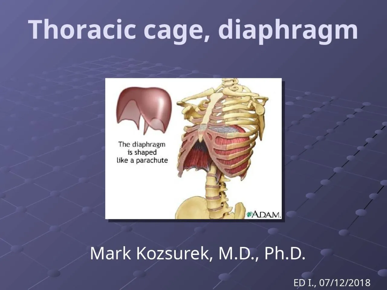
Mark Kozsurek MD PhD ED I 07122018 Questions to be answered How are the bones of the thorax connected together the joints of ID: 913288

rib intercostal thoracic ribs intercostal rib ribs thoracic costal head part joints diaphragm artery internal joint lateral transverse articular

Share:

Link:

Embed:

Download Presentation from below link

Download Presentation The PPT/PDF document "Thoracic cage, diaphragm" is the property of its rightful owner. Permission is granted to download and print the materials on this web site for personal, non-commercial use only, and to display it on your personal computer provided you do not modify the materials and that you retain all copyright notices contained in the materials. By downloading content from our website, you accept the terms of this agreement.


Presentation Transcript

Slide1

Thoracic cage, diaphragm

Mark Kozsurek, M.D., Ph.D.

ED I., 07/12/2018

Slide2

Questions

to be

answered:How are the bones of the

thorax

connected

together

?

the

joints

of

the

thoracic

cage

Which

muscles

act

upon

these

joints

?

the

muscles

of

the

thoracic

cage

How

does

the

thoracic

cage

move

?

the

mechanism

of

breathing

Slide3

1)

How

are

the

ribs connected to the vertebrae?

2)

How

do

the

ribs

attach

to

the

sternum

?

Slide4

1)

How

are

the

ribs

connected

to the vertebrae?

JOINT OF THE HEAD OF THE RIB

- costal facet of the vertebra- articular facet of the head of the rib

COSTOTRANSVERSE JOINT- costal fovea of the transverse process articular facet of the the costal tubercle

COSTOVERTEBRAL JOINTS:

Slide5

Articular surfaces of vertebrae

„TYPICAL VERTEBRAE”

TII-IX present a superior and an inferior costal demifacet.

„ATYPICAL VERTEBRAE”

- TI bears a complete

superior costal facet

for the head of the first rib and an inferior costal demifacet for the head of the second rib.

TX only has a superior costal demifacet. TXI-XII express complete costal facets for the ribs with the same number. They have no costal facets on their transverse processes.TYPICAL

Slide6

Rib I

Ribs II-X

Ribs XI-XII

Ribs

I and XI-XII

articulate

with

only

one vertebra, so they have continuous facets on their heads. The articular surfaces of the heads of ribs

II-X are divided into two by a crest, ribs

XI-XII have no tubercle and do not articulate with transverse processes.Articular surfaces of the ribs

Slide7

Joint of the head of the rib

Costotransverse joint

Slide8

Joint of the head of the rib

Costotransverse joint

Radiate ligament of the head of the rib (not shown)

Slide9

Costotransverse ligament

attaches the neck of the rib to the

transverse process.

Lateral costotransverse ligament

connects the tip of the transverse process to the roughened nonarticular part of the tubercle of the rib.

Superior costotransverse

ligament attaches the neck of the rib to the transverse process of the vertebra above.Intra-articular ligament connects the crest of the head of the rib to the intervertebral disc. Radiate ligament of the head of the rib reinforces the capsule of the joint of the head of the rib.

Slide10

How are the ribs connected to the sternum?

Manubriosternal and xiphisternal joints:

synchondroses, with age they become ossified.

Sternocostal joints:

Rib I: synchondrosis

Ribs II-VII: synovial joints

Intercostal joints:

Ribs VI-Xsynovial joints

Slide11

Thoracic muscles – the intercostal ones

Slide12

vertebra

sternum

rib

innermost intercostal m.

transversus thoracis m.

external intercostal m.

external intercostal membrane

internal intercostal m.

internal intercostal membrane

Slide13

Blood supply and innervation of the thoracic wall

Slide14

Posterior intercostal arteries are direct branches of the thoracic aorta, while the anterior intercostal arteries arise from the internal thoracic artery, a branch of the subclavian artery. Supreme intercostal arteries of the two upper intercostal spaces also come from the subclavian artery.

Posterior intercostal veins drain into the azygos vein (on the right) or into the hemiazygos/ accessory hemiazygos veins (on the left). Anterior intercostal veins open into the internal thoracic vein, which empties into the brachiocephalic vein.

Intercostal nerves exit from the spinal cord and leave the vertebral canal through the intervertebral foramina. Intercostal vessels and nerves give off posterior, lateral and anterior branches to supply the external layers of the thoracic wall.

Slide15

1.

2.

3.

4.

5.

Pectoralis major (and minor)

Serratus anterior

Abdominal muscles

Trapezius (Levator scapulae, Rhomboids, Serratus posterior)

Latissimus dorsi

Slide16

a

b

b

c

c

According to the site of origin muscle fibres constitute the:

a, sternal part

b, costal part

c, lumbar part

superior aspect

Central tendon with the opening for the inferior vena cava

IVC

esophagus

aorta

Diaphragm

Slide17

inferior view

Slide18

sternal part

costal part

lumbar part

sternocostal triangle (of LARREY)

lumbocostal triangle (of BOCHDALEK)

Slide19

Blackboard!

Slide20

L3

L1

L2

lateral, medial, median

arcuate ligaments

medial part

lateral part

Slide21

Caval opening

: IVC, right phrenic nerve, central tendon, T8

Slide22

Esophageal opening

: esophagus, ant. et post. vagal trunk, T10

Slide23

Aortic hiatus:

aorta, thoracic duct (the greatest lymph vessel), L1

Slide24

Sternocostal triangle of Larrey:

internal thoracic artery and vein *

* they are called superior epigastric a. et v. bellow the diaphragm

Slide25

Between the medial and lateral parts

(variable!!!):

greater splanchnic nerve, azygos/hemiazygos veins, lesser splanchnic nerve and sympathetic trunk

Slide26

Slide27

Innervation, blood supply of the diaphragm

Slide28

Internal thoracic artery (ITA) is one of the first branches of the subclavian artery. ITA gives off the pericardiacophrenic artery first which descends together with the phrenic nerve between the pleura and the pericardium. Before reaching the trigone of Larrey, ITA gives off the musculophrenic artery contributing to the blood supply of the diaphragm.

Already from the abdominal aorta the inferior phrenic arteries ascend to the inferior surface of the diaphragm.Phrenic nerve arises mainly from the C4 segment, this is why injuries of the neck around or above this level may cause sudden death due to the palsy of the diaphragm.

Slide29

Ribs rotate around an axis defined by the two costovertebral joints.

Respiratory movements

Slide30

During inspiration both the anteroposterior and lateral diameters of the chest increase!

Slide31

Slide32

Inspiration (active)

Expiration (passive)

Quiet breathing

Note:

The position of the central tendon and the heart resting on it is not changing during quiet breathing!!!

In midposition (green) the right dome of the diaphragm reaches the level of the 5th rib,

while on the left it is in the 5th intercostal space.

Slide33

External and internal intercostal muscles are perpendicular to each other!

90

°

Slide34

external intercostal

internal intercostal

Slide35

Slide36

Take a deep breath and relax!

It’s over!