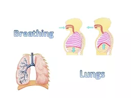
/
Bronchi, lungs, pleura and diaphragm Bronchi, lungs, pleura and diaphragm

Bronchi, lungs, pleura and diaphragm - PowerPoint Presentation

skylar
skylar . @skylar
Follow
0 views
Uploaded On 2024-03-13

Bronchi, lungs, pleura and diaphragm - PPT Presentation

Dr Paul Strutton Department of Surgery amp Cancer Faculty of Medicine Monday 18th February 2013 Bronchial tree Trachea Extends from vertebral level C6 to T45 Held open by Cshaped cartilage rings ID: 1047864

bronchi lung pleural lungs lung bronchi lungs pleural diaphragm cavity pleura left surface ribs thoracic costal anterior abdominal main

Share:

Link:

Embed:

Download Presentation from below link

Download Presentation The PPT/PDF document "Bronchi, lungs, pleura and diaphragm" is the property of its rightful owner. Permission is granted to download and print the materials on this web site for personal, non-commercial use only, and to display it on your personal computer provided you do not modify the materials and that you retain all copyright notices contained in the materials. By downloading content from our website, you accept the terms of this agreement.


Presentation Transcript

1. Bronchi, lungs, pleura and diaphragmDr Paul StruttonDepartment of Surgery & Cancer Faculty of MedicineMonday 18th February 2013

2. Bronchial treeTrachea Extends from vertebral level C6 to T4/5Held open by C-shaped cartilage ringsLowest ring has a hook – carina (keel of ship) Primary (main) bronchi (left and right)Formed at T4/5Right wider and more vertical than leftLobar (secondary) bronchi Formed within the lungsSupply the lobes of the lungsSegmental (tertiary) bronchiSupply the bronchopulmonary segments

3. Bronchial treeTrachea divides into two main bronchi at vertebral level T4/5: Right and left main (primary) bronchi divide into:Lobar (secondary) bronchi - supplying the lobes (2 left, 3 right)The lobar bronchi further subdivide into segmental (tertiary) bronchi supplying self-contained independent units of lung tissue named bronchopulmonary segments

4. Bronchial tree:Branching pattern of trachea into main, lobar and segmental bronchi

5. Segmental (tertiary) bronchi

6. Pattern of bronchopulmonary segments: lateral views of both lungsRightLeft10 segments in each lung – smallest, functionally independent region

7. The lungsEssential organs of respiration Situated in the thoraxSeparated from each other by heart and other contents of the mediastinumEach lies freely in its pleural cavity - apart from its attachment to the heart (via pulmonary vessels) and trachea at the lung root (hilum)

8. From the mediastinum, vessels, nerves and bronchi pass though the lung roots into the lungs

9. The LungsConical in shape Apex thoracic inlet oblique - apex rises 3-4 cm above level of first costal cartilageBase concaverests on convex surface of diaphragm3 borders - (edges) - anterior, posterior, inferior 3 surfaces - costal, medial (mediastinal), inferior (diaphragmatic)Diaphragm separates right lung from right lobe of liverleft lung from left lobe of the liver, stomach & spleen

10. Posterior part in contact with thoracic vertebrae Anterior part - deeply concave - accommodates the heart - cardiac impression larger on L than R because of position of heart Above and behind cardiac impression - hilum of the lung where vessels, bronchi & nerves enter & leave the mediastinum Mediastinal surface of lung: general features

11. Left lung – mediastinal aspect

12. The left lungTwo lobes: Superior Inferiorseparated by oblique fissureSuperior lobe lies above the fissure – includes:ApexMost of anterior part of lung

13. Right lung – mediastinal aspect

14. The right lungThree lobes: superiormiddle inferiorSeparated by 2 fissuresoblique fissure - separates inferior lobe from the other 2 lobeshorizontal fissure - separates superior from middle lobeThe right lung is slightly larger than the left

15. The root (hilum) of the lungConnects mediastinal surface to heart and tracheaformed by structures that enter or leave hilum:Principal (primary) bronchusPulmonary artery (deoxygenated blood from RV)2 pulmonary veins (oxygenated blood to LA)Bronchial arteries (oxygenated blood from descending aorta) and veins Pulmonary plexus of nerves (autonomic)Lymph vessels and nodesall enveloped in pleura

16. Hilum of left lungPulmonary arteryPulmonary vein (upper lobe)Primary bronchusBronchial arteryLymph NodePulmonary ligament (inferior fold of pleura)

17. Anterior dissection showing pulmonary vessels and bronchi passing from mediastinum to lungs

18. The Pleura A thin layer of flattened cells supported by connective tissue that lines each pleural cavity and covers the exterior of the lungs2 layersvisceral pleura - covers surface lungs and lines fissures between the lobesparietal pleura - lines inner surface of chest walls Visceral and parietal pleura are continuous with each other around the root of the lung – this is the hilumIn health: Pleural cavity is collapsed, but moist surfaces allow lungs to glide as they expand and collapse

19. Pleural origin:Pleural cavities inside chest wallLined by parietal pleuraLung buds grow into themWithin covering of visceral pleura

20. Anterior view of surface markings of the lungs (purple) and pleural cavity (pale blue)

21. Anterolateral view of surface markings of the lungs (purple) and pleural cavity (pale blue) Note the large costo-diaphragmatic recess of pleura – free of lung except in maximal inspiration

22. BreathingControlled by nervous system and produced by skeletal muscle Brings about inhalation and exhalation of air into/out of the lungs, to ventilate the gas exchange areas - alveolar sacscapacity of thoracic cavity can be increased: by movements of the diaphragmby movements of the ribs

23. Pleural cavity is expanded by muscles in wallsElastic lungs expand with the pleural cavity, sucking air down trachea and bronchi into lungs Mechanism of breathing

24. The diaphragm: the main inspiratory muscleContraction of the diaphragm increases the vertical dimension of the thoracic cavity. When it contracts, the diaphragm presses on the abdominal viscera which initially descend (because of relaxation of the abdominal wall during inspiration)Further descent is stopped by the abdominal viscera, so more diaphragm contraction raises the costal marginIncreased thoracic capacity produced by diaphragm and rib movements in inspiration, reduces intrapleural pressure, with entry of air through respiratory passages and expansion of the lungs

25. Thoracic surface of diaphragmSkeletal muscle from costal marginSheet-like central tendonPericardial sacIVCAOe

26. The margin of the diaphragm (black) is attached to the:costal margin (lower border of the rib cage)xiphoid processends of ribs 11 and 12lumbar vertebrae

27. But:The dome of the diaphragm (red) bulges high inside the rib cage.So high abdominal organs such as liver are covered by diaphragm, pleura and lung

28. The ribs in breathingRibs elevated - anterior ends thrust forward and upwards - increases antero-posterior dimension of thoracic cavity. At same time ribs are everted, increasing transverse diameter of thoracic cavityInternal and external intercostal muscles stiffen the rib cage to increase efficiency of diaphragm

29. Raising the costal margin widens the pleural cavities by raising drooping lateral parts of ribs

30. Another view of the bucket-handle movement of ribs to widen the pleural cavities (red arrows)

31. Raising the costal margin also raises drooping anterior ends ribs, tilting sternum upwards to increase antero-posterior diameter of pleural cavities (pump-handle action – blue arrow)

32. Breathing outQuiet expiration is a passive activity not requiring musclesIt depends on elastic recoil in the elastic tissue throughout the lungs and in the rib cageIn deep or forced expiration, this is assisted by the muscles of the abdominal walls that squeeze the abdominal organs against the diaphragm and pull the lower ribs downward