/
Cardiovascular Disease Cardiovascular Disease

Cardiovascular Disease - PowerPoint Presentation

olivia-moreira
olivia-moreira . @olivia-moreira
Follow
664 views
Uploaded On 2017-10-30

Cardiovascular Disease - PPT Presentation

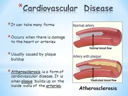
It can take many forms Occurs when there is damage to the heart or arteries Usually caused by plaque buildup Atherosclerosis is a form of cardiovascular disease It is when plaque builds up on the inside walls of the ID: 600829

carbon oxygen lungs dioxide oxygen carbon dioxide lungs breathe alveoli breathing air system blood respiratory bronchi muscles chest asthma cells airways diaphragm

Share:

Link:

Embed:

Download Presentation from below link

Download Presentation The PPT/PDF document "Cardiovascular Disease" is the property of its rightful owner. Permission is granted to download and print the materials on this web site for personal, non-commercial use only, and to display it on your personal computer provided you do not modify the materials and that you retain all copyright notices contained in the materials. By downloading content from our website, you accept the terms of this agreement.


Presentation Transcript

Slide1

Cardiovascular Disease

It can take many formsOccurs when there is damage to the heart or arteriesUsually caused by plaque buildupAtherosclerosis is a form of cardiovascular disease. It is when plaque builds up on the inside walls of the arteries.

AtherosclerosisSlide2

Causes of Plaque Buildup

High fat dietHigh salt dietLow amount of exerciseSmokingFamily historyStressBeing overweightSlide3

Demo

What can happen to our cells if there is too much plaque build up?Slide4

Prevention

We can prevent this by…Slide5

The Respiratory SystemSlide6

The Respiratory System

Respiratory System Note Activity: How do you breathe?(Model lungs and diaphragm) Slide7

The respiratory system is an ­organ system that allows humans to

breatheThe purpose of the respiratory system is to provide oxygen for the cells and remove carbon dioxideThe respiratory system is made up of the lungs, trachea, bronchi, and other structures that help the body intake oxygen and release carbon dioxideSlide8
Slide9

Mouth or Nasal Cavity  Pharynx

Trachea  Bronchi  Bronchioles  AlveoliSlide10

Cilia

and mucus line the walls of the trachea and bronchi and trap bacteria and dirt that are in the air. They sweep them upwards into the throat where the mucus is either swallowed or coughed upSlide11

Video

http://www.youtube.com/watch?v=sU_8juD3YzQSlide12
Slide13

Breathing

Breathing involves inhaling (breathing in) and exhaling (breathing out) When you breathe in, muscles attached to your ribcage and a large muscle called the diaphragm contract which makes the chest largerSlide14

When the chest is larger, air filled with

oxygen is pulled into the lungsWhen the ribcage muscles and diaphragm relax, the chest becomes smaller and the air and carbon dioxide are pushed out (exhaled) Slide15

Gas Exchange

The oxygen in the air we breathe needs to get to our cells. It does this through gas exchange. When the air enters your lungs, it travels to very small air sacs called alveoli which are surrounded by tiny blood vessels called capillariesSlide16

When we breathe in, the amount of oxygen

in the alveoli is greater than the amount of oxygen in the capillaries. The oxygen moves by diffusion into the blood. At the same time, the blood has more carbon dioxide than the alveoli so the carbon dioxide moves into the alveoliThe oxygen that entered the blood moves around the body to the cells that need it. The carbon dioxide that entered the alveoli is released when we exhale. Slide17

THE EXCHANGE OF CARBON DIOXIDE FOR OXYGEN IN THE BLOOD IS THE REASON WHY WE BREATHE!

Video: http://www.youtube.com/watch?v=EFCj9STCvdISlide18

Controlling Breathing

Breathing happens naturally but you can also control itMany people can hold their breath, but once there’s too much carbon dioxide in the blood, you HAVE to breathe!Slide19

HOLD YOUR BREATH!

When carbon dioxide builds up in the blood, it sends a message to the brain. The brain then sends a message to the ribcage muscles and diaphragm to contract. This causes the chest to get bigger and allow oxygen filled air into the lungs!Slide20

Asthma

Asthma is a ­­­­­­­­­­­­­respiratory condition which makes the airways extra sensitive. Certain particles in the air can cause the airway to become red and swollen. When this happens, a lot of ­­mucus­ is produced which makes the airways narrower. This makes breathing difficult!The muscles around your airway tighten and make it even more difficult to breathe. Medication can help to reduce the swelling which makes breathing easier.

Normal airwaysAirways of a person without asthma

Airways of a person with asthma - redness and swelling (inflammation)

Airways of a person with asthma - tightened muscles (bronchospasm)Slide21

Bronchitis

Bronchitis occurs when the bronchi become swollen. This means that the walls of your bronchi are swollen and filled with extra sticky mucus. Airflow into and out of your lungs is partly blocked because of the swelling and extra mucus in your bronchi.Bronchitis can go away on its own or can be treated with medication. Slide22

Smoking

Smoking destroys the tiny hairs (cilia), which line the upper airways and protect against infection. When your lungs' natural cleaning and repair system is damaged, germs, dirt and chemicals from cigarette smoke stay inside your lungs. This puts you at risk for chronic cough, chest infections, and lung cancer. Slide23

The alveoli

are like stretchy balloons. As you breathe in, your alveoli help you absorb oxygen into your body, and as you breathe out, alveoli help get rid of the waste gas carbon dioxide. Smoke damages your lungs so much that your alveoli become less stretchy. This means it's harder for your lungs to take in the oxygen you need and harder to get rid of carbon dioxide.