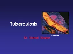
/
1. TUBERCULOSIS 2. CANCER OF THE LUNG 1. TUBERCULOSIS 2. CANCER OF THE LUNG

1. TUBERCULOSIS 2. CANCER OF THE LUNG - PowerPoint Presentation

cady
cady . @cady
Follow
64 views
Uploaded On 2024-01-29

1. TUBERCULOSIS 2. CANCER OF THE LUNG - PPT Presentation

Repiratory block SECOND PRACTICAL Respiratory Block Pathology Dept KSU TUBERCULOSIS Epithelioid and giant cell Granuloma Ghons complex or caseation is present Complications of TB are ID: 1041330

cell lung dept blockpathology lung cell blockpathology dept ksu carcinoma cells respiratory small squamous granulomas large tumor adenocarcinoma metastatic

Share:

Link:

Embed:

Download Presentation from below link

Download Presentation The PPT/PDF document "1. TUBERCULOSIS 2. CANCER OF THE LUNG" is the property of its rightful owner. Permission is granted to download and print the materials on this web site for personal, non-commercial use only, and to display it on your personal computer provided you do not modify the materials and that you retain all copyright notices contained in the materials. By downloading content from our website, you accept the terms of this agreement.


Presentation Transcript

1. 1. TUBERCULOSIS2. CANCER OF THE LUNGRepiratory blockSECOND PRACTICALRespiratory BlockPathology Dept. KSU

2. TUBERCULOSIS Epithelioid and giant cell Granuloma, Ghon’s complex or caseation is present Complications of TB are: - Amyloidosis - Tuberculous pneumonia - Miliary tuberculosis - Tuberculous meningitis - Addison disease Respiratory BlockPathology Dept. KSU

3. On closer inspection, the granulomas have areas of caseous necrosis. This pattern of multiple caseating granulomas primarily in the upper lobes is most characteristic of secondary (reactivation) tuberculosisPulmonary TB – Caseous Necrosis – GrossRespiratory BlockPathology Dept. KSU

4. Extensive caseation and the granulomas involve a larger bronchus causing soft, necrotic center to drain out and leave behind a cavity. Cavitation is typical for large granulomas with TB. Cavitation is more common in the upper lobes.Pulmonary TB – Caseous Necrosis – GrossRespiratory BlockPathology Dept. KSU

5. The Ghon’s complex is seen here at closer range. Primary tuberculosis is the pattern seen with initial infection with tuberculosis in children. Reactivation, or secondary tuberculosis, is more typically seen in adults.Pulmonary TB - Ghon’s Complex – Gross PathologyRespiratory BlockPathology Dept. KSU

6. Miliary TB can occur when TB lung lesions erode pulmonary veins or when extrapulmonary TB lesions erode systemic veins. This results in hematogenous dissemination of tubercle bacilli producing myriads of 1-2 mm. lesions throughout the body in susceptible hosts. Miliary spread limited to the lungs can occur following erosion of pulmonary arteries by TB lung lesions.Miliary TB of the LungsRespiratory BlockPathology Dept. KSU

7. Miliary TB of the Lungs – Cut sectionThis is a "miliary" pattern of granulomas because there are a multitude of small tan granulomas, about 2 to 4 mm in size, scattered throughout the lung parenchyma. The miliary pattern gets its name from the resemblance of the granulomas to millet seeds.Respiratory BlockPathology Dept. KSU lesions resembling millet seed.

8. Miliary TB of the Lungs – X-RayThis chest x-ray shows a patient with miliary TB.Respiratory BlockPathology Dept. KSU

9. At low magnification, this micrograph reveals multiple granulomas. Granulomatous disease by chest radiograph appear as reticulonodular densities.Tuberculous Granulomas - LPFRespiratory BlockPathology Dept. KSU

10. Well-defined granulomas are seen here. They have rounded outlines. The one toward the center of the photograph contains several Langhan’s giant cells. Granulomas are composed of transformed macrophages called epithelioid cells along with lymphocytes, occasional PMN's, plasma cells, and fibroblastsTuberculous Granulomas - HPFRespiratory BlockPathology Dept. KSU

11. The pyknotic nuclei of epithelioid cells in the center of the granuloma (apoptotic bodies) are a precursor of necrosis with focal caseation necrosis.Pulmonary TB - Granuloma with central early necrosisRespiratory BlockPathology Dept. KSUGranulomacentral early necrosiscaseation necrosisepithelioid cells Langhan’s giant cellsLymphocytes

12. The edge of a granuloma is shown here at high magnification. At the upper is amorphous pink caseous material [1] composed of the necrotic elements of the granuloma as well as the infectious organisms. This area is ringed by the inflammatory component [2] with epithelioid cells, lymphocytes, and fibroblasts.12Tuberculous Granulomas - HPFRespiratory BlockPathology Dept. KSU

13. At high magnification, the granuloma demonstrates that the epithelioid macrophages are elongated with long, pale nuclei and pink cytoplasm. The macrophages organize into committees called giant cells. The typical giant cell for infectious granulomas is called a Langhan’s giant cell and has the nuclei lined up along one edge of the cellEpithelioid & Giant cell Granulomas in Tuberculosis Respiratory BlockPathology Dept. KSU

14. A stain for Acid Fast Bacilli is done (AFB stain = Ziehl–Neelsen stain) to find the mycobacteria . The mycobacteria stain as red rods, as seen here at high magnification.Acid Fast bacilli of Mycobacterium TB in the Lung Respiratory BlockPathology Dept. KSU

15. LUNG CARCINOMARespiratory BlockPathology Dept. KSU

16. TWO TYPES OF LUNG CARCINOMANON-SMALL CELL CARCINOMASQUAMOUS CELL CARCINOMAADENOCARCINOMALARGE CELL CARCINOMASMALL CELL CARCINOMAThe NON-small cell cancers behave and are treated similarly, the SMALL cell carcinomas are WORSE than the non-small cell carcinomas, but respond better to chemotherapy, often drastically!Respiratory BlockPathology Dept. KSU

17. 1. Squamous Cell Carcinoma of the lung Most commonly found in men and correlated with smoking.Pathology: more differentiated, more cytoplasm, keratin whorls. Transforms to carcinoma in situ. Grading is based on the amount of keratin & cytoplasm. Respiratory BlockPathology Dept. KSU

18. This is a squamous cell carcinoma of the lung that is arising centrally in the lung (as most squamous cell carcinomas do). It is obstructing the main bronchus. The neoplasm is very firm and has a pale white to tan cut surface.Squamous Cell Carcinoma of the Lung - GrossRespiratory BlockPathology Dept. KSU

19. This is a larger squamous cell carcinoma in which a portion of the tumor demonstrates central cavitation, probably because the tumor outgrew its blood supply. Squamous Cell Carcinoma of the Lung - GrossRespiratory BlockPathology Dept. KSU

20. This chest CT scan view demonstrates a large squamous cell carcinoma of the right upper lobe that extends around the right main bronchus and also invades into the mediastinum and involves hilar lymph nodes.Squamous Cell Carcinoma of the Lung – CT scan Respiratory BlockPathology Dept. KSU

21. Microscopic appearance of squamous cell carcinoma with nests of polygonal cells with pink cytoplasm and distinct cell borders. The nuclei are hyperchromatic and angular.Squamous Cell Carcinoma of the Lung - HPFRespiratory BlockPathology Dept. KSU

22. In this squamous cell carcinoma at the upper right is a squamous eddy with a keratin pearl. At the left, the tumor is less differentiated and several dark mitotic figures are seenSquamous Cell Carcinoma of the Lung - HPFRLRespiratory BlockPathology Dept. KSUkeratin pearl

23. Squamous Cell Carcinoma of the Lung - HPFNeoplastic squamous cells show pleomorphism, hyperchromatism, individual cell keratinization, mitoses and areas of necrosis.Respiratory BlockPathology Dept. KSUkeratin pearl

24. The pink cytoplasm with distinct cell borders and intercellular bridges characteristic for a squamous cell carcinoma of the lungSquamous Cell Carcinoma of the Lung - HPFRespiratory BlockPathology Dept. KSU

25. 2. Adenocarcinoma of the lungThe most common type of lung cancer, making up 30-40% of all cases. Glandular differentiation by tumor cells and 80% of those cells produce mucin. Not as strongly associated with a smoking history as compared to Squamous or Small Cell Carcinomas Adenocarcinoma in situ - called bronchoalveolar carcinoma Early and distant metastases Respiratory BlockPathology Dept. KSU

26. Adenocarcinoma of the Lung – GrossA peripheral adenocarcinoma of the lung. Adenocarcinomas and large cell anaplastic carcinomas tend to occur more peripherally in lung. Adenocarcinoma is the one cell type of primary lung tumor that occurs more often in non-smokers and in smokers who have quit.Respiratory BlockPathology Dept. KSU

27. Adenocarcinoma of the LungCT scans in a 61-year-old man with adenocarcinoma of the lungRespiratory BlockPathology Dept. KSUCT scanX-RayA peripheral adenocarcinoma of the lung appears in this chest radiograph of an elderly non-smoker woman.

28. Microscopically, the Adenocarcinoma in Situ ( Previously named Bronchioloalveolar Carcinoma) is composed of columnar cells that proliferate along the framework of alveolar septae. The cells are well-differentiated. Adenocarcinoma of the Lung – LPFRespiratory BlockPathology Dept. KSU

29. Section of the tumor shows moderately differentiated malignant glands lined by pleomorphic and hyperchromatic malignant cells showing conspicuous nucleoli . Note the presence of tissue desmoplasia around the neoplastic glands . Adenocarcinoma of the Lung – HPFRespiratory BlockPathology Dept. KSU

30. Adenocarcinoma of the Lung – HPFDifferentiated malignant glands lined by pleomorphic and hyperchromatic malignant cells showing conspicuous nucleoliRespiratory BlockPathology Dept. KSU

31. 3. Large Cell Carcinoma of the lung Can be a neuroendocrine carcinoma. Probably represents undifferentiated SCC and adenocarcinomas. Large nuclei, prominent nucleoli. Variation in size and shape. Nuclei normally do not touch due to more cytoplasm. Moderate amount of cytoplasm. Early and distant metastases, sometimes cavitating. Respiratory BlockPathology Dept. KSU

32. Large Cell Carcinoma of the Lung – GrossRespiratory BlockPathology Dept. KSU

33. Undifferentiated Large Cell Carcinoma of the Lung – GrossRespiratory BlockPathology Dept. KSU

34. Large Cell Carcinoma of the Lung – HPFPleomorphic carcinoma of lung (large cell and giant cell subtype). It shows mixed composition of large cell carcinoma and pleomorphic multinucleated giant cells (arrows). (H and E, ×200)Respiratory BlockPathology Dept. KSU

35. Large Cell Carcinoma of the Lung – HPFThis section from lower respiratory tract shows neoplastic cells with abundant pale eosinophilic cytoplasm and a surrounding infiltrate of inflammatory cellsRespiratory BlockPathology Dept. KSU

36. Large Cell Carcinoma of the Lung – HPFThis section shows neoplastic cells with abundant pale eosinophilic cytoplasm and pleomorphic multinucleated giant cells

37. Small cell carcinoma of the lung Highly Malignant Tumor. Cells are small, with scant cytoplasm, ill-defined borders, finely granular chromatin (salt & pepper pattern) and absent or inconspicious nucleoli. High mitotic count and often extensive necrosis. Typically not graded as all SCLC are considered High Grade. Very strong relationship with smoking. Often lead to paraneoplastic syndromes. Respiratory BlockPathology Dept. KSU

38. Arising centrally in this lung and spreading extensively is a small cell anaplastic (oat cell) carcinoma. The cut surface of this tumor has a soft, lobulated, white to tan appearance. The tumor seen here has caused obstruction of the main bronchus to left lung so that the distal lung is collapsedSmall Cell Carcinoma of the Lung “Oat cell” – GrossRespiratory BlockPathology Dept. KSU

39. Small Cell Carcinoma of the Lung “Oat cell” – Gross Oat cell carcinoma which is spreading along the bronchi. The speckled black rounded areas represent hilar lymph nodes with metastatic carcinomaRespiratory BlockPathology Dept. KSU

40. This chest radiograph demonstrates a mass lesion in the right upper lobe. This was an oat cell carcinoma (yellow arrow). It obstructed the right main bronchus, leading to atelectasis on the right, evidenced by a raised right hemidiaphragm. The patient aspirated gastric contents, producing a diffuse pneumonia (blue arrow) on the left (since aspirated material could not pass the obstruction on the right).Small Cell Carcinoma of the Lung “Oat cell” : X-Ray

41. This is the microscopic pattern of a small cell anaplastic (oat cell) carcinoma in which small dark blue cells with minimal cytoplasm are packed together in sheets.Small Cell Carcinoma of the Lung “Oat cell” – HPFRespiratory BlockPathology Dept. KSU

42. Small cell carcinoma “Oat cell” of the lung - HPF Section of the tumor shows clusters of malignant cells which are small , round , ovale , or spindle shaped with prominent nuclear molding , finely granular nuclear chromatin (salt and pepper pattern ) , high mitotic count and focal necrosis Respiratory BlockPathology Dept. KSU

43. Metastatic tumours of the lungRespiratory BlockPathology Dept. KSU

44. METASTATIC TUMORSLUNG is the MOST COMMON site for all metastatic tumors, regardless of the site of origin.It is the site of FIRST CHOICE for metastatic sarcomas for purely anatomic reasons !Respiratory BlockPathology Dept. KSU

45. Multiple variably-sized masses are seen in all lung fields. These tan-white nodules are characteristic for metastatic carcinoma. Metastases to the lungs are more common even than primary lung neoplasms Chest X-ray showing multiple cannon ball opacities in both lung fields. Metastatic Tumors of the Lung – Gross & X-rayRespiratory BlockPathology Dept. KSU

46. Here are larger but still variably-sized nodules of metastatic carcinoma in lung.CT Lung shows Cannonball Metastases-large, hematogenously spread metastatic lesions in the lungs of varying sizes most often from colon, breast, renal, thyroid primariesMetastatic Tumors of the Lung – Gross & CT scanRespiratory BlockPathology Dept. KSU

47. A nest of metastatic infiltrating ductal carcinoma from breast is seen in a dilated lymphatic channel in the lung. Carcinomas often metastasize via lymphatics.Metastatic Tumors of the Lung – LPFRespiratory BlockPathology Dept. KSU

48. A focus of metastatic carcinoma from breast is seen on the pleural surface of the lung. Such pleural metastases may lead to pleural effusions, including hemorrhagic effusions, and pleural fluid cytology can often reveal the malignant cellsMetastatic Tumors of the Lung – LPFRespiratory BlockPathology Dept. KSU

49. Mesothelioma of the lungRespiratory BlockPathology Dept. KSU

50. The dense white encircling tumor mass is arising from the visceral pleura and is a mesothelioma. These are big bulky tumors that can fill the chest cavity. The risk factor for mesothelioma is asbestos exposure.Mesothelioma of the Lung – GrossRespiratory BlockPathology Dept. KSU

51. RESPIRATORY: Pleura: Mesothelioma: Gross natural color external view of lung with nodules of tumor on pleuraMesothelioma of the Lung – GrossRespiratory BlockPathology Dept. KSU

52. Mesotheliomas have either spindle cells or plump rounded cells forming gland-like configurations, as seen here at high power microscopically. They are very difficult to diagnose cytologically.Mesothelioma of the Lung – MPFRespiratory BlockPathology Dept. KSU

53. Mesothelioma of the Lung – HPFMesothelioma: Micro epithelial pattern spindle cells or plump rounded cells forming gland-like configurationsRespiratory BlockPathology Dept. KSU