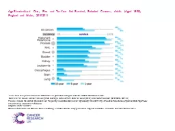
Reasons why lung cancer survival is still variable and poor Late presentation Deprivation not just smoking but mainly Lack of advocacy amp research Stigma Access to staffdiagnostics and treatment ID: 911941
Download Presentation The PPT/PDF document "Lung Cancer Age-Standardised Ten-Year Su..." is the property of its rightful owner. Permission is granted to download and print the materials on this web site for personal, non-commercial use only, and to display it on your personal computer provided you do not modify the materials and that you retain all copyright notices contained in the materials. By downloading content from our website, you accept the terms of this agreement.
Slide1
Lung Cancer
Slide2Age-Standardised Ten-Year Survival for Common Cancers in Males and Females, England and Wales, 2010-2011
Slide3Reasons why lung cancer survival is still variable and poor?
Late presentation
Deprivation (not just smoking, but mainly)Lack of advocacy & researchStigmaAccess to staff,diagnostics and treatment
Slide4Symptoms in patients who turn out to have lung cancer
Slide5Red flags are not always reliable but……NICE says
Any haemoptysis
Three weeks of unexplained clubbing or…..CoughBreathlessnessChest or shoulder painWeight lossHoarsenessChest signsOr just because smokes and tired? Unclear. But probably.Don’t wait for antibiotics to work
Slide6Causes and Risk factors of Lung Cancer
Diagnostic Tests
CXR
CT ScansMRISputum cytologyFibreoptic bronchoscopyTransthoracic fine needle aspiration
Slide9Laboratory Tests
Blood Tests
*CBC-to check red/white blood cell & platelets -to check bone marrow and organ function *Blood Chemistry Test-to assess how organs are functioning such as liver and kidneyBiopsy-to determine if the tumor is cancer or not -to determine the type of cancer -to determine the grade of cancer (slow
or fast)
Slide10Biopsy
Endoscopy
Bronchoscopy
MediastinoscopyVATS (video assisted thoracoscopic surgery)
Slide12Bronchoscopy
Slide13Mediastinoscopy
Slide14VATS (video assisted
thoracoscopic
surgery)
Slide15Slide16DIAGNOSTIC WORKUP
History:
metastasis symptomsPE: H & N lymph nodesChest X-rayCT: the most valuable radiologic study for evaluation, staging, and therapeutic planning of lung cancerMRI: mediastninum or paravetebral region
Bone scans: stage III before curative therapy
Slide17PET
influenced radiation delivery in 65% for definitive radiotherapy (Kalff et al.).
Brain CT scan: small cell carcinoma.Pulmonary function tests: ability to undergo surgical resection or withstand irradiation
Slide18Sputum cytology
:
20% to 30% sensitivityBronchoscopic examination: 90% positive CT-guided Bx: 95% positiveBx: Primary tumor lesion, scalene node
Slide19Pathology
Sputum cytology
: 20% to 30% sensitivityBronchoscopic examination: 90% positive CT-guided Bx: 95% positiveBx: Primary tumor lesion, scalene node
Slide20Incidence
Taiwan (TCOG)
USA
NSCLC
85-88 %
80 %
SCLA
12-15 %
20 %
Slide21Lung Cancer Re-cap
Small Cell Lung Cancer
Non-Small-Cell Lung Cancer
Squamous cell
Adenocarinoma
Slide22Squamous cell carcinoma
Moderate to poor differentiation
makes up 30-40% of all lung cancersmore common in malesmost occur centrally in the large bronchiUncommon metastasis that is slow effects the liver, adrenal glands and lymph nodes.Associated with smokingNot easily visualized on xray (may delay dx)Most likely presents as a Pancoasts tumor
Slide23Adenocacinoma
Increasing in frequency. Most common type of Lung cancer (40-50% of all lung cancers).
Clearly defined peripheral lesions (RLL lesion)Glandular appearance under a microscopeEasily seen on a CXRCan occur in non-smokersHighly metastatic in nature Pts present with or develop brain, liver, adrenal or bone metastasis
Slide24Large cell carcinomas
makes up 15-20% of all lung cancers
Poorly differentiated cellsTends to occur in the outer part (periphery) of lung, invading sub-segmental bronchi or larger airwaysMetastasis is slow BUTEarly metastasis occurs to the kidney, liver organs as well as the adrenal glands
Slide25TMN Staging system for Lung Cancer
T
= Tumors : tumor size, (local invasion) N= Node : node involvement (size and type)M= Metastasis : general involvement in organs and tissues
Slide26Lung Cancer Staging Continued
T:
Tx, T0, Tis, T1-T4 (T3-tumors greater than 7cm, T4 is a tumor of any size)N: N0, N1, N2, N3M: M0, M1a, M1b
Slide27Stage grouping (AJCC 2002)
T1 T2
T3 T4N0 IA IB IIB IIIBN1 IIA IIB IIIA IIIBN2 IIIA IIIA IIIA IIIBN3 IIIB IIIB IIIB IIIB
Slide28Man, age: 76, cough and BWL
Slide29Man, age: 72, LLL
Slide30Small cell lung Ca
Limited stage
Slide31Woman, age: 68
SVC syndrome
Slide32Treatment
Surgery is preferred radical option
‘Resectable’ versus ‘operable’Radical RT (or SBRT) should be considered even if patient not fit for surgery (‘operable’)Performance status at diagnosis is crucial:
Grade
Explanation of activity
0
Fully active, able to carry on all pre-disease performance without restriction
1
Restricted in physically strenuous activity but ambulatory and able to carry out work of a light or sedentary nature, e.g., light house work, office work
2
Ambulatory and capable of all selfcare but unable to carry out any work activities. Up and about more than 50% of waking hours
3
Capable of only limited selfcare, confined to bed or chair more than 50% of waking hours
4
Completely disabled. Cannot carry on any selfcare. Totally confined to bed or chair
5
Dead
Slide33Medical Management
The three main cancer treatments are:
*surgery (lung resections) *radiation therapy *chemotherapyOther types of treatment that are used to treat certain cancers are hormonal therapy, biological therapy, Immunotherapy, targeted chemotherapy or stem cell transplant.
Slide34Prognostic Factors
The best estimate on how a patient will do based on:
*type of cancer cells *grade of the cancer *size or location of the tumor *stage of the cancer at the time of diagnosis *age of the person *gender *results of blood or other tests *a persons specific response to treatment
*overall health and physical condition