/
TIMOTHY OLADOSU DISORDERS OF THE DIGESTIVE SYSTEM TIMOTHY OLADOSU DISORDERS OF THE DIGESTIVE SYSTEM

TIMOTHY OLADOSU DISORDERS OF THE DIGESTIVE SYSTEM - PowerPoint Presentation

caitlin
caitlin . @caitlin
Follow
342 views
Uploaded On 2022-06-11

TIMOTHY OLADOSU DISORDERS OF THE DIGESTIVE SYSTEM - PPT Presentation

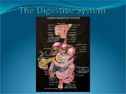
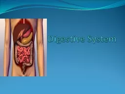
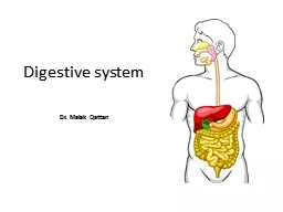
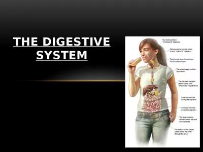
OUTLINE Overview of anatom y amp physiology Disorders of the mouth and esophagus Stomatitis Hiatal hernia amp reflux esophagitis Achalasia Diverticulum OVERVIEW OF ANATOMY amp PHYSIOLOGY OF THE DIGESTIVE SYSTEM ID: 916475

amp esophageal esophagus food esophageal amp food esophagus mouth alimentary management foods patient diverticulum meals reflux organs pressure abdominal

Share:

Link:

Embed:

Download Presentation from below link

Download Presentation The PPT/PDF document "TIMOTHY OLADOSU DISORDERS OF THE DIGESTI..." is the property of its rightful owner. Permission is granted to download and print the materials on this web site for personal, non-commercial use only, and to display it on your personal computer provided you do not modify the materials and that you retain all copyright notices contained in the materials. By downloading content from our website, you accept the terms of this agreement.


Presentation Transcript

Slide1

TIMOTHY OLADOSU

DISORDERS OF THE DIGESTIVE SYSTEM

Slide2

OUTLINE

Overview of anatom

y & physiology

Disorders of the mouth and esophagus

Stomatitis

Hiatal hernia &

reflux

esophagitis

Achalasia

Diverticulum

Slide3

OVERVIEW OF ANATOMY & PHYSIOLOGY OF THE DIGESTIVE SYSTEM

Digestive system is the collective name used to describe the

alimentary canal, accessory organs and variety of digestive processes

which take place at different levels in the canal

The alimentary canal is a long tube where food passes. The alimentary canal begins at the mouth and terminates at the anus

The parts of the alimentary tract are;

Mouth

Pharynx

Oesophagus

Stomach

Small intestine

Large intestine

Rectum

Anal canal

Slide4

The accessory organs of the digestive system are;

3 pairs of salivary glands

Pancreas

Liver

Biliary tract

The accessory organs secretes juices which passes through ducts to enter into the alimentary tract

The activities in the digestive system can be grouped under 5 main headings:

Ingestion: the process of taking food into the alimentary tract

Propulsion: this moves the contents along the alimentary tract

Slide5

Digestion: this consists of;

Mechanical breakdown of food by mastication (chewing)

Chemical digestion of food by enzymes present in the secretions produced by glands and accessory organs of the digestive system

Absorption: this is the process by which digested food substances pass through the walls of some organs of the alimentary canal into the blood and lymph capillaries for circulation round the body

Elimination: food substances which have been eaten but cannot be digested and absorbed are excreted by the bowel as feces

Slide6

Structure of the Alimentary canal

The layers of the

walls in the

different organs from the esophagus onwards follow a consistent pattern with some slight modifications depending on the structure and functions of the organs

the walls of the alimentary tract are formed by 4 layers of tissue

Adventitia or outer covering

Muscle layer

Submucosal

layer

Mucosa lining

Slide7

Adventitia: this consists of

loose fibrous tissue

in the thorax; while in the abdomen the organs are covered by a serous membrane called

peritoneum

Muscle layer: with some exceptions, this consists of two layers of smooth (involuntary) muscle

Submucosa layer: is consists of loose connective tissue with some elastic fibers

Mucosa: this consists of three layers of tissue; mucous membrane; lamina

propria

;

muscularis

mucosa

Nerve supply: the alimentary tract is supplied by both divisions of autonomic nervous system

i.e

sympathetic and

parasymphatetic

Slide8

DISORDERS

OF MOUTH AND ESOPHAGUS

Slide9

STOMATITIS

Stomatitis is inflammation of the mouth and mucous

membrane

It is the term generally applied to variety of mouth disorder characterized by mucosal cell destruction and disruption of the mucosal

lining

It mostly

occur to the following:

secondary to systemic disease and

infection

nutritional and fluid

deficiencies

poorly fitting

derivatives

neglect of oral

hygiene

as side effect of irritants and drugs

Slide10

Clinical manifestations

Oral pain

Sensitivity to hot, spicy

foods

Foul

taste

Oral bleeding or

drainage

Fever

Xerostomia

(

dry mouth

)

Difficulty chewing or

swallowing

Poorly fitting dentures

Slide11

Assessment & diagnostic findings

Diagnosis is mostly

by physical examination

The

oral mucosa will appear swollen, red and

ulcerated

The lymph glands may be

swollen

The breath is often

foul-smelling

The lips may have cracks, fissures, blisters, ulcers, and

lesions

The tongue may appear dry, cracked, and contain

masses

, lesions or

exudates

Microscopic culture

and sensitivity of the lesion or drainage to identify offending

organism

Platelet count

: this is done if bleeding

is

present in the

immunosuppressed

patient

Slide12

Management

Treatment varies, depending on the type of impairment and its cause;

Identify and

/or attempt to control or remove causative factor(s

)

Oral hygiene

/mouth

irrigations

Pharmacotherapy

local/systemic analgesics for relief of pain

Topical/systemic steroids to reduce inflammation and promote

healing

Antibiotics, antifungals and antiviral agents to combat

infection

Vitamins:

to correct deficiencies (

e.g.

vitamin C to strengthen connectives tissues in the

gums; and

niacin and riboflavin to promote efficient cellular

growth)

Artificial saliva product: to maintain a normal fluid and electrolyte environment in the mouth

Slide13

Dietary management:

food

high in protein,

high

in calories, and high in vitamins is given to promote wound healing, correct the specific deficiency.

Hot

and spicy foods are restricted, and the consistency of the food can ranges from liquid to regular, as

tolerated

Cauterization of ulceration; if necessary

Dental restoration and repair: if

indicated

Adequate rest for optimal tissue repair

Slide14

Nursing interventions

Inspect the mouth for inflammation,

lesions,

and bleeding. Record observation and reports

appropriately

Administer analgesics, corticosteroids, anesthetics, and mouth washes as

prescribed

Provide mouth care quarter or even hourly if

indicated

Instruct the patient to brush teeth after meals and at night, using a soft-bristled toothbrush and nonabrasive

toothpaste

Dietary

alterations

may be necessary for

example

changing to a full liquid or

pureed diet

Keep the lips moist with emollients such as lanolin or petroleum

jelly

Advise patient to avoid irritants, including smoking and foods that are hot, spicy and rough in

texture

Explain the importance of meticulous, frequent oral hygiene and periodic dental exams

Slide15

HIATAL HERNIA

&

REFLUX

OESOPHAGITIS

Hiatal hernia occurs when there is a weakening of the muscular collar around the esophageal and diaphragmatic function, permitting a portion of the lower esophagus and stomach to rise up into the chest during an increase in intra-abdominal

pressure

Slide16

Causative factors

Degenerative

changes (

aging

)

Trauma

Esophageal

neoplasms

Kyphoscoliosis

(a curvature of the spine

)

Surgery

Slide17

Increase abdominal pressure can occur with

Coughing;

Straining;

Bending;

Vomiting;

Obesity;

Pregnancy;

Trauma;

Ascites;

Severe physical exertion

The incidence of hiatal hernia increases with

age

Women and obese individuals are more often

affected

Many patients are asymptomatic unless

esophageal

reflux is also present

Slide18

Clinical manifestations

Reflux

esophagitis;

1-4 hours after eating, possibly aggravated by reclining, stress, and increased intra-abdominal

pressure

Heart burn

Belching

Regurgitation

Vomiting

Retrosternal or

substernal

chest pain (dull, full, heavy

)

Hiccups

Mild

anemia

Dysphagia can also occur

Slide19

Assessment & diagnostic findings

Physical examination

reveals;

Abdominal

distention

Pressure of

palpitation

Auscultation of peristaltic sounds in the

chest

Chest x-ray ; may reveal a large

hernia

Barium swallow; will reveal

gastroesophageal

and diaphragmatic

abnormalities

Oesophagoscopy

and

biopsy –

this

helps to differentiate between hiatal hernia,

varices

and

gastroesophageal

lesions

Determine the extent of

esophagitis

or

ulceration

Detect organic

stenosis

Rule out

malignancies

Esophageal function slides

identify

primary and secondary motor dysfunction before surgical

repair

Slide20

Gastric analysis

To assess for bleeding, if ulceration is present

CBC – may reveal anemic conditions if bleeding ulcers are present

Stool guaiac test – this will be positive if bleeding occurs

EKG – to rule out cardiac origin of pain

Slide21

Management

The goals of management are to;

prevent or reduce gastric reflux caused by increased intra-abdominal pressure

Prevent or reduce increased gastric acid production

Encourage limitation of activities that increase intra-abdominal pressure, e.g. coughing, bending, staining & physical exertion

Restrict or limit gastric acids stimulants e.g. caffeine & nicotine

Dietary management;

Small, frequent meals

Bland foods

Weight reduction for obese patients

Food restriction 2-3 hours before reclining

Elevate head of bed to prevent postural reflux at night

Restrict tight, waist-constricting clothing

Slide22

Pharmacotherapy

Antiemetics

, cough suppressants, and stool softeners to prevent increased intra-abdominal pressure from vomiting, coughing, and straining with bowel movements

Antacids to neutralize gastric acid

Cholinergics

to promote motility and prevent reflux

Histamine H2 receptor blockers to suppress acid secretion

Slide23

Nursing interventions

Assess & document the amount and character of the discomfort

Administer medications as prescribed

Encourage the patient to follow dietary and activity restrictions

Assess the patient’s knowledge of the disorder, its treatment, and the methods use to prevent symptoms & their complications….. Provide instructions as appropriate

Advise the patient to drink water after eating to cleanse the esophagus of residual food which can be irritating to the esophageal lining

Explain the effect of alterations in body positions and activities;

Avoid using supine

position 2-3 hours after eating

Sleeping on the right side with the head of bed elevated to promote gastric emptying

Avoid bending, coughing, lifting heavy objects, straining with bowel movements, strenuous activities, clothing that is too tight around the waist

Slide24

Explain the need to follow dietary management;

Eat a low-fat, high-protein diet

Eat small, frequent meals

Eating slowly

Chewing well to avoid reflux

Avoiding extremely hot or cold foods

Limiting gastric acid stimulants e.g. alcohol, caffeine, spices, fruit juices, nicotine

Losing weight, if applicable and appropriate

Slide25

ACHALASIA

This is a chronic, progressive motor disorder that affects the lower two thirds of the esophagus

Characterized by;

Ineffective peristalsis

Hypertonic lower esophageal sphincter = does not relax in response to swallowing

E

sophageal dilation

Cause is idiopathic

Complications include;

esophagitis with edema and hemorrhage

Respiratory complications caused by aspiration of esophageal contents

Malnutrition

Predisposition to esophageal carcinoma

Slide26

Clinical manifestations

Dysphagia of both liquids and solids

esp

with cold liquids

Halitosis

Feeling of fullness in the chest

Heartburn (

pyrosis

)

Weight loss

Retrosternal pain during or after meals that radiates to the back, neck and arms

Sometimes regurgitation of esophageal contents

Nocturnal choking

Slide27

Assessment & Diagnostic findings

Diagnstic

x-ray reveals esophageal dilatation above the narrowing

Barium swallow

Esophageal function studies

Esophagoscopy

biopsy

Slide28

Management

Medical management is geared towards relieving symptoms caused by LES obstruction and emptying esophageal contents

Activity/positional alterations:

patient to remain upright after meals

Wait 2-4hours after a meal before lying down

Sleep with the head elevated

Patients may arch their back, flex the chin towards the chest, strain (

vasalva

maneuver) while swallowing

Dietary management:

Small, frequent meals

Eat and drink slowly in a relaxed environment

Avoid rough foods and foods that can cause discomfort e.g. spices

Drink fluids with meals to enhance movement of food into the stomach

Slide29

Pharmacotherapy:

Steroids & NSAIDs are contraindicated as it can cause ulceration

Vitamins & iron supplements – to treat malnutrition and anemia

Antacids – to reduce the amount of gastric acid and relieve pain

Local anesthetics/analgesics – to minimize discomfort and promote esophageal relaxation

Mechanical esophageal dilatation with the use of inflatable tube into the esophagus – for temporary symptomatic relief

Surgical interventions –

esophagomyotomy

; it enables the mucosa under the muscular layers to expand and allow food to pass into the stomach unobstructed

Either an abdominal or thoracic approach may be used to provide access to the lower esophagus

Slide30

Nursing interventions

Monitor intake & output

Document daily weight

Administer local anesthetics/analgesics before meal to relax the esophagus and aid ingestion

Monitor and document what patient can/cannot swallow

Provide oral hygiene before and after meals and at bedtime

Advise patient to avoid smoking and constrictive clothing

Restrict foods and substances that decrease LES pressure, e.g. fats, refined carbohydrates, alcohol, tobacco

Restrict foods that can irritate the esophageal lining e.g. coffee, citrus juices, tomato juice

Administer vitamin and iron supplements if prescribed

Educate the patient on effect of stress and how to reduce stress

Emphasize the importance of increased nutritional intake and precautions to take while eating

Slide31

DIVERTICULUM

It is an

outpouching

of mucosa and submucosa that protrudes through a weak portion of the musculature

Diverticula may occur in one of the three areas of the esophagus;

Pharyngoesophageal

or upper part of the esophagus

Midesophageal

part

Epiphrenic

or lower part of the esophagus

Pharyngoesophageal

diverticulum (

Zenker’s

diverticulum)

It is the most common type of diverticulum

It is 3 times more commoner in males than females

It is seen in people older than 60years

It occurs posteriorly through the

cricopharyngeal

muscle in the midline of the neck

Slide32

Midesophageal

diverticula;

are uncommon

Symptoms are less acute

It does not require surgery

Epiphrenic

diverticula

Usually larger

Found in the lower esophagus just above the diaphragm

Believed to be related to improper functioning of the lower esophageal sphincter

Slide33

Clinical manifestations

Difficulty swallowing

Fullness in the neck

Belching

Regurgitation of undigested food

Gurgling noises after eating

Irritation of the trachea

Halitosis or sour taste following decomposition of retained food in the diverticulum

1/3 of patients with

epiphrenic

diverticula are asymptomatic while 2/3 complains of dysphagia and chest pain

Slide34

Assessment & Diagnostic findings

Barium swallow to determine the exact nature and location of a diverticulum

Esophagoscopy

is contraindicated because of the danger of perforating the diverticulum, with resulting

mediastinitis

Slide35

Management

The only means of cure is surgical removal of the diverticulum –

diverticulectomy

Myotomy

of the

cricopharyngeal

muscle is done to relieve spasticity of the musculature

Surgery is indicated for

epiphrenic

and

midesophageal

diverticula if the symptoms are troublesome and worse

Intramural diverticula usually regress when the esophageal stricture is dilated