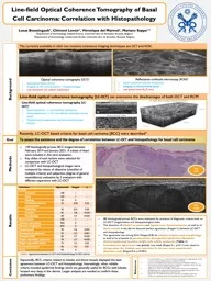
/
Line-field Optical Coherence Tomography of Basal Cell Carcinoma: Correlation with Histopathology Line-field Optical Coherence Tomography of Basal Cell Carcinoma: Correlation with Histopathology

Line-field Optical Coherence Tomography of Basal Cell Carcinoma: Correlation with Histopathology - PowerPoint Presentation

candy
candy . @candy
Follow
342 views
Uploaded On 2022-02-14

Line-field Optical Coherence Tomography of Basal Cell Carcinoma: Correlation with Histopathology - PPT Presentation

Lucas Boussingault 1 Clément Lenoir 1 Véronique del Marmol 1 Mariano Suppa 12 1 Department of Dermatology Hôpital Erasme Université Libre de Bruxelles Brussels Belgium ID: 908641

resolution oct coherence optical oct resolution optical coherence tomography criteria histopathology field agreement line kappa imaging bcc cell basal

Share:

Link:

Embed:

Download Presentation from below link

Download Presentation The PPT/PDF document "Line-field Optical Coherence Tomography ..." is the property of its rightful owner. Permission is granted to download and print the materials on this web site for personal, non-commercial use only, and to display it on your personal computer provided you do not modify the materials and that you retain all copyright notices contained in the materials. By downloading content from our website, you accept the terms of this agreement.


Presentation Transcript

Slide1

Line-field Optical Coherence Tomography of Basal Cell Carcinoma: Correlation with Histopathology

Lucas Boussingault1, Clément Lenoir1, Véronique del Marmol1, Mariano Suppa1,2 1Department of Dermatology, Hôpital Erasme, Université Libre de Bruxelles, Brussels, Belgium2Department of Dermatology, Institut Jules Bordet, Université Libre de Bruxelles, Brussels, Belgium 

Background

Goal

Methods

Conclusions

The currently available in vivo non invasive cutaneous imaging techniques are OCT and RCM.

Reflectance

confocale

microscopy [RCM]2 Good resolution (cellular resolution)Imaging on the horizontal plane Low penetration (0,25 mm)

Optical coherence tomography [OCT]1Good penetration (2mm)Imaging on the vertical plane ( ~ histopathology)Low resolution (no cellular resolution)

Line-field optical coherence tomography [LC-OCT] can overcome the disadvantages of both OCT and RCM

Line-field optical coherence tomography

[LC-OCT]3Good resolution : ± 1 µm (cellular resolution)Good penetration : ± 0,5 mm (dermis reticularis can be seen)Tridimensional exploration (vertical and horizontal planes)

Recently, LC-OCT based criteria for basal cell carinoma [BCC] were described4

To assess the existence and the degree of correlation between LC-OCT and histopathology for basal cell carcinoma

149 histologically-proven BCC imaged between February 2019 and January 2021. A subset of them were included in this pilot evaluation

Key slides of each lesions were selected for comparison with LC-OCT

LC-OCT and histopathological images were compared by means of objective (checklist of multiple criteria) and subjective (degree of general resemblance) evaluation by 3 evaluators with different experience with LC-OCT

Expectedly, BCC criteria related to lobules and blood vessels displayed the best agreement between LC-OCT and histopathology. Interestingly, other reliable criteria included epidermal findings which are generally useful for BCCs with lobules located very deep in the dermis. Larger analyses are needed to confirm these preliminary findings.

Key slide

References

Ferrante di Ruffano L et al. Optical coherence tomography for diagnosing skin cancer in adults. Cochrane Database Syst Rev. 2018 Dec 4;12(12)Lan J, et al. The diagnostic accuracy of dermoscopy and reflectance confocal microscopy for amelanotic/hypomelanotic melanoma: a systematic review and meta-analysis. Br J Dermatol. 2020 Aug;183(2):210-219.Ogien J et al. . Dual-mode line-field confocal optical coherence tomography for ultrahigh-resolution vertical and horizontal section imaging of human skin in vivo. Biomed Opt Express. 2020 Feb 10;11(3):1327-1335. Suppa M et al. Line-field confocal optical coherence tomography of basal cell carcinoma: a descriptive study. J Eur Acad Dermatol Venereol. 2020 Dec 7


Key image

Results

20 histologically-proven BCCs were evaluated for presence of diagnostic criteria both on LC-OCT images/videos and histopathological slidesThe detection of lobule’s presence and typical core characterization, as well as of blood vessels in the dermis featured perfect agreement (Kappa=1) between LC-OCT and histopathology.The agreement was strong (0.61<Kappa<0.8) for lobule’s location and morphology, as well as for presence of parakeratosis, disorganised epidermis, disrupted dermal-epidermal junction, bright cells within epidermis (Table 1).Interobserver agreement was globally very weak (Kappa 0.1, p=0.11) but raised to strong when the analysis was restricted to the two most experienced observers only (Kappa=0.6, p<0.001)

CriterionAgreementKappapLobule   Presence1001-Core1001-Clefting500-Perilobular stroma compression350.040.25Palisading450.010.20Location850.8<0.001Morphology800.7<0.001Blood vessels1001-Stroma’s stretching90%0-Stroma’s brightness65%0-Parakeratosis900.60.003Disorganised epidermis950.70.001Disrupted DEJ850.7<0.001Bright cells within epidermis900.7<0.001Bright cells within lobule750.30.02Erosion/ulceration850.50.01Crust900-

Table 1.

Agreement between LC-OCT and histopathology for BCC criteria