/
Selective Reinnervation of Intercostal Muscles Transplanted from Diffe Selective Reinnervation of Intercostal Muscles Transplanted from Diffe

Selective Reinnervation of Intercostal Muscles Transplanted from Diffe - PDF document

cappi
cappi . @cappi
Follow
342 views
Uploaded On 2022-10-29

Selective Reinnervation of Intercostal Muscles Transplanted from Diffe - PPT Presentation

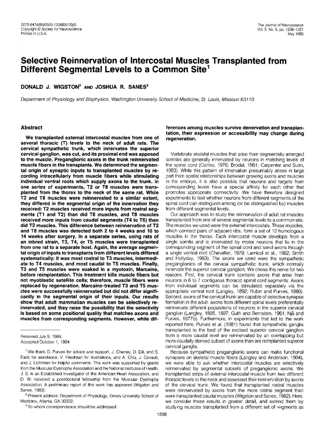
DONALD J WIGSTON AND JOSHUA R SANES3 Depaltment of Physiology and Biophysics Washington University School of Medicine St Louis Missouri 63110 Abstract We transplanted external intercostal musc ID: 961839

muscles muscle fibers intercostal muscle muscles intercostal fibers transplanted inputs synaptic axons reinnervation cervical external fig transplantation ventral trunk

Share:

Link:

Embed:

Download Presentation from below link

Download Pdf The PPT/PDF document "Selective Reinnervation of Intercostal M..." is the property of its rightful owner. Permission is granted to download and print the materials on this web site for personal, non-commercial use only, and to display it on your personal computer provided you do not modify the materials and that you retain all copyright notices contained in the materials. By downloading content from our website, you accept the terms of this agreement.


Presentation Transcript

Selective Reinnervation of Intercostal Muscles Transplanted from Different Segmental Levels to a Common Site’ DONALD J. WIGSTON AND JOSHUA R. SANES3 Depaltment of Physiology and Biophysics, Washington University School of Medicine, St. Louis, Missouri 63110 Abstract We transplanted external intercostal muscles from one of several thoracic (T) levels to the neck of adult rats. The cervical sympathetic trunk, which innervates the superior cervical ganglion, was cut, and its proximal end was apposed to the muscle. Preganglionic axons Received July 9, 1984; Accepted October 1, 1984 ’ We thank D. Purves for advice and support, J. Cheney, D. Dill, and S. Eads for assistance, V. Friedman for illustrations, and A. Chiu. J. Covault, and J. Lichtman for helpful comments. This work was supported by grants from the Muscular Dystrophy Association and the National Institutes o

f Health. J. S. is an Established Investigator of the American Heart Association, and D. W. received a postdoctoral fellowship from the Muscular Dystrophy Assocration. A preliminary report of this work has appeared (Wigston and ferences among muscles survive denervation and transplan- tation, their expression or accessibility may change during regeneration. Vertebrate skeletal muscles that arise from segmentally arranged somites are generally innervated by neurons in matching levels of the spinal cord (Corliss, 1976; Brodal, 1981; Carpenter and Sutin, 1983). While this pattern of innervation presumably arises in large part from spatial relationships between growing axons and muscles in the embryo, it is also that neurons and targets from corresponding levels have a special affinity for each other that promotes appropriate connectivity. We have therefore designed experiments t

o test whether neurons from different 1208 T2 SUPERIOR CERVICAL GANGLION i + CERVICAL&Y’$lKPATHETIC COMMUNICATING RAMUS DORSAL ROOT GANGLION DORSAL ROOT VENTRAL ROOT C8 TI T2 Figure 7. Diagram of the preparation used to compare reinnervation of muscles from different segmental levels. A strip of Materials and Methods Dawley, obtained from Eldndge, St. Louis, MO) was anesthetized with chloral hydrate (350 mg/kg, i.p.), and incisron was made on hgure 2 0 b 1 ‘0 Figure 3. Synaptic potentrals recorded intracellularly from transplanted intercostal muscles upon stimulation of the indicated ventral roots. a, This muscle fiber received one input from bar indicates 40 msec for a b and msec for c. The vertical bar indicates 25 mV for the action potential in a and mV for all other traces. muscle is composed of a thicker, internal and thinner, external musc

le, separated by loose connective tissue. The internal intercostal was peeled away, leaving a sheet of external intercostal muscle fibers attached to ribs at both ends. The rat’s Figure 2. Cross-sections of external intercostal muscles, before and after transplantation. a and b, Normal external intercostal is 10 to 20 fibers thick. The adherent internal intercostal muscle has been peeled away. B, blood vessel; N, intramuscular nerve; S, muscle spindle. c and d, 6 days after transplantation, Fibers on the surface have survived, while fibers in (d) has already begun, e f, 25 days after transplantation. Surviving (surface) fibers have atrophied, while regenerated central fibers have matured, reducing obvious differences between surface and central areas. The bar is 60 wrn for a e, 80 pm for c, and pm for b, and f. Wigston and Sanes Vol. 5, No. 5, May 1985

T f + f T f I I 1 I 1 14-17 B-20 21-2526-X) 31-35 Days after transplantation Figure 4. Average number of synaptic inputs per innervated muscle fiber (a), and the percentage of inputs that exceeded threshold and initiated action potentials (b) in muscles tested at varying times after transplantation. Sym- bols indicate mean; n = 10, 7, and muscles for the six intervals shown. Bars, SE. In some experiments using inbred animals, intercostal muscles were bathed for 20 to 60 min in a solution of Marcaine (bupivicaine; 0.75% in isotonic NaCI; Sterling Drugs) Instead of saline while being prepared for implantation. Marcaine kills intact muscle fibers Results Transplantation and reinnervation of external intercostal muscles. Strips of external intercostal muscle were transplanted to the neck from the 2nd, 3rd, 4th, or 8th thoracic interspace (Fig. 1). Muscle fibers near the

surfaces of the transplants survived, while fibers in the center died and were replaced by regeneration (Fig. 2, a to d). Regenerated myotubes matured and surviving fibers atrophied, so f). This sequence of events is similar to described by Hansen-Smith and Carlson (1979) in transplanted rat extensor digitorum longus muscle, except that since the external intercostal is a thinner muscle, a larger fraction of its fiber are near a free surface and thus survive. Axons in the cervical sympathetic trunk reinnervated muscle fibers in the transplants within 2 weeks of surgery. Muscles twitched in response to stimulation of the cervical trunk or of individual ventral roots, and postsynaptic potentials could be recorded intracellularly from muscle fibers (Fig. 3). Since sympathetic preganglionics are the only axons in the cervical trunk that run through the ventral roots (Brooks-

Fournier and Coggeshall, 1981; Bowers and Zigmond, 1981), we assume that responses evoked by stimulating ventral roots represent synapses made by PM), indicating that transmission was cholinergic. Synaptic potentials varied greatly in amplitude, sometimes initiated action potentials, and generally fatigued rapidly on repetitive stimulation at 0.5 Hz. About 60% (1194 of 1975) of the innervated fibers in transplanted intercostals were polyneuronally innervated. Individual muscle fibers could receive inputs from more than one segment (Fig. 3a) and/or more than one input from a single segment (Fig. 36). We detected an average 4a); the maximum was 7 (11 fibers). Rise times of synaptic potentials recorded from a single fiber were usually indistinguishable (Fig. 3c), indicating that the inputs were approximately equidistant from the recording electrode and thus probably closely

spaced. The average number of inputs per innervated fiber changed little between 2 weeks after transplantation (Fig. 4a), while the percentage of inputs that were large enough to initiate an action potential doubled (Fig. 4b). It therefore seems likely that muscle fibers acquired nearly their full complement of inputs early in reinnervation and that individ- ual then became stronger during the subsequent few Electron microscopy of reinnervated transplanted intercostals re- vealed conventional neuromuscular synapses, in which vesicle-laden nerve terminals were separated from the muscle fiber membrane by a -50-nm synaptic cleft which was traversed by basal lamina 5a). In many cases, nerve terminals abutted but did not completely cover infolded regions (Fig. 5b) and thus had presumably partially reinnervated a preexisting synaptic site. Since original synaptic sites occupy o

nly a tiny fraction (-0.1%) of the muscle fiber surface, this observation indicates that preganglionic axons preferentially formed synapses at such sites. In some cases, however, nerve terminals apposed uninvaginated stretches of mus- cle membrane (Fig. 5c), indicating that some completely new syn- apses were formed during reinnervation of the transplants. Thus, muscle fibers in transplanted intercostals were reinnervated prefer- entially, although not exclusively, at original synaptic sites. Segmental origin of inputs to transplanted T2 and T8 intercostal muscles. In one series of experiments, strips of external intercostal muscle were excised from either T2 and T8 and implanted in the The Journal of Neuroscience Segmentally Selective Reinnervation 1213 F/gure 5. Electron mrcrographs of neuromuscular junctrons In rernnervated, transplanted intercostals, 3 to 4 weeks after

surgery. Several terminals share an infolded synaptic area in a. The nerve termrnal in b covers part of a folded patch and thus has apparently reinnervated an original synaptic site. The terminal in c abuts undifferentiated postsynaptic membrane and thus presumably has established a new synaptic site. Black spots are reaction product of the hrstochemrcal stain for cholrnesterase, which was used to facilitate localization of synaptrc sites. Bar, 1 pm. TABLE I Vol. 5, No. 5, May 1985 Reinnervation of transplanted T2 and T8 external intercostal muscles by the cervical sympathetic 346 789 2.2 + a TI T2 Ventral root stimulated neck of the same rat. Transplanted T2 and T8 muscles were and T8 muscles received a larger fraction of their inputs from reinnervated to a similar extent (Table I, left co/umns) but differed in segments T3 to . 4 T2 Segmentally Selective Reinner

vation 1215 A : A I A 1 A A T2 Muscles Figure 7. Average segmental innervation of transplanted T2 and T8 intercostal% This index, calculated as described under “Results,” is the segmentally weighted average of all synaptic inputs recorded intracellularly from the muscle fibers of a transplant. Thus, the higher a muscle’s value, the more caudal, on average, was its innervation. Small symbols represent individual muscles, and large symbols show mean of groups; bars, SE. a, Muscles studied 2 to 4 weeks after transplantation (see Table I, leff columns). b, Muscles studied 10 to 14 weeks after transplantation a Vol. 5, No. 5, May 1985 Figure 8. Segmental origin of synaptic inputs to T2 and T8 intercostals studied 10 to 14 weeks after transplantation (see Table I, right columns). a, Distri- bution of all I b Ventral root stimulated TABLE II Reinnewat

ion of transplanted T3, and T.5 external intercostal muscles by the cervical sympathetic trunk Muscle T3 T4 Number of muscles 11 Time after surgery (days) 27 + 2 f 1 + 3 Innervated fibers studied 320 Total inputs 706 however, no marked histological differences between untreated and Marcaine-treated transplants by 3 to 4 weeks after surgery. Marcaine-treated transplants were successfully reinnervated by preganglionic axons in the cervical trunk. Marcaine-treated and untreated transplants differed little The Journal of Neuroscience Segmentally Selective Reinnervation 1217 I a 40 - A VI f .E 30 - z E g - z a IO - transplanted T3, and intekokal mwzles de- in Table II. a, Distribution of all inputs recorded intracellularly. b, Distribution of suprathreshold inputs. Figwe 9. Segmental origin of synaptic inouts to the scribed TI T2 Ventral root stimulated Wigston and Sa

nes Vol. 5, No. 5, May 1985 along pathways to the muscle may have masked selectivity within the muscle itself. In contrast, in our experiments, implantation of the cervical trunk provided axons from many segments with direct access to the target muscle. Third, our experiments made use of, and our results may have depended on, the juxtaposition of sympathetic preganglionic axons with skeletal muscles. Preganglionic axons are known to selectively reinnervate subsets of sympathetic ganglionic neurons (see the introduction) and may reveal differences among muscles that adult motor axons ignore. Furthermore, while preganglionic axons are able 0 . T3 Transplanted muscle While reinnervation of intercostal muscles was demonstrably se- lective, there was no absolute preference of axons from any ventral root for muscles from any thoracic level. There are, however, two factors that

may have reduced the degree of selectivity manifested in our experiments. First, while the rat’s cervical trunk contains axons from six to eight levels (Rando 7 7 a and e and a and and 20 b and T3 T5 9 10 1 33 265 219 491 390 0.1 1.6 0.19 2.28 0.05 2.55 and one 1981 and 1 1 1 1 T2 T3 T4 T5 T6 J. J. T3 T4 T5 Bryant (1981) Duron, B. (1981) Intercostal and diaphragmatic muscle endings and afferents. In Regulation of Breathing, T. F. Hornbein, ed., Part 1, pp. 473-540, Marcel Dekker, Inc., New York. Gerding, Ft., N. Robbins, and J. Antosiak (1977) Efficiency of reinnervation of neonatal rat muscle by original and foreign nerves. Dev. Biol. 67: 177- 183. Gillespie, M. J., T. Gordon, and P. R. Murphey (1983) Random reinnervation of LG and SOL muscles by their common nerve? Sot. Neurosci. Abstr. 9: 11. Gordon, T., N. Niven-Jenkins, and G. Vrbova (1980) Observations on neu- ro