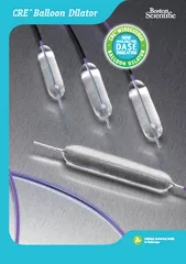
/
GatewayTMPTA Balloon CatheterInstructions for UseBostonScientificManu GatewayTMPTA Balloon CatheterInstructions for UseBostonScientificManu

GatewayTMPTA Balloon CatheterInstructions for UseBostonScientificManu - PDF document

caroline
caroline . @caroline
Follow
342 views
Uploaded On 2022-08-16

GatewayTMPTA Balloon CatheterInstructions for UseBostonScientificManu - PPT Presentation

GatewayTMPTA Balloon CatheterInstructions for UseHumanitarian Device The Wingspan Stent System with Gateway PTA Balloon Catheter is Authorizedby Federal law for use in improving cerebral artery lumen ID: 937253

balloon stent catheter system stent balloon system catheter delivery wingspan device patients stroke body vessel gateway lesion guidewire pta

Share:

Link:

Embed:

Download Presentation from below link

Download Pdf The PPT/PDF document "GatewayTMPTA Balloon CatheterInstruction..." is the property of its rightful owner. Permission is granted to download and print the materials on this web site for personal, non-commercial use only, and to display it on your personal computer provided you do not modify the materials and that you retain all copyright notices contained in the materials. By downloading content from our website, you accept the terms of this agreement.


Presentation Transcript

GatewayTMPTA Balloon CatheterInstructions for Use,BostonScientificManufactured By:Boston Scientific Corporation47900 Bayside ParkwayFremont, CA 94538-6515USAUSA Customer Service 1-888-272-100190158894.B Page Ia3 GatewayTMPTA Balloon CatheterInstructions for UseHumanitarian Device: The Wingspan Stent System with Gateway PTA Balloon Catheter is Authorizedby Federal law for use in improving cerebral artery lumen diameter in patients with intracranialatherosclerotic disease, refractory to medical therapy, in intracranial vessels with �50% stenosis that areaccessible to the system.The effectiveness of this device for this use has not been demonstrated.DEVICE DESCRIPTIONThe GatewayTMPTA Balloon Catheter is an over-the-wire co-axial catheter with a balloon near the distaltip. The outer lumen is used for inflation of the balloon, and the wire lumen permits the use ofguidewires(0.014 in./0.36 mm) to facilitate advancement of the catheter to and through the stenosis to be dilated.The balloon is designed to provide an inflatable segment of known diameter and length at recommendedpressures. The catheter is coated on the exterior with BIOSLIDETM, a hydrophilic surface that reducesfriction during manipulation. The catheter includes a tapered tip to facilitate advancement of the catheter toand through the stenosis. Marker bands, in conjunction with fluoroscopy, aid in the placement of thecatheter balloon segment. The proximal portion of the catheter includes one Luer-lock side port connectedto the outer lumen. The wire lumen has a port for use with appropriate neurovascular guidewires. Thecatheter is advanced through the neurovascular anatomy for dilating stenotic portions of intracranialarteries.INDICATION FOR USEThe Gateway PTA Balloon Catheter is indicated for balloon dilation of the stenotic portion of intracranialarteries prior to stenting for the purpose of improving intracranial perfusion.CONTRAINDICATIONSThe Gateway PTA Balloon Catheter is contraindicated for use in:* Patients in whom antiplatelet and/or coagulation therapy is contraindicated.* Patients who are jud

ged to have a lesion that prevents effective angioplasty.WARNINGS* Since the use of this device carries the associated risk of subacute thrombosis, vascularcomplication and/or bleeding events, judicious selection of patients is necessary.* Only physicians who have received appropriate training should perform intracranial angioplasty.* Angioplasty and stenting procedures should only be performed at hospitals where emergencyintracranial surgery can be readily performed in the event of a potentially injurious or life-threatening complication.PRECAUTIONSGeneral PrecautionsStore in a dry, dark, cool place. Do riot resterilize.* Note product "Use By" date.Preparation Precautions* Follow the Gateway PTA Balloon Catheter preparation and use instructions carefully, as describedon page 6, Preparation.90158894.B Page 2 * Do not prepaie or pre-inllate the balloon other thain as directed. Use the balloon purgingtechnique described in this Insrlrcltionsfir Use.* Typical antiplarelet and anticoagulation regimen used for interventional intracranial procedure isan important adjunct to balloon angioplasty treatment. Do not use the Gateway PTA BalloonCatheter in patients in whom anliplatelet and/or anticoagulation therapy is contraindicated. Vesselthrombosis may occur during the procedure if proper antiplatelet and anticoagulation therapy isnot administered.Procedure Precautions* Angioplasty may lead to dissection of the vessel and may cause other complications(vasospasm/acute closure) of the vessel requiring additional intervention (i e., further dilation,placement of stents).* Use only the appropriate balloon inflation mcdia. Do not use air or any gaseous medium to inflatetile balloon because it may cause uneven inflation and complications.* If unexpected difficulty is experienced during infiation, do not continue; remove the device and donot attempt to use it.* To reduce the potential for vessel damage, the inflated diameter of the balloon should approximatethe lesser of the vessel diameters just proximal and distal to the stenosis.* Do not use a guidewire having a diameter greater

than 0.014 in/0.36 mm.* When the delivery catheter is exposed to the vascular system, it should be manipulated whileunder high-quality fluoroscopic observation. If resistance is met during manipulation, determinethe cause of the resistance before proceeding.* Infusion of any medium other than a flush of heparinized normal saline through the guidewirelumen may compromise device performance.D Do not attempt to reposition a partially deployed balloon. Attempted repositioning ofa partiallydeployed balloon may result in severe vessel damage.* Balloon pressures should be monitored during inflation. l)o not exceed rated burst pressureindicated on the product label. Use of pressuies higher than those specified on the product labelmay result in a ruptured balloon and potential intimal damage and dissection. The rated burstpressure is based on the results of it vilro testing. At least 99.9% of the balloons (with a 95%confidence interval) will not burst at or below their rated burst pressure. Use ofa pressuremonitoring device is recommended to prevent over pressurization.* Before withdrawing the device, visually confirm complete balloon deflation by fluoroscopy. Iftheballoon has already been inflated and difficulty is experienced deflating, connect a large-barrelsyringe and manually attempt to deflate the device.Table 1: Typical Gateway System Balloon CompliancePressure liallooti Size(atm/bar) 1.50mm 2.0amr 2.25mm 2.50amm 2.75mnm 3.0lmm 3.25mm 3.S0unm 3.75nim 4.0mm1.0 I 39 185 2.09 2.29 2.55 2.77 3 02 3.24 3.50 3752.0 1.41 1.88 2.12 2.34 2 59 2.82 3.07 3.29 3.55 3803.0 143 1.91 2.15 2.38 2.63 2.86 3A.1 335 3.60 3854.0 1.46 1.94 2.19 2.42 2 67 2.91 3.16 3.40 3.65 3.905.0 148 1.97 2.22 2.46 2.71 2.95 3 20 3.45 3.70 3956.0 Nominal IS.0 2.00 2.25 2_50 2.75 3.001 3.25 3.50 3.75 4.007.0 1.52 203 2.28 2.54 2 79 3.05 3.30 355 3.80 4 0580 1.55 2.06 2.31 2.58 2.83 3.09 3 34 3.60 3.85 4 109.0 1.57 2 09 2.35 2.62 2 87 3.14 3 39 3.65 3.90 4A1510.0 1 59 2.12 2.38 2.67 2.91 3 18 3.43 3.71 395 4.2011.0 1.61 2 15 2.41 2.71 2 95 32 34 3.76 4.00 4 2512.0 I 63* 2 19' 2.44 2)75 2 99 3 28 352 3.81

* 4.05* 4.3113.0 1.66 2.22 2.47 2.7') 3.0 33' 3.57 3.86 4.10 43614.0 1.68 2.25 2,51 2.83* 3 07* 3 61 3.91 4.15 44115.0 1.70 2.28 2 54 2 87 3.1 3A1 3.66 3.96 4.20 4 4616.0 2 57 2.91 3.15 3 46 3 7017.0 F 2.60 295 3 V} 510 3 7518.0 2 63 2.99 3 2x3,_-3 79* Rated Burst Pressulc. Do not exceed.90158894.B Page 3 POTENTIAL ADVERSE EVENTSAdverse events (in alphabetical order) may be associated jith the use of an intracranial angioplasty instenotic lesions of the intracranial arteries:* Death* Dissection* Drug reactions to antiplatelet agents/conlrast reediumn* Distal emboli (air, tissue, or thrombotic emboli)* Hematorna* Hemorrhage, requiring transfusion* Hypotension/Hypertension* Infection and pain at insertion sitelschemia/infarct* Perforation* Pseudoaneurysm, (femoral and intracranial)* Restenosis of the dilated vessel* Spasm* Stroke/TIA* Total occlusion of tile intracranial arteryPROCEDURE STEPSPrior to angioplasty, carefully examine all equipment to be used during the procedure, including the ballooncatheter, to verify proper function. Verify that the catheter, and sterile packaging have not been damagedin shipment and that the catheter size is suitable for the specific procedure for which it is intended.Materials Required (Not included in the Gateway Package)Material* Appropriate guide catheter(s) (0.064 in/I.63 uni minimum ID)* 1000U/500mI (cc) Heparinized Normal Saline (HepNS)* 0.014 in./O.36 mm, neurovascular exchange length guidewire(s)·Rotating Hemostasis Valve (RHV) and adapter* 60% contrast diluted 1:i with normal saline* Appropriate arterial sheath and dilator set and guide catheters for femoral approach* 20 ml (cc) syringe with Luer-lock* Inflation device with manometer* Three-way stopcock90158894.B Page 4 CAUTIONIn order to achieve optimal performance of CauewayI"catheters and Boston Scientificsteerable gnidewires and to maintain tile Ihrhricity of the BIOSLIDETM surface, it is criticalthat a continuous flow of appropriate fluqh solution be maintained between a) the Gatewaycatheter and guide catheter, and b) the Gatewkay catheter and any intraluminal device.

Inaddition, flushing aids in preventing contrast crystal formation and/or clotting on both theguidewire and inside the catheter lunelThe recommended continuous flush set up as illustrated requires two stopcocks and two rotatinghemostatic valves (RHV); the RHV's provide a fluid tight seal and are attached to the guidecatheter Gateway catheter. The stopcocks attach to the RHV sidearms, which become infusionports for appropriate flush or contrast medium injection.PREPARATIONStopco3 to 0 23 Un toflshs5ugionInfallati'pHyr Torue OevevPreGIreing St-oPcctkBaCatheter Eb Por e to flushI aea~ Eoc~zoo S'rvic Tarq.:Inflation Device PreparationStep ActionI Prepare the inflation device accot-ding to (he IannUfacturer 's instruactions.Balloon Catheter SelectionStep ActionThe inflation diameter of the balloon must not exceed the diameter of the artery proximal anddistal to the stenosis. If the stenosis cannot be crossed with the desired balloon catheter, use asmaller diameter catheter to pre-dilate the lesion to facilitate passage of a more appropriate sizedballoon catheter.Balloon Catheter PreparationStep ActionI Remove the catheter from the protectiv e hoop.2 Remove the balloon protector and stylet by grasping the balloon catheter just proximal to theballoon and with the other hand, gentl) grasp the proximal section of the balloon protector andslide distally.3 Prepare the balloon catheter for purging. Fill a Luer-lock syringe wvith 3 ml (cc) of contrastmedium. Use only appropriate balloon inflation medium. Do not use air or any gaseousmedium to inflate the balloon.4 Connect a three-way stopcock to the port filling oil the balloon catheter. Flush through thestopcock.5 Connect the syringe to the stopcock.90158894.B Page 5;a- 6 If bId the syr in eewith the nio~zzle pointing. do ~tiiward a nd as pirate for 5 seconds RelIease thI eplunger.7 Remove the syr inge and evacuiate all a ir tiorn lie bharrelI.8 Reconnect the sringe and aspirate tint i I bubbles to lon~ger appear dluring~ aspiration. [f air bubblespersist, do not use thle device.9 Disconnect the syringge.1 0 Carefully wet the h

ydrophilic outer shaft of the bal loon catlheter,Inflation Device Connection to CatlieterStep ActionI To remove any air lodged in the distal Ltter fittin~g of the inflation device, purgec approximatelylint (cc) of contrast medium.2 By applying positive pressure to the balloon before discontiection the syringe used in preparation,a meniscus will appear in the balloon port when thle syringe is removed. Verify that a meniscus of'contrast medium is evident in both the catheter balloon port and the inflation device connection.Securely couple the inflation device to the balloon port of-the balloon catheter.DEPLOYMENT PROCEDUREStep ActionI Prepare the vascular site according to standard practice.2 Prepare the balloon catheter and guidewire as follows:a) Flush the balloon catheter gulidewire lumlen.b) Introduce the guidewire, flexible end first, into the straight (back) port of the manifold.To avoid kinking. advance the g-Uidewire slowly in small increments to the end of theballoon catheter. If desired, leave the distal end of thle guidewire inside the catheterlumien for protection.3 Loosen the knob onl the hemostatic adapter.4 With the balloon fully deflated, carefully insert the balloon catheter throLugh the hemostatic adaptervalve into the Luer fitting of the guidle catheter .Tighten thle hemostatic adapter knob to create aseal around the balloon catheter without inhibiting- movement of the catheter; this will allowvcontinuous recording, of the proximal arterial pressure. Caution should be taken not to over-tighten thle hemostatic adapter around the balloon catheter shaft as lumen constriction may Occur,affecting inflation/dleflation of the balloon.5 Advance the balloon catheter and guidewire assembly until the proximal marks align with thehemostatic adapter hub. This indicates that the balloon catheter tip has reached the guide cathetertip.6 Slowly rotate the gutidewite while advancing, thtrot.Ig thle artery until crossing of the stenosis byangiographic assessment.7 Advance the balloon catheter over the guwidewire and position the balloon relative to the lesion tobe dil

ated; inflate tile balloon to the appropriate preCssIre. If difficulty is experienced duringballoon inflation, do tint Contitnue use; reinove ili e deice and do nlot attempt to use it.BALLOON PRESSURE MUST NOT EXCEED THIE RATED BURST PRESSURE.Note: Refer to Table I or to the balloon cotipliance cheart included with labeling.REMOVAL PROCEDUREStep ActionI After angioplasty is complete, deflate tile balloon. Before withdrawing thle device, visuallyconfirm coniplete balloon deflation by fluoroscopiv. If difficttltv is experienced deflating, connecta large-barrel syringe and manually deflate thle device.2 Withdraw the balloon catheter until it i~s clear of'the lesion. Maintain the guidlewire across thledilated stenosis.3 Perform ang-iography using" the guinde catheter vis an a ngiog~raph ic catheter to con firm dilation.90158894.B Page 6 4 After angiography has confirmed that the Lumen of the dilIated artery has not acutely occluded,slowly withdraw the guidewire and the deflated balloon from tile g uide catheter and through theadapter. Tighten the hemostatic adapter knob.Note: After the deflated balloon catheter is withidiawni. if ieinsertion is necessary, it shotlid be wiped clearwith sterile heparinized normal saline and stored in a hasiin of tie same solution until reinsertion.WARRANTYBoston Scientific Corporation (BSC) warrants that reasonable care has been used in the design andmanufacture of this device. This warranty is in lieu of and excludes all other warranties not expressly setforth herein, whether expressed or implied by operation of law~ or otherwise, including, but not limited to,any implied warranties of merchantability or fitness for a particular use. Handling, storage, cleaning andsterilization of the device as well as other factors relating to tile patient, diagnosis, treatment, surgicalprocedures, and other matters beyond BSC's control directly affect the device and tile results obtained fromits use. BSC's obligation under this warranty is limited to the repair or replacement of this device and BSCshall not be liable for any incidental or consequential

loss, damage, or expense directly or indirectly arisingfrom the use of this device. BSC neither assumes, nor authorizes any other person to assume for it, anyother or additional liability or responsibility in connection with this device. BSC assumes no liability withrespect to device reused, reprocessed or resterilized and makes no warranties, expressed or implied,including but not limited to merchantability or fitness foi intended use with respect to such device.Svmhl lr I aiIsittio. Ket':CONT: ContentThis Prodliet Contains No Detectable Latex.90158894.B Page 7,3%~ Gateway and Bioslide are trademarks of Boston Scientific Corporation or its affilIiates.Patent PendingICopyright © 2005 by Boston Scientific Corporation or its affiliates. All rights reserved.Boston,Scietctific90158894.B PageS8'so WingspanTMStent System withGatewayTMPTA Balloon CatheterInstructions for UseBostonScientificManufactured By:Boston Scientific Corporation47900 Bayside ParkwayFremont, CA 94538-6515USAUSA Customer Service 1-888-272-100190163966-01 Rev. B WingspanTMStent System withGatewayTMPTA Balloon CatheterInstructions for UseHumanitarian Device: The Wingspan Stent System with Gateway PTA Balloon Catheter is Authorized by Federal lawfor use in improving cerebral artery lumen diameter in patients with intracranial atherosclerotic disease, refractory to medical therapy,in intracranial vessels with �50% stenosis that are accessible to the system.The effectiveness of this device for this use has not been demonstrated.DEVICE DESCRIPTIONThe Wingspan Stent System includes:A self-expanding, nitinol Stent with four radiopaque markerbands on each end (distal and proximal).A flexible over-the-wire Stent Delivery System (Inner Body and Outer Body) with pre-loaded StentThe Wingspan Stent System is used in conjunction with the Gateway PTA Balloon Catheter.otanmg DistlH ono0asls FleXibe onVa.e Snat Stent Ted________________________ _____________________ /Tip~~~' ~~~~ /L~~~j t D I~~~~~~~fl ~~Proximael proximal DlItIner OuterMO~~ ~ ~akr Mar~rHub HuteHMbINDICATION FOR USEThe Wingspan Stent System with

Gateway PTA Balloon Catheter is indicated for use in improving cerebral artery lumen diameter inpatients with intracranial atherosclerotic disease, refractory to medical therapy, in intracranial vessels with �500/% stenosis that areaccessible to the system.CONTRAINDICATION* Patients in whom antiplatelet and/or anticoagulation therapy is contraindicated.Lesions that are highly calcified or otherwise could prevent access or appropriate expansion of the Stent.WARNINGSThe Wingspan Stent System with Gateway PTA Balloon Catheter should only be used by physicians who have receivedappropriate training in interventional neuroradiology and treatment of intracranial atherosclerotic disease.The Wingspan Stent System is not designed or intended for contrast injections or injections other than heparinized saline.If excessive resistance is encountered during the use of the Wingspan Stent System or with the Gateway PTA BalloonCatheter at any time during the procedure, discontinue use of the System. Movement of the System against resistance mayresult in damage to the vessel, or a System component.In animal evaluations, the severity of vessel stenosis/neointimal thickness appears to be correlated with the degree oftrauma inflicted on the arterial walls by Stent placement or Stent radial expansion.Experience with stent implants indicates that there is a risk of restenosis. Subsequent restenosis may require repeat dilationof the vessel segment containing the stent. The risks and long-term outcome following repeat dilation of endothelializedstents is unknown at presentIf the stent is implanted adjacent to or contacting other implanted metal, such as another stent or embolic coil, the metalsshould be of similar composition to avoid galvanic corrosion potential.2N; PRECAUTIONSGeneral Precautions* The Wingspan Stent System and the Gateway PTA Balloon Catheter are provided STERILE for single use only. Do norresterilize. Store in a cool, dry place.* Use the Wingspan Stent System and Gateway PTA Balloon Catheter prior to the "Use By" date printed on the package.*Select aStent size (len

gth and diameter) that extends a minimum of 3mm on both sides of the lesion.Prena~ration Precaut~ions* Carefully inspect the sterile package and Wingspan Stent System prior to use to verify that neither has been damagedduring shipment. Do not use kinked or damaged components.* Typical antiplatelet and anticoagulation regimen used for interventional intracranial procedures'is an important adjunct toStent treatment. Patients must be advised to take their prescribed medications after the Stent is implanted and should becounseled on the risk of not complying with medical therapy. In-stent thrombosis may occur during the procedure ifproper anitiplatelet and anticoagulation therapy is not administered.* Do not steram shape the tip of the Wingspan Stent System because it could damage the Stent or Delivery System..Procedure Precautions* Implanting a Stent may lead to dissection of the vessel distal or proximal to the Stent and may cause other complications(vasospasim/acute closure) of the vessel requiring additional intervention (i.e., further dilation, placement of stents).* Do not deploy the Stent if it is not properly positioned in the vessel.* Placement of the Stent may compromise side branch patency.* Follow the Wingspan Stent System preparation and use instructions carefully.* Previous studies have shown that some metal stents may be incompatible with MRI scanning. The Wingspan Stent Systemhas been shown to be MR1 compatible in MRI systems operating at field strengths of 3.0 Tesla or lower. MR1 laboratoryevaluation demonstrated that no significant image distortion or heating was created by the presence of the Sterns atscanning sequences commonly used during MRI procedures.* Do not use the Wingspan Stent System or the Gateway PTA Balloon Catheter for repositioning or recapturing the Stent.* Exercise caution when crossing the deployed Stent with guidewvires or other devices.* In tortuous vessels, a stiff guidewire may cause binding within the Wingspan Stent System or the Gateway BalloonCatheter during deployment. In such cases, use only soft guidewires, and position the

floppy section of the guidewirewithin the Stent.*After deployment, the Stent may foreshorten up to 2.4% in 2.5 mm Stents and up to 7. 1% in 4.5 mm Stents.* Stent retrieval methods (use of additional wires, snares and/or forceps) may result in additional trauma to the vasculatureand/or the vascular access site. Complications may include bleeding, hematoma, or pseudoaneurysm.ADVERSE EVENTSObserved Adverse EventsA clinical study was conducted on 45 patients with intracranial atherosclerotic disease at 12 international sites. Data are presented on44 patients through 30 days and on 42 patients who have reached the 6-month follow-up visit. Table I summarizes the adverse eventsobserved in the clinical study.3 Table I -Adverse EventsN=5 Time of OccurrenceEvent N % ProcedureO)days 0 0;30 daysInfection 9 20.0 0 7 2TIA 7n15.6 0 I 6Stroke 5 1W1.1°0 2t4) 3t'Hematoma 6 13.3 3 2 IVasospasm 5 11.1 5 0 0Hemorrhagic Event 4 8.9 0 2 2Hypertension 4 8.9 3 0 1Peripheral vascular diseases 4 89 0 0 4Neurological symptoms 3 6.7 I I IPain 3 6.7 0 3 0AMI 2 4.4 0 I IAngina 2 4.4 0 2 0Arrhythmia 2 4.4 I 0 1Creatinine increase 2 4.4 0 I 1Hematuria 2 4.4 0 2 0Hypoglycemialhyperglycemia 2 4.4 1 I 0Asymptomatic Thromboembolic Event 1 2.2 I 0 0Bradycardia (35 min) 1 2.2 0 0Broken middle-foot leftV-fracture I 2.2 0 0 IChronical antrum gastritis I 2.2 0 0 IDeath I 2.2 0 1 0Elevated bili0bin, GOT, GUPT'6I 2.2 0 I 0Fever I 22 t 0 0Hiatus hernia 1 2.2 0 0 1Hypervolemia I 2.2 I 0 0New distal in stent stenosist~I 2.2 0 0 1Pulmonary edema I 2.2 0 I 0Respiratory failuret"' I 2.2 I 0 0Seizure I 2.2 0 t 0Syncope I 2.2 0 I 0(1) Procedural events were those occuring within 24 hours of the procedure (day 0)(2) Seven TIAs occurred in 6 patients(3) Five strokes occurred in four patients. Four strokes were adjudicated as isehemic stroke, and one as a hemotrhagi¢ stroke(4) Both events were adjudicated as major ipsilateral stroke. One of these was a hemorrhagic stroke, and the patient later died. Theother was an ischemic stroke from which the patient recovered.(5) All three events were ischemic strokes One even

was adjudicated as ipsilateral and minor. The remaining two events wereadjudicated as contralateral, one major and the other minor.(6) Due to unknown reasons(7) This patient was implanted with a coronary stent after experiencing TIA but without CT scan evidence of anew infarction. Angiographic results indicated an in-stent stenosis 0 0;of90% distal to the previously treatedlesion.(8) Due to epiglottic edema caused by an unknown allergic reaction.Potential Adverse EventsPotential adverse events that were not observed in the clinical study, but that, may be associated with the use of the Wingspan StentSystem with Gateway PTA Balloon Catheter or with the procedure include:Cerebral aneurysm Stent migration Vessel perforationCoagulopathy Stent misplacement Vessel ruptureEmboli (air, tissue, or thrombotic tissue) Stent occlusion Vessel thrombosisIntimal dissection Stent embolization Vessel trauma requiring surgicalPseudoaneurysm Stent thrombosis repair or intervention4W-tA CLINICAL EXPERIENCEThis study was a prospective, multi-center, single-arm trial of 45 patients enrolled at 12 international centers Patients were consideredeligible if they had presented with evidence of recurrent stroke, refractory to medical therapy and thought to be secondary tointracranial stenosis �50%. For the purpose of the study inclusion criteria, recurrent stroke was defined as patients with stroke history,treated with medical therapy, who remain symptomatic at enrollment screening. The study did not include a control group because noalternative standard therapy was readily available for this disease state. The results from this study were compared with historicalcontrols based on literature published in peer-reviewed journals pertaining to a similar cohort of patients.The objective of the study was to evaluate the safety and feasibility of the Wingspan Stent System with Gateway PTA BalloonCatheter for the treatment of symptomatic atherosclerotic lesions in the intracranial arteries. Patients were evaluated with aneurological examination and cerebral angiography preoperatively, with

a cerebral angiography immediately postoperatively, with aneurological examination prior to hospital discharge and at 30-day follow-up, and with a neurological examination and cerebralangiography at 6 months post-procedure.The primary safety endpoint was composite ipsilateral stroke/death at 30 days. Changes in the target vessel were evaluatedangiographically. Procedure success was defined as Stent success without stroke or death at discharge. Safety was evaluated by theincidence of adverse events at discharge, 30-day follow-up, and 6-month follow-up.The study was considered complete, with respect to the primary endpoint, after 30 evaluable patients completed the 30-day follow-upevaluation. However, all enrolled patients were to have a follow-up digital subtraction angiogram and neurological exam at 6 months.Evaluable patients were those who met eligibility requirements for primary endpoint assessment and who received a Stent.Patient Data AvailableOf the 45 patients enrolled, 44 were treated with the Wingspan Stent System with Gateway PTA Balloon Catheter andwere considered evaluable patients. All 45 patients were followed through discharge. One patient was enrolled but nottreated due to problems with access through the patient's tortuous anatomy. One patient died ten days post-procedure fromcerebral hemorrhage, and 44 were followed through 30-day follow-up. Of these, 42 patients were followed through 6months with clinical and neurological examinations, and 40 patients were followed through 6 months with post-operativeangiographic assessment of the treated lesions. Patient demographics are listed in Table 2, patient neurological history islisted in Table 3, and patient medical history is listed in Table 4.Table 2- Patient DemographicsPatient Characteristics N-45Age (Years)Mean + SD 66_+8Median 65Range (r i, max) 47, 81Male 73.3% (33/45)EthnicityCaucasian 73.3% (33/45)Asian 26.7% (12/45)Table 3 -Neurological HistoryNeurological History N-5Stroke 43 95.6Transient Ischemic Attacks 13 28.9Other Neurological Diseases 35 77.85 Table 4- Medical HistoryMedical History N=45

N %Hypertension 41 91 1Hypercholesterolemia/Hyperlipidemia 26 57.8Smoking 24 53 3Diabetes 24 53.3Angina/Coronary Artery Disease to 22.2Peripheral Artery Disease 6 13.3Arrhythmia 4 8.9Congestive Heart Failure 3 6.7Renal Failure 2 44Myocardial Infarction I 2.2Liver Dysfunction I 2.2Table 5 summarizes the data from the investigators regarding lesion locations. A total of 44 intracranial atheroscleroticlesions were treated in 45 patients. Twenty-three (51.1%) of the lesions were located in the anteriorcirculation, and 22(48.9%) were located in the posterior circulation.Table 5 -Lesion LocationLocation N=45N %Carotid petrous artery 5 11.1Carotid cavernous artery 4 8.9Carotid ophthalmic artery I 2.2Posterior communicating artery I 2.2Supraclinoid carotid artery I 2.2Carotid bifurcation I 2.2Middle cerebral artery (MI ) 10 22.2Vertebral artery 13 28.9Basilar trunk 9 20Total 45 1006 Primary Safety EndpointsThe results of the study indicated that the Gateway PTA Balloon Catheter could be inflated safely to dilate the lesion, andthe Stent could be deployed safely across the target lesion (44/45 lesions, 97.8% successfully accessed). The primaryendpoints for safety were composite ipsilateral stroke or death at 30 days. The data are presented below for the evaluablepatient populations (N=44) in Table 6Table 6 -Primary Endpoints: Stroke or Death (Evaluable Patients)Endpoints (30 Day)*Death or lpsilateral stroke** (composite) 2 4.5Major I silateral stroke 2 4.5Death I 2.3Results were based on adjudication by the Clinical Events Committee (CEC)* Ipsilateral stroke is defined as events that occurred in the same hemisphere ofthe target lesionMajor stroke is defined as NIHSS �15. MRS �4 or BI where NIHSS is the National Institute of HealthStroke Scale, MRS is Modified Rankin Scale, and RI is Barthel IndexSecondary EndpointsThe secondary endpoints in this study include incidence of parent vessel dissection, symptomatic restenosis, Stentmigration, access site complications requiring treatment, and clinical outcomes of stroke and death at 6 months. No parentvessel dissecti

ons or Stent migration were reported at immediate post-implant or at 6-month follow-up. There were fourreported incidents of access site complications requiring treatment. Five patients developed seven access site-relatedadverse events, but only four events required treatment.7 Table 7 summarizes the secondary endpoints for safety of composite ipsilateral stroke or death at 6-month follow-up. Atotal of 42 patients had 6-month follow-up and are included in this analysis.TABLE 7- Incidence of Stroke or Death at 6-Month Follow-Up (Clinical Follow-Up)Endpoints at 6 Months (N = 42)**(Evaluable Patients)* N %Death or ipsilateral stroke (coiposite) 3 7 AIpsilateral stroke* 3 7.1Major ipsilateral stroke' 2 4.8Minor ipsilateral stroke I 2.4Contralateral stroke I 2.4Major contralateral stroke' I 24Minor contralateral stroke 0 0.0Death I 2.4All-cause stroke 4 9.5Major all-cause stroke 3 7.1Minor all-cause stroke I 2.4Results were based on adjudication by the CECAt 6 months. 2 of the 44 patients were lost to follow-upIpsilateral stroke is defined as events that occurred in the same hemisphere of the targetlesionMajor stroke is defined as NIHSS �15, MRS �4, or BI where NIHSS is the National Institute of HealthStroke Scale, MRS is Modified Rankin Scale, and BI is Barthel IndexTable 8 below compares the angiographic results between treatment and 6-month follow-up. At trial's end, 40 patientswere examined angiographically at 6 months.Table 8 -An iographic Treatment Results at 6-Month Follow-UpMeasure Baseline Post PTA Post Stent 6 Months.(N-45) (N=44) (N=44) N- 40Reference Vessel Diameter (mm)Mean±SD 3.1±0g8 3.2±08 31.±0.8 31 08Median 3.1 3.2 3.2 31Range (min, max) (1.3,4.) (I 3,48) (1.3 ,4.) (13 ,48)MLD at Target Lesion (mum)Mean±SD 0.8±0.6 1 6±0,6 2.1 ±0.5 22_+0.8Median 08 16 2.0 21Range (rain, max) (00 ,2.0) (05 29) (1.3 32) (0.4,4.0)Gain in MLD f-ore Baseline (mmn)Mean ± SD .-08 ± 06 -1.3 ± 06 -1.4 ± 07Median 4)-7 -1.2 -1.4Range (min, max) (-3.0 0.2) (-3.5 -0.2) (-3.5 -0.0)% Stenosis 7 001Mea±:SD 3974 ±98 50.0 ± 13 6 28.0 ± 23.2Median 75.0 530 33.0 30.0Range (ain,

max) (57.0, 99.0) (0.0 , 79.0) (-.0,49.0) (-33.0, 81.0)Change in % Stenosis from BaselineMean ± SD 24 8 ± 19.5 43.0 ± 18 6 47.8 ± 25.6Median 22.5 39.0 42.0Rang~e (rain, max) ___________ (-5.0,88.0) ( 18.0 107.0) (2.0, 116.0)0.0% 7.5%` 0;-50% Stenosis 100% (45/45) 54.5% (24/44) (04)340(0/44) (3/40)* Of the 44 evaluable patients, 40 patients were available for angiographic follow-up* MLD -Minimum Lumen Diameter A comparison of the stroke rates in the SSYLVIA study to those in the Wingspan study are summarized in Table 9. The SSYLVIAstudy was a prospective, single arm study of angioplasty and balloon expandable stenting of intracranial atherosclerotic disease inpatients with a history of stroke or TIA. From the small number of patients studied, it appears that the Wingspan study results aresimilar to those reported for the SSYLVIA study.Table 9 -Stroke Rate Comparison (SSYLVIA* vs. Wingspan)Clinical Follow-Up All S apsilateralStudy and Death StrokeSSYLVIA Mean: 216 days 13.1% 66% 13.1% 11.5%n=61 (n=48 at 6 months) (8/61) (4/61 ) (8/61) (7/61)Wingspan Mean: 174 days 9.5% 2.4% 9.5% 7.1%n=45 (n=42 at 6 months) (4/42) (1/42) (4/42) (3/42)Food and Drug Administration, CDRH SSYLVIA Study NEUROLINKI® System Summary of Safety andProbable Benefit page. Available at: http://www.fda.bov/cdrh/pdf/HO10004b.pdf. Accessed January 19, 2005.PROCEDURE STEPSAngiographic Assessment of Lesion and Stent SelectionI. Using angiography, determine the location and size of the lesion and vessel diameter. Careful Stent sizing is important tosuccessful Stenting. In general, the Stent size should be chosen to match the normal vessel diameter adjacent to the lesion.2. Select a Stent length that is at least 6mrm longer than the lesion to extend a minimum of 3 mm on both sides of the lesion.Stent sizing guidelines for each Stent diameter are given in Table 10.3. Select a Balloon size to match the lesion length and no more than 80%/ of the reference vessel diameter, allowing for vesseldilation up to but no more than the vessel diameter proximal and distal to the lesion. (See Gateway PTA Balloon Ca

theterInstructions for Use.)Wingspan Stent System PreparationI .Open the pouch to remove the packaging tray, and inspect for compromised packaging.2. Flush the dispenser hoop with sterile heparinized saline, carefully pull out the proximal hub assemblies from tray, tightenthe rotating hemostasis valve onto the Inner Body, and remove the Delivery System. Inspect Delivery System for damage,such as kinks. The Stent should be preloaded into the distal tip of the Delivery System.3. Connect a rotating hemostasis valve to the hub of the Delivery System Inner Body, and flush the lumen of the DeliverySystem Inner Body with sterile, heparinized saline.4. Loosen the Delivery System Outer Body rotating hemostasis valve, flush the Delivery System Outer Body withheparinized saline, and tighten the hemostasis valve onto the Delivery System Inner Body.5 Continue to flush the Delivery System Outer Body to purge air from the system.6 Connect the hemostasis valve side port of the Delivery System Outer Body and Delivery System Inner Body to apressurized sterile heparinized saline flush.7 Loosen the hemostasis valve on the Delivery System Outer Body that is locked onto the Delivery System Inner Body, andgently retract the Delivery System Inner Body so that there is a 1-2 mm gap between the proximal end of the dual taperedtip and the distal end of the Outer Body. This should result in a rapid saline drip from the Outer Body tip.Note: Do not use excessive force or lodge the Inner Body tip inside the Delivery System.8 Tighten the Delivery System Outer Body hemostasis valve around the Delivery System Inner Body to hold the DeliverySystem Inner Body in place during advancement of the Wingspan Stent System.Gateway PTA Balloon Catheter PreparationI. Prepare the Gateway PTA Balloon Catheter as outlined in the Gateway PTA Balloon Catheter Instructions for Use.9 Guidewire PositioningI. Position an access guidewire across the lesion using standard microcatheter and guidewire techniques. Recommendedguide catheter specifications include a minimum 90cm length and 0.064in ID.2. Replace the access guid

ewire with an exchange length 0.014in guidewire, and remove the microcatheter. Leave theexchange length guidewire across the lesion. Soft guidewires are recommended rather than support guidewires.Balloon DeploymentI. Insert the Gateway PTA Balloon Catheter over the guidewire and pre-dilate the lesion as described in the InstructionsforUse. (See Gateway PTA Balloon Catheter Instructionsfor Use). Ensure that Balloon inflation does not exceed 80% of thereference vessel diameter (proximal or distal to the lesion, whichever is smaller).Stent Positioning and DeploymentI. Carefully backload the Wingspan Stent System onto the 0.0 14in guidewire through the Delivery System,2. Carefully advance the Wingspan Stent System into the guide catheter.3. Open the guide catheter hemostasis valve. Under fluoroscopic guidance, advance the Wingspan Stent System over theguidewire until the Stent is slightly distal to the target lesion site (use the four distal radiopaque markerbands to identify theStent position). See Figure 1, Delivery Catheter Advancement.Guidewire Guidewire oSte DistalStent Distal Markerbands~ J Markerbands ~ " akradStentArtery ..- StentPlaqueFig 1 -Delivery Fig 2 -Stent Pre-DeploymentCatheter Advancement Alignment4. Loosen the Delivery System Outer Body rotating hemostasis valve, and advance the Delivery System Inner Body until theproximal radiopaque markerband bumper is just proximal to the Stent. Tighten the Outer Body rotating hemostasis valve.See Figure 2, Stent Pre-Deployment Alignment.5. Slightly withdraw the hub of the Delivery System Outer Body until the Stent is directly aligned with the target lesion site.Pull back on the Delivery System to make the final adjustment for Stent positioning. This will ensure that slack has beenremoved from the Delivery Systemrjust prior to deployment See Figure 3, Stent Positioning.6. The Stent is now ready to be deployed.NOTE: The best fluoroscopic view for positioning the Stent for deployment is the view that shows the vessel distal tothe lesion. This view may not be the same view as that used as the working position for Sten

t deployment.10SO GuidewireArtery ArteryWall Wal lpi, Stent~~~~~~~~~'Q4 StnPlaque nt PlaqueDeliveryCatheterFig. 3 -Stent Positioning Fig. 4 -Deployed Stent7. Loosen me rotating nemostasis varve on me Delivery System Outer Body Deploy the Stent by holding the DeliverySystem Inner Body Hub stationary with one hand while continuing to carefully withdraw the hub of the Delivery SystemOuter Body hub with the other hand. This will deploy the Stent.8. As the Stent deploys, you will see the markerbands on the distal end of the Stent spread out from one another. This is theStent opening. Continue deploying the Stent in a continuously smooth motion. Do not attempt to move the Stent afterdeployment has begun. Be careful to not advance the Delivery System Outer Body as the Stent is deploying. See Figure 4,Deployed Stent.9. After the Stent is completely deployed, tighten the Delivery System Outer Body rotating hemostasis valve, and gentlyremove the Wingspan Stent System. If excessive friction is experienced during removal of system, loosen the DeliverySystem Outer Body rotating hemostasis valve, and pull the Delivery System Inner Body Hub back so the tip is in contactwith the Delivery System Outer Body tip. Tighten the rotating hemostasis valve and remove the Delivery System.TABLE 10 -Wingspan Stent SystemRecommended Sizing GuidelinesLabeled Labeled Self- Recommended Delivery maximum MinimumStent Stent Expanded Diameter3System Guidewire GuideLength' Stent UseableDiameter (mm) Diameter Catheter ID(mm) Diameter2_ ______ Length ______ ______9 mm2.5 mm 15 mm 2.8 mm �2.0 and 2.520 mm9 mm3.0 mm 15 mm 3.4 mm �2.0 and 20 mm9 mm3.5 mm 15 mm 3.9 mm �.00;3.5 and135 cm 0.014 in 0.064 in20 mm9 mm4.0mm 15 mm 4.4 mm &#xJ5 0;3.5 and 20 mm __ __9 mm4.5 mm 15 mm 4.9 mm .00;4.0 and 4.520 mmSelect a Stent length that is at least 6mm longer than the lesion to extend a ninimurn of 3mm on bolh sides ofihe Iesion'Stent will not expand beyond the self-expanding diameter.'Select a Stent diameter based both on the sizing recommendations in this table and on the larger vessel diame

ter (proximal or distalreference vessel diameter).71) QUESTIONS AND ANSWERSQ: The Wingspan Stent System seems to be binding with the guidewire, making it difficult to advance the System What should / do2A: Use soft guidewires rather than support guidewires because soft guidewires facilitate maneuverability of the Wingspan StentSystem and deployment of the Stent. Excess tension can build up in the guidewire resulting in increased friction in the System.Alleviate the friction by slightly retracting the guidewire and Delivery System to remove any accumulated tension If excessivefriction continues, confirm that the Delivery System saline flush is functioning. With use, guidewires call become kinked and losetheir lubricious coatings. If excessive friction persists, consider removing and discarding the guidewire and Wingspan Stent Systemand replacing them with new devicesGenerally, once the Wingspan Stent System is tracking forward over the guidewire, take advantage of the momentum and continuetracking to a target site that is distal to target lesion. It is easier to move the Wingspan Stent System from a distal to proximal locationacross the target lesion instead of trying to reposition it by advancing the Wingspan Stent System.Q: Which Stent size should I chose ifl/intend to place the Stent in a vessel that has a d4ferent dtameter betveen the proximal anddistal ends of the Stent? Example: Vessel increases from 2mm PCA (posterior communicating artery) to a 3.4mm basilar.A: Choose the Stent sized for the larger vessel. In this example, choose the 3.5mm Stent This Stent can be deployed safely in thesmaller PCA and will be well anchored in the basilar artery.Q: I have accidentally started to deploy the Stent, but it is not in the location that I wanted What should I do?A: The safest course of action generally is not to try repositioning the Stent, but to continue to deploy the Stent where it is, and thendeploy a second Stent at the desired location. Safely deploying a Stent, even in an undesired location will minimize vascular injury.Animal studies have demonstrated that the

Stent endothelializes in less than 30 days.Q: I misjudged the positioning of the Stent and have deployed it with one end adjacent to the target lesion rather than in the normalpart of the parent vessel? What should I do?A: Leave the guidewire in place, remove the Delivery System, and insert and deploy a second Stent starting from inside from the firstStent to the normal portion of the parent vessel (telescoping Stents). The second Stent should be of the same diameter or larger thanthe first.WARRANTYBoston Scientific Corporation (BSC) warrants that reasonable care has been used in the design and manufacture of this device. Thiswarranty is in lieu of and excludes all other warranties not expressly set forth herein, whether expressed or implied byoperation of law or otherwise, including, but not limited to, any implied warranties of merchantability or fitness for aparticular purpose. Handling, storage, cleaning and sterilization of this device as well as other factors relating to the patient,diagnosis, treatment, surgical procedures, and other matters beyond BSC's control directly affect the device and the results obtainedfrom its use. BSC's obligation under this warranty is limited to the repair or replacement of this device and BSC shall not be liable forany incidental or consequential loss, damage, or expense directly or indirectly arising from the use of this device. BSC neitherassumes, nor authorizes any other person to assume for it, any other or additional liability or responsibility in connection with thisdevice. BSC assumes no liability with respect to devices reused, reprocessed, or resterilized and makes no warranties,expressed or implied, including but not limited to merchantability or fitness for a particular purpose with respect to suchdevice.Symbol Translation Key:CONT: ContentThis Product Contains No Detectable Latex.Wingspan and Gateway are trademarks of Boston Scientific Corporation or its affiliatesCopyright © 2005 by Boston Scientific Corporation or its affiliates All rights reserved.12 BostonScientific90163966-01II lIII~II~IIIIIII IIIblllllIIIllIl