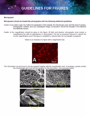
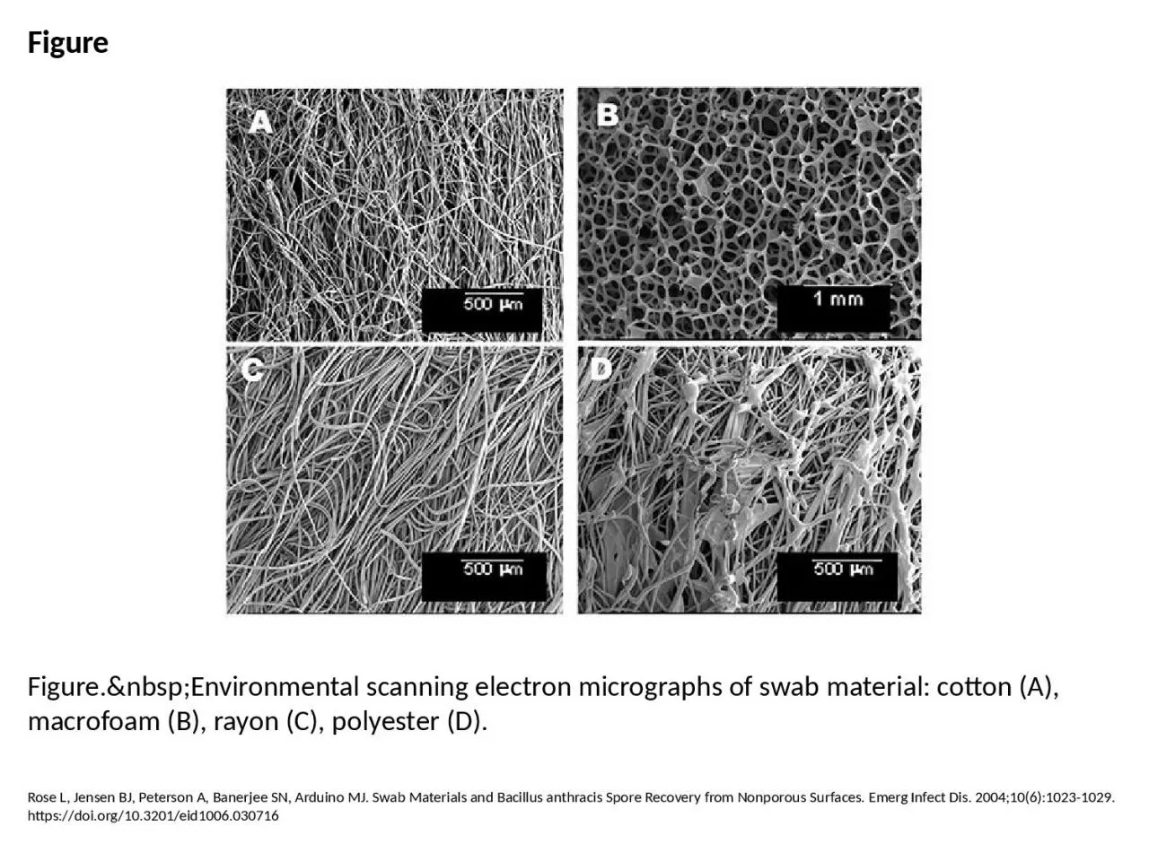
PPT-Figure Figure. Environmental scanning electron micrographs of swab material:

Author : cecilia | Published Date : 2023-07-08

Rose L Jensen BJ Peterson A Banerjee SN Arduino MJ Swab Materials and Bacillus anthracis Spore Recovery from Nonporous Surfaces Emerg Infect Dis 200410610231029

Presentation Embed Code

Download Presentation

Download Presentation The PPT/PDF document "Figure Figure. Environmental sc..." is the property of its rightful owner. Permission is granted to download and print the materials on this website for personal, non-commercial use only, and to display it on your personal computer provided you do not modify the materials and that you retain all copyright notices contained in the materials. By downloading content from our website, you accept the terms of this agreement.

Figure Figure. Environmental scanning electron micrographs of swab material:: Transcript


Rose L Jensen BJ Peterson A Banerjee SN Arduino MJ Swab Materials and Bacillus anthracis Spore Recovery from Nonporous Surfaces Emerg Infect Dis 200410610231029 httpsdoiorg103201eid1006030716. QUALITY STANDARDS As per Customers specification PRODUCTION CAPACITY PM MONTH YEAR OF PREPAR ATION December 2010 PREPARED BY MSME Development Institute Ministry of Micro Small Medium Enterprises Government of India 107 Industrial Estate Kal This information can be found on all micrographs together with the magnification size. If necessary, please contact the morphologist who took the micrographs for this information. See examples below. www.liofilchem.net 1PYR2SGA3STA4HAE5NES6STG7STR8PSE9PRO10ESC11IND12KES13VP14CAN15CN16NET17AZM18CLA19AUG20AMS21TZP22CRO23CFP24CAZ25CXM26PEF27LEV28OFX29RD30MRP31SXT32Growth PATHOGENIC SYSTEM AST **** 1P Scanning Electron Microscopy (SEM). Uses. Sample Preparation. Instrument. Principles. Micrographs. Transmission Electron Microscopy (TEM). Uses. Sample Preparation. Instrument. Principles. Micrographs. Specimen for COVID-19 TestingUse only an authorized specimen collection kit given to you by your healthcare provider or personnel at the testing center or purchased over the counter in a pharmacy or o IN0146Rev004For diagnostic useNOTEIf you have any questions about this procedure please ask your doctor nurse or care-providerWash your hands before startingIn the privacy of the examination room or See Us at M&M 2020 in Milwaukee Altekruse SF, Stern NJ, Fields PI, Swerdlow DL. Campylobacter jejuni—An Emerging Foodborne Pathogen. Emerg Infect Dis. 1999;5(1):28-35. https://doi.org/10.3201/eid0501.990104. Holmes AH, Govan J, Goldstein R. Agricultural Use of Burkholderia (Pseudomonas) cepacia: A Threat to Human Health?. Emerg Infect Dis. 1998;4(2):221-227. https://doi.org/10.3201/eid0402.980209. Ng M, Lee J, Leong M, Ling A, Tan H, Ooi E. Topographic Changes in SARS Coronavirus–infected Cells during Late Stages of Infection. Emerg Infect Dis. 2004;10(11):1907-1914. https://doi.org/10.3201/eid1011.040195. The aims of this course are:. ● . to introduce the principles of scanning electron microscopy. ● . to describe the components of the microscope and explain how they work. ● . to highlight some of the problems which can arise during imaging. Read the laundry guide on cleaning cotton clothes at home, and spare a few hours on the weekend to get your laundry work done in no time. Donlan RM. Biofilms and Device-Associated Infections. Emerg Infect Dis. 2001;7(2):277-281. https://doi.org/10.3201/eid0702.700277. Jabra-Rizk MA, Falkler WA, Meiller TF. Fungal Biofilms and Drug Resistance. Emerg Infect Dis. 2004;10(1):14-19. https://doi.org/10.3201/eid1001.030119.

Download Document

Here is the link to download the presentation.
"Figure Figure. Environmental scanning electron micrographs of swab material:"The content belongs to its owner. You may download and print it for personal use, without modification, and keep all copyright notices. By downloading, you agree to these terms.

Related Documents