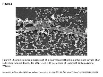
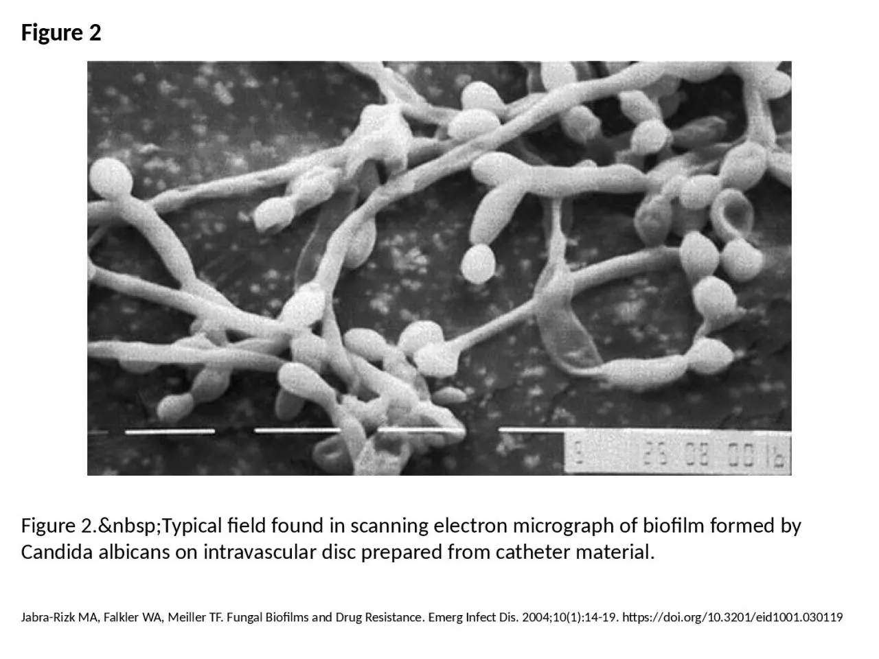
PPT-Figure 2 Figure 2. Typical field found in scanning electron micrograph of biofilm
Author : melody | Published Date : 2024-01-29
JabraRizk MA Falkler WA Meiller TF Fungal Biofilms and Drug Resistance Emerg Infect Dis 20041011419 httpsdoiorg103201eid1001030119
Presentation Embed Code
Download Presentation
Download Presentation The PPT/PDF document "Figure 2 Figure 2. Typical fiel..." is the property of its rightful owner. Permission is granted to download and print the materials on this website for personal, non-commercial use only, and to display it on your personal computer provided you do not modify the materials and that you retain all copyright notices contained in the materials. By downloading content from our website, you accept the terms of this agreement.
Figure 2 Figure 2. Typical field found in scanning electron micrograph of biofilm: Transcript
JabraRizk MA Falkler WA Meiller TF Fungal Biofilms and Drug Resistance Emerg Infect Dis 20041011419 httpsdoiorg103201eid1001030119. Figure C28. SEM micrograph of uncompressed mature yellow-poplar (4mm), TRT1. Figure C29. SEM micrograph of compressed mature yellow-poplar (4mm), TRT1. Figure C30. SEM micrograph of compressed mature Figure C80. SEM micrograph of compressed mature southern pine, TRT1. Figure C81. SEM micrograph of compressed mature southern pine, TRT1, showing multiplefractures in earlywood tracheids. Figure C82. Goosney DL, Knoechel DG, Finlay BB. Enteropathogenic E. coli, Salmonella, and Shigella: Masters of Host Cell Cytoskeletal Exploitation. Emerg Infect Dis. 1999;5(2):216-223. https://doi.org/10.3201/eid0502.990205. Altekruse SF, Stern NJ, Fields PI, Swerdlow DL. Campylobacter jejuni—An Emerging Foodborne Pathogen. Emerg Infect Dis. 1999;5(1):28-35. https://doi.org/10.3201/eid0501.990104. Holmes AH, Govan J, Goldstein R. Agricultural Use of Burkholderia (Pseudomonas) cepacia: A Threat to Human Health?. Emerg Infect Dis. 1998;4(2):221-227. https://doi.org/10.3201/eid0402.980209. Rose L, Jensen BJ, Peterson A, Banerjee SN, Arduino MJ. Swab Materials and Bacillus anthracis Spore Recovery from Nonporous Surfaces. Emerg Infect Dis. 2004;10(6):1023-1029. https://doi.org/10.3201/eid1006.030716. Donlan RM. Biofilms: Microbial Life on Surfaces. Emerg Infect Dis. 2002;8(9):881-890. https://doi.org/10.3201/eid0809.020063. Adeleke A, Pruckler J, Benson R, Rowbotham T, Halablab M, Fields B. Legionella-Like Amebal Pathogens––Phylogenetic Status and Possible Role in Respiratory Disease. Emerg Infect Dis. 1996;2(3):225-230. https://doi.org/10.3201/eid0203.960311. Ng M, Lee J, Leong M, Ling A, Tan H, Ooi E. Topographic Changes in SARS Coronavirus–infected Cells during Late Stages of Infection. Emerg Infect Dis. 2004;10(11):1907-1914. https://doi.org/10.3201/eid1011.040195. Donlan RM. Biofilms: Microbial Life on Surfaces. Emerg Infect Dis. 2002;8(9):881-890. https://doi.org/10.3201/eid0809.020063. Sonntag M, Mühldorfer K, Speck S, Wibbelt G, Kurth A. New Adenovirus in Bats, Germany. Emerg Infect Dis. 2009;15(12):2052-2055. https://doi.org/10.3201/eid1512.090646. Goosney DL, Knoechel DG, Finlay BB. Enteropathogenic E. coli, Salmonella, and Shigella: Masters of Host Cell Cytoskeletal Exploitation. Emerg Infect Dis. 1999;5(2):216-223. https://doi.org/10.3201/eid0502.990205. Donlan RM. Biofilms and Device-Associated Infections. Emerg Infect Dis. 2001;7(2):277-281. https://doi.org/10.3201/eid0702.700277. Kim MJ, Bancroft E, Lehnkering E, Donlan RM, Mascola L. Alcaligenes xylosoxidans Bloodstream Infections in Outpatient Oncology Office. Emerg Infect Dis. 2008;14(7):1046-1052. https://doi.org/10.3201/eid1407.070894.
Download Document
Here is the link to download the presentation.
"Figure 2 Figure 2. Typical field found in scanning electron micrograph of biofilm"The content belongs to its owner. You may download and print it for personal use, without modification, and keep all copyright notices. By downloading, you agree to these terms.
Related Documents