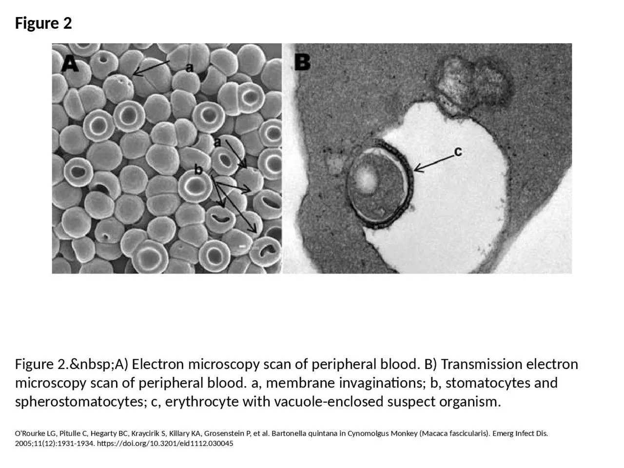
PPT-Figure 2 Figure 2. A) Electron microscopy scan of peripheral blood. B) Transmission

Author : emma | Published Date : 2023-07-08

ORourke LG Pitulle C Hegarty BC Kraycirik S Killary KA Grosenstein P et al Bartonella quintana in Cynomolgus Monkey Macaca fascicularis Emerg Infect Dis 2005111219311934

Presentation Embed Code

Download Presentation

Download Presentation The PPT/PDF document "Figure 2 Figure 2. A) Electron ..." is the property of its rightful owner. Permission is granted to download and print the materials on this website for personal, non-commercial use only, and to display it on your personal computer provided you do not modify the materials and that you retain all copyright notices contained in the materials. By downloading content from our website, you accept the terms of this agreement.

Figure 2 Figure 2. A) Electron microscopy scan of peripheral blood. B) Transmission: Transcript


ORourke LG Pitulle C Hegarty BC Kraycirik S Killary KA Grosenstein P et al Bartonella quintana in Cynomolgus Monkey Macaca fascicularis Emerg Infect Dis 2005111219311934 httpsdoiorg103201eid1112030045. Jim Atherton. Development of Light Microscope. 1590 Hans Zacharias Janssen. 1660 Robert Hook. Onserved. . cells (cork). 1. Klein, Aaron E. The Electron Microscope: A Tool of Discovery. New . Y. ork,McGRaw. By Austin Avery. Overview. What is Transmission Electron Microscopy?. History of TEM. Theory of TEM. The instrumentation. How is TEM useful?. Pros/Cons. Summary. Transmission Electron Microscopy. TEM is an instrumental technique that uses a tiny focused beam of electrons.. -grown . InGaAs. quantum dot chains using transmission electron microscopy. Tyler Park. John Colton . Haeyeon. Yang. *. Jeff . Farrer. APS March Meeting 2013 Baltimore, MD. * South Dakota School of Mines. This information is provided by Cleveland Clinic and is not intended to replace the medical advice of your doctor or health care provider. Please consult your health care provider for advice about a s Peter Harris. Transmission . electron . microscopy. ► What . is transmission electron microscopy?. . ► TEM in biological science . ► CfAM’s TEMs. ► TEM in physical . science . ► Recent developments in TEM. precocious puberty . Gonadotropin-independent . precocious . puberty. Dr. . Payam. . Samei. References. . 1. . Williams 2016. 2. Sperling 2014. 3. Up to . Date 2017. INTRODUCTION. . Electron microscopy (EM) . is an electron beam which is focused into a small probe across the surface of a specimen . . The first electromagnetic lens was developed in 1926 by . Hans Busch. .. . Glynnis Ingall MD, PhD. Hantavirus Cardiopulmonary Syndrome . Hantavirus . Cardiopulmonary . Syndrome (. HCPS. ) is a severe, sometimes fatal, respiratory disease . caused . by infection . with hantavirus. (SEM) Electron Microscopy (SEM) and TEM Scanning electron microscopy is used for inspecting topographies of specimens at very high magnifications using a piece of equipment called the scanning electr Theoretically a microscope is an array of two lenses Need of M icroscopy Living organisms, cells and their organelles which are too small to be seen with the human eye, can only be seen with the aid The theory of image formation in electron microscope s is very similar to image formation in light microscope s. However, the practice and design are completely different : The most crucial Riquelme C, Escors D, Ortego J, Sanchez CM, Uzelac-Keserovic B, Apostolov K, et al. Nature of the Virus Associated with Endemic Balkan Nephropathy. Emerg Infect Dis. 2002;8(8):869-870. https://doi.org/10.3201/eid0808.020042. Holmes AH, Govan J, Goldstein R. Agricultural Use of Burkholderia (Pseudomonas) cepacia: A Threat to Human Health?. Emerg Infect Dis. 1998;4(2):221-227. https://doi.org/10.3201/eid0402.980209. 10. Learning Objectives—Level l . At the end of this unit of study, the student should be able to:. List the assays typically included in the complete blood count (CBC).. Describe how to properly identify a patient prior to blood collection..

Download Document

Here is the link to download the presentation.
"Figure 2 Figure 2. A) Electron microscopy scan of peripheral blood. B) Transmission"The content belongs to its owner. You may download and print it for personal use, without modification, and keep all copyright notices. By downloading, you agree to these terms.

Related Documents