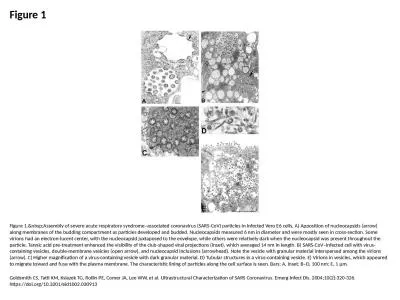
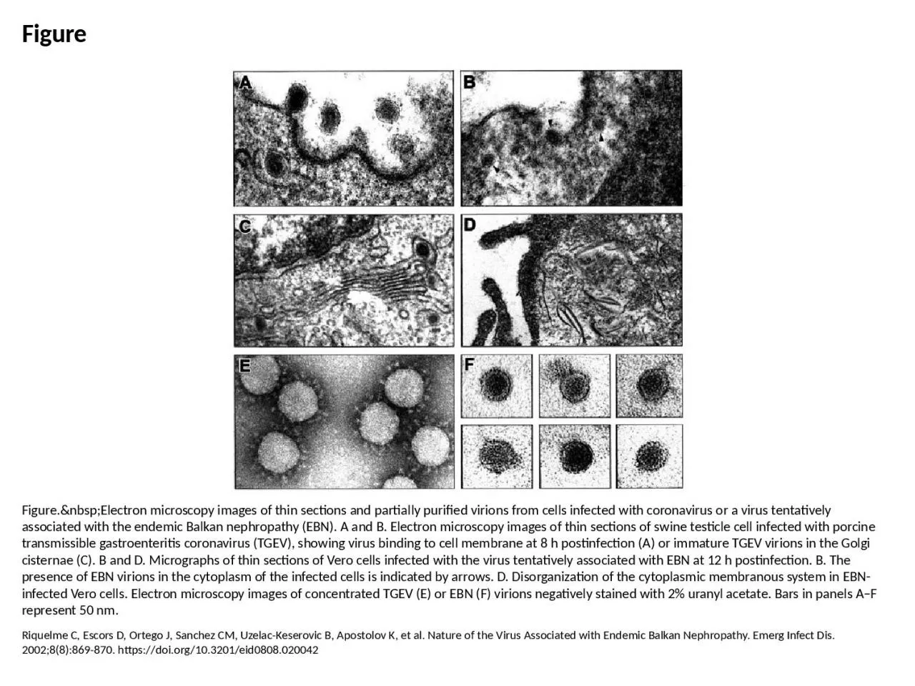
PPT-Figure Figure. Electron microscopy images of thin sections and partially purified

Author : faith | Published Date : 2023-05-31

Riquelme C Escors D Ortego J Sanchez CM UzelacKeserovic B Apostolov K et al Nature of the Virus Associated with Endemic Balkan Nephropathy Emerg Infect Dis 200288869870

Presentation Embed Code

Download Presentation

Download Presentation The PPT/PDF document "Figure Figure. Electron microsc..." is the property of its rightful owner. Permission is granted to download and print the materials on this website for personal, non-commercial use only, and to display it on your personal computer provided you do not modify the materials and that you retain all copyright notices contained in the materials. By downloading content from our website, you accept the terms of this agreement.

Figure Figure. Electron microscopy images of thin sections and partially purified: Transcript


Riquelme C Escors D Ortego J Sanchez CM UzelacKeserovic B Apostolov K et al Nature of the Virus Associated with Endemic Balkan Nephropathy Emerg Infect Dis 200288869870 httpsdoiorg103201eid0808020042. Chelsea Aitken. Peter Aspinall. http://www.zyvexlabs.com/EIPBNuG/EIPBN%20images/05Ion.jpg. Intro to Electron Microscopy. Similar to optical microscopy except with electrons rather than photons. Used to image samples with a resolution of 10 Å. By Austin Avery. Overview. What is Transmission Electron Microscopy?. History of TEM. Theory of TEM. The instrumentation. How is TEM useful?. Pros/Cons. Summary. Transmission Electron Microscopy. TEM is an instrumental technique that uses a tiny focused beam of electrons.. Microscopy is the technical field using microscopes to view samples and objects that can not be seen without unaided eye (objects that are not within the resolution range of the normal eye). There are three well-known branches of microscopy: optical, electron and scanning probe microscopy.. Kumari. Department of Veterinary Microbiology. Bihar Veterinary College, BASU, Patna. Term “Pox” derives from . English . pocks, blister like . skin . lesions. Smallpox . vaccine, introduced by Edward Jenner in . . Electron microscopy (EM) . is an electron beam which is focused into a small probe across the surface of a specimen . . The first electromagnetic lens was developed in 1926 by . Hans Busch. .. . AGENTS. . THAT. . INACTIVATE. . VIRUS INFECTIVITY. . 1. . Physical. . agents. . . 1.a. . Temperature. All. . infective. . viruses. . lose. . infectivity. . over. . time,. . AIM : T THEORY: Microscope is the most commonly used piece of apparatus in the laboratory. It produces greatly enlarged images of minute objects. LIGHT MICROSCOPE A Light Microscope can be simple or Theoretically a microscope is an array of two lenses Need of M icroscopy Living organisms, cells and their organelles which are too small to be seen with the human eye, can only be seen with the aid The theory of image formation in electron microscope s is very similar to image formation in light microscope s. However, the practice and design are completely different : The most crucial Goldsmith CS, Tatti KM, Ksiazek TG, Rollin PE, Comer JA, Lee WW, et al. Ultrastructural Characterization of SARS Coronavirus. Emerg Infect Dis. 2004;10(2):320-326. https://doi.org/10.3201/eid1002.030913. Ng M, Lee J, Leong M, Ling A, Tan H, Ooi E. Topographic Changes in SARS Coronavirus–infected Cells during Late Stages of Infection. Emerg Infect Dis. 2004;10(11):1907-1914. https://doi.org/10.3201/eid1011.040195. Hodo CL, Mauldin MR, Light JE, Wilkins K, Tang S, Nakazawa Y, et al. Novel Poxvirus in Proliferative Lesions of Wild Rodents in East Central Texas, USA. Emerg Infect Dis. 2018;24(6):1069-1072. https://doi.org/10.3201/eid2406.172057. Rock thin sections. Rock thin sections are slices of rock glued onto microscope slides and polished until they are about 30μm thick (about the thickness of human hair).. The microscope used to study minerals in rock thin sections.. The aims of this course are:. ● . to introduce the principles of scanning electron microscopy. ● . to describe the components of the microscope and explain how they work. ● . to highlight some of the problems which can arise during imaging.

Download Document

Here is the link to download the presentation.
"Figure Figure. Electron microscopy images of thin sections and partially purified"The content belongs to its owner. You may download and print it for personal use, without modification, and keep all copyright notices. By downloading, you agree to these terms.

Related Documents