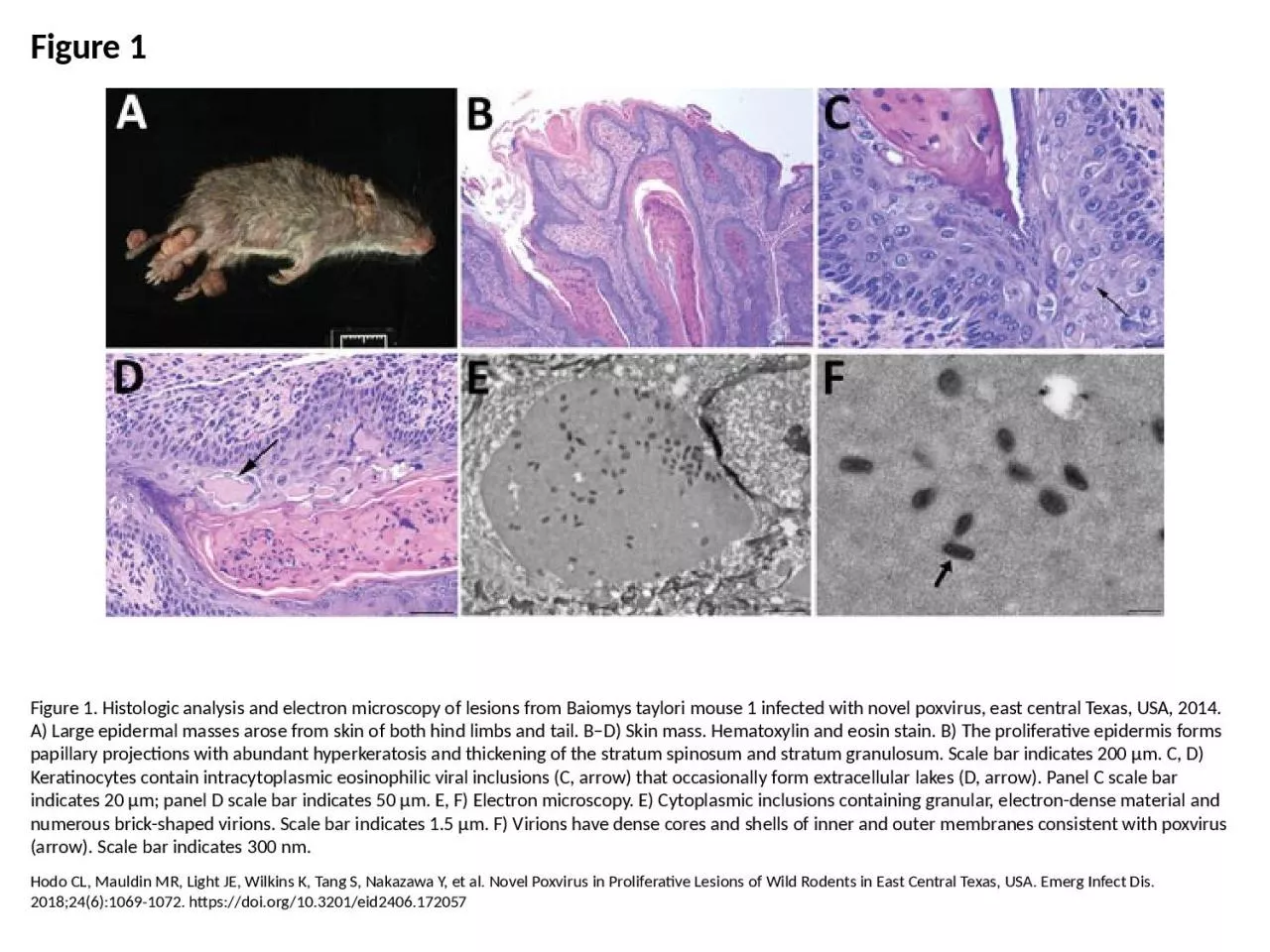
PPT-Figure 1 Figure 1. Histologic analysis and electron microscopy of lesions from Baiomys

Author : cora | Published Date : 2023-07-27

Hodo CL Mauldin MR Light JE Wilkins K Tang S Nakazawa Y et al Novel Poxvirus in Proliferative Lesions of Wild Rodents in East Central Texas USA Emerg Infect Dis

Presentation Embed Code

Download Presentation

Download Presentation The PPT/PDF document "Figure 1 Figure 1. Histologic analysis a..." is the property of its rightful owner. Permission is granted to download and print the materials on this website for personal, non-commercial use only, and to display it on your personal computer provided you do not modify the materials and that you retain all copyright notices contained in the materials. By downloading content from our website, you accept the terms of this agreement.

Figure 1 Figure 1. Histologic analysis and electron microscopy of lesions from Baiomys: Transcript


Hodo CL Mauldin MR Light JE Wilkins K Tang S Nakazawa Y et al Novel Poxvirus in Proliferative Lesions of Wild Rodents in East Central Texas USA Emerg Infect Dis 201824610691072 httpsdoiorg103201eid2406172057. Lecture . 07: . Confocal Microscopy. Adding the Third Dimension. Lecture . 7: Confocal Microscopy. Optical Sectioning: adding the third dimension. Wide-field . Imaging. Point . Spread Function. Deconvolution. Chelsea Aitken. Peter Aspinall. http://www.zyvexlabs.com/EIPBNuG/EIPBN%20images/05Ion.jpg. Intro to Electron Microscopy. Similar to optical microscopy except with electrons rather than photons. Used to image samples with a resolution of 10 Å. Lecture 13:. Super-resolution microscopy: Part I. Lecture 13: . Fluorescent labeling, multi-. sprectral. imaging and FRET. Review of previous lecture. FRET. FLIM. Super resolution . microscopy. NSOM. By Austin Avery. Overview. What is Transmission Electron Microscopy?. History of TEM. Theory of TEM. The instrumentation. How is TEM useful?. Pros/Cons. Summary. Transmission Electron Microscopy. TEM is an instrumental technique that uses a tiny focused beam of electrons.. Fire Protection Laboratory Methods Day. June 25, 2014. Paul M. Anderson. Graduate Research Assistant. University of Maryland. Department of Fire Protection Engineering. Transmission Electron Microscopy. Tutor: Peter Harris. Click on the buttons to move between slides. .. (Make sure you are in “Slide Show” mode). The aims of this course are:. ● . to explain how X-rays are generated in the electron microscope. Jenny . Malmstrom. AFM invented by Binning and co-workers in 1986. . Belongs to the Scanning Probe Microscopy family. Binning et al., Physics Review Letters 1986. AFM invented by Binning and co-workers in 1986. . Peter Harris. Transmission . electron . microscopy. ► What . is transmission electron microscopy?. . ► TEM in biological science . ► CfAM’s TEMs. ► TEM in physical . science . ► Recent developments in TEM. AIM : T THEORY: Microscope is the most commonly used piece of apparatus in the laboratory. It produces greatly enlarged images of minute objects. LIGHT MICROSCOPE A Light Microscope can be simple or The theory of image formation in electron microscope s is very similar to image formation in light microscope s. However, the practice and design are completely different : The most crucial O'Rourke LG, Pitulle C, Hegarty BC, Kraycirik S, Killary KA, Grosenstein P, et al. Bartonella quintana in Cynomolgus Monkey (Macaca fascicularis). Emerg Infect Dis. 2005;11(12):1931-1934. https://doi.org/10.3201/eid1112.030045. Martin Whiteside, DC, PhD, MSPH. Director, Tennessee Cancer Registry. NAACCR Annual Conference. June 14. th. -16. th. , 2016. Harold N. Lovvorn, III, MD, co-Author. This study was supported in part through a cooperative agreement obtained from the Centers for Disease Control & Prevention (CDC), 5 NU58DP003901.. A . fluorescence microscope.  is an optical microscope that uses fluorescence and phosphorescence instead of, or in addition to, reflection and absorption to . study . organic . or inorganic substances. ELECTRON. . MICROSC. O. PY. . . . Electron. . micros. c. opy. . (EM). . is a technique. . u. s. ed. . for. . obtaining. . high re. s. olu. t. ion. . images. . of. . biological and non-biologi.

Download Document

Here is the link to download the presentation.
"Figure 1 Figure 1. Histologic analysis and electron microscopy of lesions from Baiomys"The content belongs to its owner. You may download and print it for personal use, without modification, and keep all copyright notices. By downloading, you agree to these terms.

Related Documents