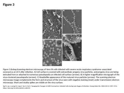
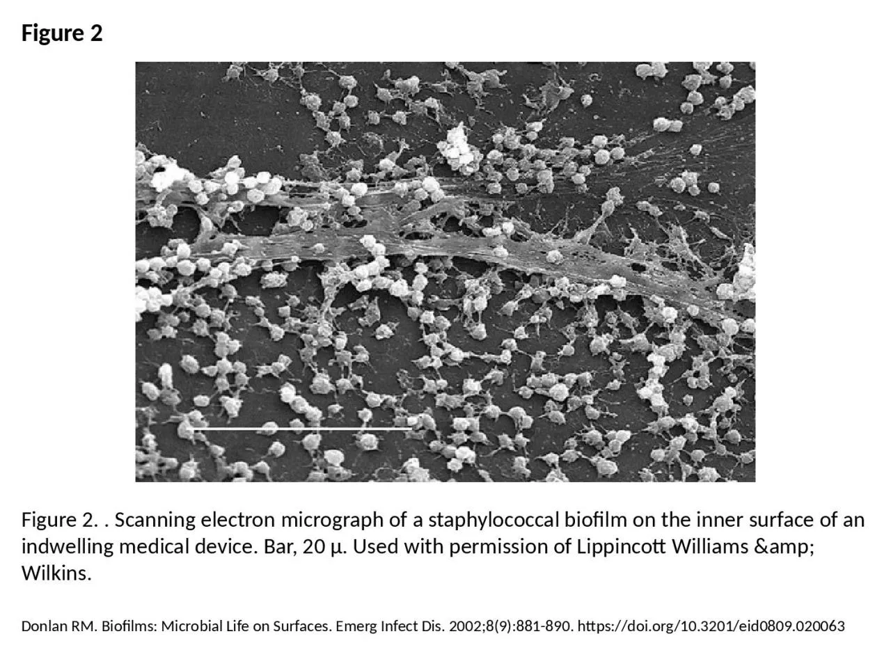
PPT-Figure 2 Figure 2. . Scanning electron micrograph of a staphylococcal biofilm on the inner
Author : patricia | Published Date : 2023-07-08
Donlan RM Biofilms Microbial Life on Surfaces Emerg Infect Dis 200289881890 httpsdoiorg103201eid0809020063
Presentation Embed Code
Download Presentation
Download Presentation The PPT/PDF document "Figure 2 Figure 2. . Scanning electron m..." is the property of its rightful owner. Permission is granted to download and print the materials on this website for personal, non-commercial use only, and to display it on your personal computer provided you do not modify the materials and that you retain all copyright notices contained in the materials. By downloading content from our website, you accept the terms of this agreement.
Figure 2 Figure 2. . Scanning electron micrograph of a staphylococcal biofilm on the inner: Transcript
Donlan RM Biofilms Microbial Life on Surfaces Emerg Infect Dis 200289881890 httpsdoiorg103201eid0809020063. Figure C54. SEM micrograph of compressed mature yellow-poplar (4mm), TRT3. Figure C55. SEM micrograph of uncompressed mature yellow-poplar (4mm), TRT3. Figure C56. SEM micrograph of uncompressed matur Figure C28. SEM micrograph of uncompressed mature yellow-poplar (4mm), TRT1. Figure C29. SEM micrograph of compressed mature yellow-poplar (4mm), TRT1. Figure C30. SEM micrograph of compressed mature Figure C80. SEM micrograph of compressed mature southern pine, TRT1. Figure C81. SEM micrograph of compressed mature southern pine, TRT1, showing multiplefractures in earlywood tracheids. Figure C82. M. aterials . U. sed . in . Water . D. istribution . A. ffect . B. iofilm . G. rowth . and . Drinking . W. ater . Q. uality. D. . . Clark, . Chemical . Engineering; . V. . Sumner, . Chemical . Engineering; . . . Conclusions. Changing the solvent that PHB-. HHx. is dissolved in, greatly alters the mechanical properties. Water replaced gels are stronger but deform more easily than solvent filled gels. Ng M, Lee J, Leong M, Ling A, Tan H, Ooi E. Topographic Changes in SARS Coronavirus–infected Cells during Late Stages of Infection. Emerg Infect Dis. 2004;10(11):1907-1914. https://doi.org/10.3201/eid1011.040195. Holmes AH, Govan J, Goldstein R. Agricultural Use of Burkholderia (Pseudomonas) cepacia: A Threat to Human Health?. Emerg Infect Dis. 1998;4(2):221-227. https://doi.org/10.3201/eid0402.980209. Rose L, Jensen BJ, Peterson A, Banerjee SN, Arduino MJ. Swab Materials and Bacillus anthracis Spore Recovery from Nonporous Surfaces. Emerg Infect Dis. 2004;10(6):1023-1029. https://doi.org/10.3201/eid1006.030716. Adeleke A, Pruckler J, Benson R, Rowbotham T, Halablab M, Fields B. Legionella-Like Amebal Pathogens––Phylogenetic Status and Possible Role in Respiratory Disease. Emerg Infect Dis. 1996;2(3):225-230. https://doi.org/10.3201/eid0203.960311. Ng M, Lee J, Leong M, Ling A, Tan H, Ooi E. Topographic Changes in SARS Coronavirus–infected Cells during Late Stages of Infection. Emerg Infect Dis. 2004;10(11):1907-1914. https://doi.org/10.3201/eid1011.040195. Donlan RM. Biofilms: Microbial Life on Surfaces. Emerg Infect Dis. 2002;8(9):881-890. https://doi.org/10.3201/eid0809.020063. Donlan RM. Biofilms and Device-Associated Infections. Emerg Infect Dis. 2001;7(2):277-281. https://doi.org/10.3201/eid0702.700277. Jabra-Rizk MA, Falkler WA, Meiller TF. Fungal Biofilms and Drug Resistance. Emerg Infect Dis. 2004;10(1):14-19. https://doi.org/10.3201/eid1001.030119. Kim MJ, Bancroft E, Lehnkering E, Donlan RM, Mascola L. Alcaligenes xylosoxidans Bloodstream Infections in Outpatient Oncology Office. Emerg Infect Dis. 2008;14(7):1046-1052. https://doi.org/10.3201/eid1407.070894.
Download Document
Here is the link to download the presentation.
"Figure 2 Figure 2. . Scanning electron micrograph of a staphylococcal biofilm on the inner"The content belongs to its owner. You may download and print it for personal use, without modification, and keep all copyright notices. By downloading, you agree to these terms.
Related Documents