/
Does upright magnetic resonance imaging of the lumbar spine Does upright magnetic resonance imaging of the lumbar spine

Does upright magnetic resonance imaging of the lumbar spine - PowerPoint Presentation

celsa-spraggs
celsa-spraggs . @celsa-spraggs
Follow
386 views
Uploaded On 2017-09-18

Does upright magnetic resonance imaging of the lumbar spine - PPT Presentation

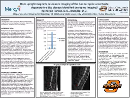
Katherine Rankin DO Brian Do DO Department of Diagnostic Radiology at Oklahoma State University Medical Center Tulsa Oklahoma OBJECTIVE The objective of this study is to determine if upright magnetic resonance imaging accentuates degenerative disc disease identified on supine imaging ID: 588822

upright imaging disease disc imaging upright disc disease supine mri degenerative lumbar position spine pain study patients exams patient

Share:

Link:

Embed:

Download Presentation from below link

Download Presentation The PPT/PDF document "Does upright magnetic resonance imaging ..." is the property of its rightful owner. Permission is granted to download and print the materials on this web site for personal, non-commercial use only, and to display it on your personal computer provided you do not modify the materials and that you retain all copyright notices contained in the materials. By downloading content from our website, you accept the terms of this agreement.


Presentation Transcript

Slide1

Does upright magnetic resonance imaging of the lumbar spine accentuate degenerative disc disease identified on supine imaging?Katherine Rankin, D.O., Brian Do, D.O.Department of Diagnostic Radiology at Oklahoma State University Medical Center, Tulsa, Oklahoma

OBJECTIVE:The objective of this study is to determine if upright magnetic resonance imaging accentuates degenerative disc disease identified on supine imaging.INTRODUCTION:Back pain is the most common cause of disability in people younger than age 45. The lifetime incidence of back pain in the United States population is 50-80%. The etiology of degenerative disc disease is multifactorial and includes manual labor, obesity, cigarette smoking, psychosocial factors, and genetics. MRI is a commonly used imaging modality to evaluate the severity of degenerative disc disease in quantifying central canal stenosis and neuroforaminal narrowing. Traditional MRI imaging of the spine is performed with the spine in a functional position of rest when the patient is lying supine. Low back pain is often exacerbated in the upright position. The size of the spinal canal and neural foramen can be falsely elevated with the patient in the supine position, which can result in an underestimation of the severity of disease or to negative findings in the presence of symptoms. Historically X-ray was the only modality with the capability of imaging in the upright position. Recent technological advances have allowed the use of open low and medium intensity magnetic resonance imaging to evaluate the spine in an orthostatic position. These open MRI devices also have the added benefit of aiding in the setting of patient claustrophobia, which can be a limiting factor in image quality.METHODS AND MATERIALS: All upright lumbar spine imaging performed between July 1, 2013 to September 30, 2013 at a single outpatient imaging center were reviewed. Approximately 280 cases were reviewed. A Paramed 0.5 Tesla open magnetic resonance scanner was used for upright imaging. Patients with prior lumbar surgical fixation were excluded because of the potential for image quality degradation. The exams were reviewed and the results were categorized into positive or negative based on accentuation of degenerative disc disease on upright imaging in comparison to the supine imaging. Accentuation of degenerative disc disease was quantified by increased anteroposterior diameter of the disc bulge, protrusion, or extrusion on T2 upright sagittal imaging.

CONCLUSION: Data from this study suggests that upright imaging does accentuate degenerative disc disease in approximately 10% of cases. Therefore, supine MRI imaging remains the current gold standard imaging modality in diagnosing lumbar degenerative disc disease in the setting of acute and chronic back pain. Factors to consider upon further research include performing upright MRI imaging on patients who specifically have pain in the upright position as well as utilizing upright MRI imaging in patients who have negative lumbar MRI exams in the supine position with suspicious clinical symptoms. This study only evaluated patients who had evidence of degenerative disc disease on supine imaging and then they underwent subsequent sagittal imaging in the upright position. Further studies could also evaluate the significance of flexion or extension of the lumbar spine while in the upright position. REFERENCES: 1. Tarantino U, Fanucci E, et al. Lumbar spine MRI in upright position for diagnosing acute and chronic low back pain: statistical analysis of morphological changes. J Orthop Traumatol. 2013 Mar; 14(1): 15–22.2. Hadjipavlou AG,et al. The pathophysiology of disc degeneration: a critical review. J Bone Joint Surg Br. 90(10):1261-70, 2008.3. Modic MT et al. Lumbar degenerative disk disease. Radiology. 245(1):43-61, 2007.4. Gallucci M, et al. Degenerative disease of the spine. Neuroimaging Clin of N Am. 2007;17:87–103.5. Claus A, et al. Sitting versus standing: does the intradiscal pressure cause disc degeneration or low back pain? J Electromyogr Kinesiol. 2008;18:550–558.

RESULTS:Upon evaluating three months data 282 exams were reviewed. Twenty-one of the cases were excluded due to the presence of spinal fixation hardware. Twenty-seven of the 261 exams used demonstrated accentuation of degenerative disc disease.

DISCUSSION: This retrospective study suggests that upright MRI of the lumbar spine only accentuates findings of degenerative disc disease identified on supine MRI imaging a small percentage of the time. Limitations of the study include lack of information as to the position in which the patient has maximal low back pain, supine versus upright. Due to the exclusion of patients with prior lumbar fixation, an entire subset of people with low back pain, specifically failed back surgeries, could not be evaluated.

Supine sagittal MRI image of the lumbar spine reveals a shallow posterior disc protrusion at L2-L3. Larger disc bulges are also identified from L3 to S1.

Upright sagittal MRI image of the lumbar spine reveals accentuation of the disc bulge at the L2-L3 level.

Research performed by Tarantino, et al, regarding supine and upright imaging utilized more specific parameters to assess morphological and anatomic changes. These parameters included the lumbosacral angle,

lordosis

angle, disc height, interspinous distance between L3-L4, and maximum anteroposterior diameter of the dural sac.

Utilizing these more specific measuring guidelines led to improved outcome in regard to the use of upright imaging, which was found to be a good adjunct technique when used in conjunction with supine MRI imaging.

Imaging example of a positive exam