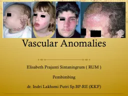
Department of diangosis imaging Childrens Hospital 2 Classification of vascular anomalies INTRODUCTION Vascular anomalies are among the most common congenital abnormalities observed in infants and children ID: 774602
Download Presentation The PPT/PDF document " Updated ISSVA (International Society fo..." is the property of its rightful owner. Permission is granted to download and print the materials on this web site for personal, non-commercial use only, and to display it on your personal computer provided you do not modify the materials and that you retain all copyright notices contained in the materials. By downloading content from our website, you accept the terms of this agreement.
Slide1
Updated ISSVA(International Society for the Study of Vascular Anomalies)
Department of diangosis imagingChildren’s Hospital 2
Classification of vascular anomalies
Slide2INTRODUCTION
Vascular anomalies are among the most common congenital abnormalities observed in infants and children.
Older nomenclature continues to cause confusion, misunderstood diagnoses, and potential mismanagement
In 1982, Mulliken and Glowacki proposed a classification system for vascular anomalies based on their clinical behavior and endothelial cell characteristics into two groups:
hemangiomas
and
vascular malformations.
This system, which was adopted by the ISSVA, has since been expanded and is now widely accepted.
Radiologists can use the ISSVA classification system by correlating imaging findings with patient history and physical findings. Consistent use of this system will help patients receive the correct diagnosis and treatment.
Slide3Traditional classification
Capillary hemangioma
Strawberry hemangioma
Strawberry nevus
Port wine stain
Flame nevus
Cavernous hemangioma
Venous
angioma
Lymphangioma
Arteriovenous malformation
Slide4CLASSIFICATION(ISSVA)
Slide5Updated ISSVA classification of vascular anomalies.
Infantile hemangiomasCongenital hemangiomas (RICH and NICH)Tufted angioma (with or without Kasabach-Merritt syndrome)Kaposiform hemangioendothelioma (with or without Kasabach-Merritt syndrome)Spindle cell hemangioendotheliomaOther, rare hemangioendotheliomas (epithelioid, composite, retiform, polymorphous, Dabska tumor, lymphangioendotheliomatosis, etc.)Dermatologic acquired vascular tumors (pyogenic granuloma, targetoid hemangioma, glomeruloid hemangioma, microvenular hemangioma, etc.)
1..Slow-flow vascular malformations:Capillary malformation (CM)Port-wine stainTelangiectasiaAngiokeratoma Venous malformation (VM)Common sporadic VMBean syndromeFamilial cutaneous and mucosal venous malformation (VMCM)Glomuvenous malformation (GVM)(glomangioma)Maffucci syndromeLymphatic malformation (LM)2. Fast-flow vascular malformations:Arterial malformation (AM)Arteriovenous fistula (AVF)Arteriovenous malformation (AVM)3.Complex-combined vascular malformations:CVM, CLM, LVM, CLVM,AVM-LM, CM-AVM
C:capillary; V:venous; L:lymphatic; AV:arteriovenous; M:malformation. RICH:rapidly involuting congenital hemangioma; NICH:noninvoluting congenital hemangioma.
Vascular tumors
Vascular malformations
Slide6Differentiating Features
Hemangioma
Vascular Malformations
True tumors, with proliferation of the vascular endothelium
>3:1
female:male
Small or absent at birth
Rapid growth during infancy
“self-limited”
Diagnosis:Clinical
history+ appearance
No tumor, Comprised of dysplastic vessels
1:1
female:male
Present at birth
Growth proportional to child
never disappear
Diagnosis:
MRI,Doppler
ultrasonography,angiography
Slide7Comparison of Previous Terminology and New ISSVA Terminology
Previous
ISSVA
Capillary or cavernous Hemangioma of any organ
Infantile hemangioma
Infantile hemangioendothelioma of the liver
Hepatic or infantile hemangioma
hepatic
hemangioma
, cavernous
hemangioma
Venous malformation
Lymphangioma,Cystic
hygroma
Lymphatic Malformation
Port-wine stain,Capillary Hemangioma
Capillary Malformation
Slide8Key Imaging Features of the Most Common Pediatric Vascular Anomalies
Slide9Differentiation between Hemangioma and Hemangioendothelioma of the liver
Slide10Classification of Vascular Tumors
Benign tumors and tumor-like conditions
Hemangiomas
Spindle cell hemangioma (‘hemangioendotheliomas’)
Epithelioid hemangioma
Low-grade malignant tumors
Retinform hemangioendotheliomas
Composite hemangioendotheliomas
Polymorphous hemangioendotheliomas
Kaposiform hemangioendotheliomas
Malignant tumors
Epithelioid hemangioendotheliomas
Angiosarcoma
Slide11HEMANGIOMA
congenital hemangioma
Arterio-Venous Malformation(AVM)
Infantile hemangioma
Slide12HEMANGIOMA
Kaposiform Hemangioendothelioma with Kasabach-Merritt Phenomenon
Slide13Hemangioma
Benign endothelial cell tumor
2 main types
1. Infantile Hemangioma
Most common tumor of infancy/childhood
Usually has overlying patch of redness
Appears weeks/months after birth
Natural course - 3 stages
1. Proliferating - first year
2. Involuting - few years
3. Involuted - most resolved by age 10
Slide14Hemangioma(cont)
2. Congenital Hemangioma
Present at birth
Rare (compared to infantile)
Blue/gray hue w/ pale halo (skin)
2 types
Non-Involuting (NICH) - persistent
Rapidly Involuting (RICH) - resolved by 1-2 yrs
Slide15Lymphatic malformations
Commonly occur in the cervicofacial region, Lymphatic malformations in an extremity can cause diffuse or localized swelling with soft-tissue and skeletal overgrowth
Slide16Venous Malformation (VM)
Present at birth
skin discoloration, local swelling, and pain
Thin-walled, dilated veins:Inadequate smooth muscle layer
Complications: Thrombosis, bleeding
Slide17Venous Malformation (VM)
Slide18Arterio-Venous Malformation (AVM)
Present at birth
Reddish vascular hue (skin), often warm pulsations, thrill, and bruit
High-flow arterio-venous communication - absence of developed capillary bed
Complications: ulceration, bleeding,pain, compression/displacement of organs, high-outputcardiac failure
Slide19Arterio-Venous Malformation (AVM)
Slide20AVM
Slide21Summary of Regional and Diffuse Syndromes Associated With Vascular Malformations
Regional syndromes with associated vascular malformationsSturge–Weber: facial capillary malformation with intracranial capillary malformation, venous malformation, or AVM.Klippel–Trenaunay: limb/trunk capillary venous lymphatic malformations with overgrowth.Parkes Weber: CAVM with overgrowth; lymphatic malformation.Diffuse syndromes associated slow-flow malformationsProteus syndrome: vascular malformations (capillary or venous), hamartomatous syndrome with overgrowth(hemihypertrophy and macrodactyly), lipomas, pigmented nevi.Blue rubber bleb nevus (Bean) syndrome: multiple cutaneous, musculoskeletal, and gastrointestinal tract venous malformations.Epidermal nevus syndrome (Solomon syndrome): vascular malformations (intracranial AVM), epidermal nevi, various developmental abnormalities of the skin, eyes, nervous, skeletal, cardiovascular, and urogenital systems.Bannayan–Riley–Ruvalcaba syndrome: vascular malformations (cutaneous, intracranial), macrocephaly, ectodermal dysplasia, lipomatous masses, and intestinal hamartomatous polyps, PTEN suppressor gene mutation association.Diffuse syndromes associated fast flow malformationsHereditary hemorrhagic telangiectasia (Osler–Weber–Rendu): telangiectasias (skin, mucous membranes, gastrointestinal mucosa) and AVMs (lungs, liver, brain, spinal cord).
AVM, arteriovenous malformation; CAVM, capillary arterial venous malformation; PTEN, phosphatase and tensin homolog.
Slide22OVERGROWTH SYNDROMES:
Klippel-Trénaunay syndrome
which is a low-flow combined vascular anomaly (capillary-lymphatic-venous malformation) usually associated with marked overgrowth of the leg and capillary stains.
Parkes-Weber syndrome
consists of an AVM-like high-flow malformation that involves the entire extremity (usually a lower limb), and it is usually associated with a capillary malformation over the enlarged limb.
Slide23Simple malformations
slow flow
capillary
lymphatic
venous
fast flow
arterial
aneurysm
coarctation
ectasia
arteriovenous fistulae
(with one ore more shunts)
arteriovenous malformations
(with a nidus of multiple shunts)
Slide24Complex malformations
regional
Sturge-Weber syndrome
Klippel-Trénaunay syndrome
F. P. Weber syndrome
diffuse
Maffucci syndrome
Solomon syndrome
Proteus syndrome
Slide25Arteriovenous malformations (AVMs)
Slide26Low flow malformations
Lymphatic Malformations
-Microcystic
-Macrocystic
Venous Malformations
Capillary Malformations
Combined types
Slide27