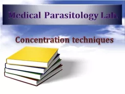
Flotation Method Concentration techniques Concentration Methods Sedimentation method Modified Formal Ether sedimentation technique Acid Ether sedimentation technique Flotation method Saturated Salt Solution technique ID: 360291
Download Presentation The PPT/PDF document "Medical Parasitology Lab." is the property of its rightful owner. Permission is granted to download and print the materials on this web site for personal, non-commercial use only, and to display it on your personal computer provided you do not modify the materials and that you retain all copyright notices contained in the materials. By downloading content from our website, you accept the terms of this agreement.
Slide1
Medical Parasitology Lab.
Flotation Method
Concentration
techniquesSlide2
Concentration MethodsSedimentation method
Modified Formal- Ether sedimentation techniqueAcid- Ether sedimentation techniqueFlotation method
Saturated Salt Solution technique
Sheather’s Sugar Centrifugal Flotation technique
Zinc Sulphate Centrifugal Flotation technique
Raed Z. Ahmed, Medical Parasitology Lab.,2012Slide3
Flotation techniqueThese method use the high specific gravity of a solution to float the lighter ova and cyst. They can be improved by centrifugation.
Advantage:Easy to perform .Disadvantage:
Delay in examination can result in
distortion.
Larvae and some fluke eggs do not concentrate.
Frequent checking of specific
gravity.
Raed Z. Ahmed, Medical Parasitology Lab.,2012Slide4
Saturated salt solution
Flotation Methods
Raed Z. Ahmed, Medical Parasitology Lab.,2012Slide5
Materials and Method Boil granular sodium chloride in excess in water to produce a saturated solution which when cooled has a specific gravity of 1.18 - 1.2.
Half fill a wide- mounted flat bottomed container with the saturated salt solution.Emulsify 1gm of feces in the solution and strain it to remove the debris from the surface.Pour the filtrate into meniscus and fill it to the top with saturated salt solution.
Lay a glass slide or large coverslip over the top, avoiding any bubbles being trapped.
Leave for 20 min before quickly inverting the slide.
Scan for ova using the 10x objectives.
Raed Z. Ahmed, Medical Parasitology Lab.,2012Slide6
Saturated Salt solution techniqueAdvantages:
It is cheap preparation using simple apparatus.It concentrates nematode ova well.Disadvantages:
It doesn’t concentrate cysts.
Raed Z. Ahmed, Medical Parasitology Lab.,2012Slide7
Sheather’s sugar centrifugal flotation technique
Flotation Methods
Raed Z. Ahmed, Medical Parasitology Lab.,2012Slide8
Materials and Method
Sheather’s sugar solution:Table sugar -------------------------------------- 500gmDistilled water ---------------------------------- 320mlPhenol crystal ( melt in hot water bath) ----- 6.5gm
Raed Z. Ahmed, Medical Parasitology Lab.,2012Slide9
Procedures Soften 1gm of feces with water to a soft.
Strain the aqueous suspension through a wire sieve.Mix 1 part aqueous suspension with 2 part of Sheather's sugar solution.Pour into a centrifuge tube, centrifugation 1500 rpm for 10 minutes.
Pour the supernatant into a meniscus and add a sufficient solution to bring the meniscus to the top.
Place a coverslip and wait for 10 minutes.
Examine under microscope.
Raed Z. Ahmed, Medical Parasitology Lab.,2012Slide10
Advantages:Reveals most nematode eggs and protozoan cyst.Disadvantages:
Flukes eggs and tape worm eggs are not demonstrate well.Also most nematode larvae are not demonstrate well.Raed Z. Ahmed, Medical Parasitology Lab.,2012
Sheather’s Sugar solution techniqueSlide11
Balantidium coli
Raed Z. Ahmed, Medical Parasitology Lab.,2012
Intestinal Protozoa Slide12
Balantidium coliIs a parasitic species of ciliate protozoa that cause
balantidiasis or Balantidium dysentery. Balantidium coli has two developmental stages, a trophozoite stage and a cyst stage.
The
cyst
is the infective stage measures about 50 to 70 µm in diameter, characterized by the presence of a large kidney-shaped macronucleus.
The
trophozoite
is oval
with 2 nuclei and
measures approximately 50 to 100 µm long and 40 to 70 µm wide.
Raed Z. Ahmed, Medical Parasitology Lab.,2012Slide13
B.coli Cyst
Raed Z. Ahmed, Medical Parasitology Lab.,2012Slide14
B. coli Trophozoite
Raed Z. Ahmed, Medical Parasitology Lab.,2012Slide15
Cryptosporidium parvum
Raed Z. Ahmed, Medical Parasitology Lab.,2012
Intestinal Protozoa Slide16
Cryptosporidium parvumInfect human and most mammals.
The infective stage is oocyst containing sporozoites measuring 5u in diameter.The diagnostics stage is sporocyst containing 4 sporozoites.Diagnosis:
Detecting oocyst in stool.
Acid-fast stain.
Raed Z. Ahmed, Medical Parasitology Lab.,2012Slide17
Cryptosporidium parvum oocyst
Raed Z. Ahmed, Medical Parasitology Lab.,2012Slide18
Toxoplasma gondii
Raed Z. Ahmed, Medical Parasitology Lab.,2012
Tissue ProtozoaSlide19
Toxoplasma gondiiHabitat:
reticulo-endothelial system, monocyte, and muscle fiber and cause toxoplasmosis.Humans can acquire Toxoplasma gondii infection by
ingestion cyst or
sporylated
oocystCyst: precipitated in flesh of cow.Sporulated oocyst: cat feces.
Intermediate host
: Human.
Definitive host
:
Cat.
The
tachyzoites
are
crescent-shaped and measure about 5 µm in
length.
Diagnostic stages:
Diagnosis also can be done by detection antibody
IgG
,
IgM
,
IgE
and IgA.
Raed Z. Ahmed, Medical Parasitology Lab.,2012Slide20
Tachyzoites
Raed Z. Ahmed, Medical Parasitology Lab.,2012Slide21
Trichomonas vaginalis
Raed Z. Ahmed, Medical Parasitology Lab.,2012
Urogenital Protozoa Slide22
Trichomonas vaginalisIs the most common cause of vaginitis,
inhabit the urogenital systemThere is no cyst stage but only have trophozoite stage.
The trophozoite
measures
about 15 x 10 µm. The trophozoite has a single nucleus and four flagella and undulating membrane.
Diagnosis:
Male
: finding trophozoite in urethral prostatic discharge
Female
: finding trophozoite in vaginal discharge.
Raed Z. Ahmed, Medical Parasitology Lab.,2012Slide23
T. vaginalis Trophozoite
Raed Z. Ahmed, Medical Parasitology Lab.,2012Slide24
Hymenolepis nana
Raed Z. Ahmed, Medical Parasitology Lab.,2012
Intestinal Cestodes Slide25
Hymenolepis nanaThis is one of the most common cestodes of humans, especially
children. And it’s the smallest tape worm in man, also called dwarf tape worm.
Eggs measures 30-37u, and have
double membrane and contain six hooked oncosphere
.The scolex of
H. nana
has four suckers and a short
rostellum
with
hooks.
Infective stage: Eggs.
Diagnosis:
Stool examination to detect the eggs.
Raed Z. Ahmed, Medical Parasitology Lab.,2012Slide26
H. nana
eggRaed Z. Ahmed, Medical Parasitology Lab.,2012Slide27
Raed Z. Ahmed, Medical Parasitology Lab.,2012H.
nana adult wormSlide28
H. nana
scolexRaed Z. Ahmed, Medical Parasitology Lab.,2012