/
Guideline Protocol for the Assessment of Aortic for Echocardiography i Guideline Protocol for the Assessment of Aortic for Echocardiography i

Guideline Protocol for the Assessment of Aortic for Echocardiography i - PDF document

claire
claire . @claire
Follow
342 views
Uploaded On 2022-08-16

Guideline Protocol for the Assessment of Aortic for Echocardiography i - PPT Presentation

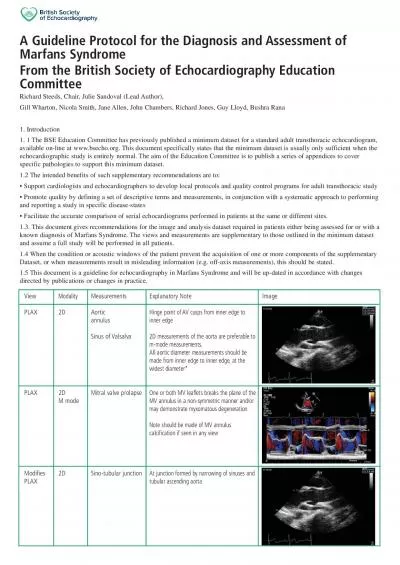
PAGE9 Facilitate the accurate comparison of serial echocardiograms performed in patients at the same or different sites13 This document gives recommendations for the image and analysis dataset requ ID: 936150

aortic lvot flow severe lvot aortic severe flow level annulus measurements ava stenosis diameter calcification velocity severity x0000 wall

Share:

Link:

Embed:

Download Presentation from below link

Download Pdf The PPT/PDF document "Guideline Protocol for the Assessment of..." is the property of its rightful owner. Permission is granted to download and print the materials on this web site for personal, non-commercial use only, and to display it on your personal computer provided you do not modify the materials and that you retain all copyright notices contained in the materials. By downloading content from our website, you accept the terms of this agreement.


Presentation Transcript

PAGE9 Guideline Protocol for the Assessment of Aortic for Echocardiography in Relation to From the British Society of Echocardiography Education CommitteeHollie BrewertonDave OxboroughMartin StoutGill Whartonand Simon Ray.available on-line at www.bsecho.orsufficient when theechocardiographic study is entirely normal. The Facilitate the accurate comparison of serial echocardiograms performed in patients at the same or different sites.1.3. This document gives recommendations for the image and analysis dataset required in patients being assessed for aortic stenosis.reconciled and important information gained to assist patient management. Astructured approach is outlined in Appendix 1.implantation of these devices, there is a further Appendix (2) relating specifically to assessment for transcatheter aortic valve1.4. The views and measurements are supplementary to those outlined in the minimum dataset and are given assuming a full studyDataset, or when measurements result in misleading information (e.g. off-axis measurements) this should be stated. PAGE10 MeasurementCusps viewed AppearanceThickeninggrade as:mild =restricted mo

tion at basal1/3 adjacent to hinge only,moderatemiddle thirdmiddle thirdsevereMild/moderate/severeDescribe severity:mild/mod/severe mild = small isolated spots ;moderate =multiple larger spots;severe = heavily calci-fied,extensive thickening and calcification ofall cusps.2,body,base (point of,aortic wall,aortic root,VOT PW Doppler velocity traceobtained from either 5CV or 3CV,see below..,mid systole,min 3 beats (5 ifAF) measure inner edge to inner edge]Zoom mode;Measure from cusp hingepoints (at point of cusp insertion into wall),ignoring all calcification.Measure maximumTry to obtain symmetrical aortic root sinusesbest in cardiac cycle.Inner edge-inner edge Bicuspid AV with asymmetric closure line PAGE11 (AV level,above,below) Mobility/thickening/cal-AV levelVOTTurbulent flow ,below) Mobility/thickening/cal-LVOT VTI(NOTE:can also beEnsure turbulent flow as expected at valvelevel.If below ?LVOT obstructionIf possible,state which leaflets are not sepa-rated.Note:location and extentAV cusps- free edge,body,insertion AV -Protrusion ofcalcification into outflow tract below level of-Aortic wall,extension into lumen,size.sin

ce ÔeffectiveÕ rather than ÔanatomicÕ ori-fice is primary predictor of outcome.mation is unreliable (e.g coexisting LVOTidentified,i.e.usually at the tips.3D willgeneralobservationSample volume positioned just at level of AVannulus and moved carefully into the LVOT ifi.e.smooth velocity curve with narrow band,well defined peak.(Typically 0.5-1.0cm fromannulus in calcific AS;AV annulus level inbicuspid AS).Trace around outer signalAV level No turbulent flow seen below or above the AVtricuspid AV AV with elliptical orifice PAGE12 AV Vmax/VTI Turbulent flow (AV level,above,below) Mobility/appearance/cawith flow across AV,measurements,asTurbulent flow level,above,below) reported.Trace around outer signal.Repeatsevere stenosis:rounded shape,peak in mid-mild stenosis:triangular,peak in early sys-Max AV velocity Mean AV gradientAVContinuity equationContinuity equationVTI ratio or velocity ratio (dimensionlessgeneralObservation bulent flow in outflow tractTurbulent flow seen within outflow tract;LVOT PAGE13 AV in short axisHunt for maximum AVR.parasternalSuprasternal LVEFLV hypertrophyadient,AVAreport window where maximum velo

city obtainedreport window where maximum velocity obtainedWhen AV parameters are discordant see appendix 1,congenital/bicuspid)Dilatation associated with AVD/bicuspid..May indicate need for early surgery.Also exclude aortic coarc-MR,functional versus degenerative diseaseIndex values as appropriate AV annulus sizing/TAsee appendix 2see appendix 2considerations Appendix 1:DISCREPANCIES IN PARAMETERS OF AORTIC STENOSIS SEVERITY Discrepancies in aortic valve (AV) parameters may occur in up to 25% of cases.The evidence base is not complete.However,the BSEEducation committee would like to provide guidance in this clinical scenario.The following is a possible approach to imaging suchases.Emphasis is placed on assessment of AV anatomy and cusp mobility.Colour flow imaging can help judge approximate orifice size.TOE may be indicated if doubt remains.Discrepancies in these parameters can be broadly divided into three categories.Before proceeding,ensure you are satisfied the meas-urements are accurate.Run through the checklist below.Then make some additional calculations where relevant and decide whichcategory the discrepancies in AV

par1-31-3Therefore: A.Checklist: LVOT diameter:compare with previous,is LVOT measurement accurate? Does resolution allow accurate measurement? Sigmoid sep-tum causing LVOT to be non-circular? Remember small error is LVOT diameter result in moderate AS becoming severe on calculationse.g.diameter 2.1= AVA 1.1cm ;1.9= AVA 0.9cm ;while diameter 1.8= AVA 0.8cm LVOT PW Doppler:has the sample volume been placed at correct level and correct distance from AV,where laminar flow isAV CW Doppler:is AV CW Doppler profile consistent? Ensure not mitral regurgitation signal!!DIDIatio:severe Although a useful additional measure,by removing the potential inaccura-cies of LVOT measurement,remember that it ignores inaccuracies due to abnormal LVOT anatomy e.g.isolated basal hypertrophy.Hence,its particular use is in the setting of serial measurements within the same individual or when assessing prosthetic valves,especially where the size of the valve is unknown. .Calculate additional parameters where relevant AVA indexed for BSA ( VAi );severe SVi Impedance (valvulo-arterial), Zva haemodynamic load,i.e.the double afterload on LV from the st

enosed AV and from the vascular system (sys-temic arterial compliance and systemic vascular resistance):Zva calculated as = mean AVG + systolic BPSViDegenerASAS.Thisis clinically manifested as systolic hypertension.This additional arterial afterload results in underestimation of AS severity;a greaterproportion of such patients may not be referred appropriately and in a timely manner for surgery.This is typically seen in patientsadient severe AS in the setting of normal LVEF or in patients with presumed moderate AS with symptoms. C.Discrepancies 6-8 in these parameters can be broadly divided into three categories 1. AVA suggests severe AS,but max velocity and mean AV gradient (AVG) do not..AVA VG a) Impaired LV function (LVEF )differential diagnosis will either be truly severe AS ormoderor less severeor less severeAS with poor valve opening due to poor cardiac outputPWD,AV CWD)Step 3:Consider low dose dobutamine stress echocardiography to determine severity of aortic stenosis 1,7 PAGE14 test terminated when LVOT VTI or strok��e volume increases 20% and/or HR increases 10-20 beats/min (or significant-sympto

ms/ LV RWMA/arrhythmias occur)contractile reserve is present if LVOT VTI or stroke volume increase by 20%AS is:severe when AVA remains AV.0 ; nd/;&#xor m;ên ;吀G increases 40pseudosevere (moderate or less) when AV.0 ; nd/;&#xor m;ên ;吀A increases 1.0 and or mean AVG remains if no contrno change in SVno change in SVAVA and mean AVG,then unable to comment on severity of AS if doubt remains then TOE may be necessary to assess A3D may be very useful3D may be very usefulb) LV function is normal :differential diagnosis will be low flow severe AS ormoderate or less severe AS for that individual i.e.smaller body habitusConfirm measurements are correct (LVOT diameter,LVOT PWD,AV CWD)Step 2:Ensure the anatomy of the Aincluding degree of calcification and cusp motionincluding degree of calcification and cusp motionStep 3:Calculate AVA index (AVAi),SV index (SVi) and Valvular-arterial Impedance (Zva)if AV�Ai is 0.6 then re-evaluate patient body size and consider if less than severe AS in setting of a smaller individual.NOTE:caution in over-weight patients where indexing for AVA (AVAi) may over estima

te aortic stenosis severity.if SVi 5 a;&#xnd/o;&#xr Zv;ᜀa 5.5,then consider low flow severe AS with normal LVEF.Look for corroboratory evidence;LVH,raised LV wall thickness to radius ratio,small LV cavity,diastolic dysfunction 2. gradient(AVG) do i.e.AV�A 1.0 cm the AVA is correct and there is moderor less severeor less severeAS with high flow states ortruly severe AS for that individual i.e.larger body habitusStep 1:Confirm measurements are correct (LVOT diameter,LVOT PWD,AV CWD)VA index (AVAi) and SV and CO if SV and CO are raised,consider non-severe AS with high flow states.Hence look for other causes e.g.severe aortic regur-gitation,haemodialysis/A-V fistula,anaemia Ai is consider severe �.6,;&#x-227;usually height 185cmusually height 185cm 3. Moderate aortic stenosis but the patient is symptomatic AS severity is under-estimated due to hypertension (pressure difference between LV and Ao).Step 1:Confirm measurements are correct (LVOT diameter,LVOT PWD,AV CWD)Step 3:Ensure measurement of BP at time of echo study.Step 4:Calculate AVAi and SVi and ZvaStep 5:BNP may be usefulBNP may be useful PAGE15 PAGE16

LVOT Measurements: Number of cusps;bicuspid may be a relativecontra-indicationProtrusion of calcification into outflow tractbelow level of annulus;above in Ao root,aortic wall,extension into lumen,best in cardiac cycle.Measure from cuspEDWARDS 23:18-22mm 29:24-27mmCORVALVE 23:17- 20mm29:23-27mm31:26-29mmbest in cardiac cycle.Inner edge-inner edgeEDWARDS although no specific measure-ments given consideration should be givencomplications e.g root rupture CORVAL�VE 26:SoV width 27mm AX AV and below AV LAX AV APPENDIX 2:TOE for TAVI assessment :specific considerations additional to TOE minimum dataset lease note:information on current TAVI valves is given,however new products are continually under development and the readershould consult with company literature with regards to current valve sizes and annulus sizes. Tricuspid AV Examples of AV annulus measurements PAGE17 AV 29 AND 31:�SoV width 29mm in systole) to AV annulus levelEDWARDS COREVALVE septum dimension should bemm;&#x ;-2;✀if 17mm at increased risk ofDescribe distribution,location and extentAV cusps- free edge,body,insertion point,Aortic wall-

include LVOT,level of annulus,aortic root,ascending aortathe way into annulus insertion into wall ÐiiiiÔlumpsÕ of calcificationin LVOT (with tendency to displace AV pros-thesis upwards during deployment) or inaortic root (with tendency to displace AVprosthesis downwards during deployment).Further clarify number of cusps.and at valve level 3D imagingmay offermation AV and sur-tures (LVOT/Ao) Solid ÔsheetÕ of calcification RCC of AV PAGE18 AV annulus(usually TOE).saggital and coronal planes).Use maximumaverage annulus size.If the annulus is toolliptical,this is a relative contra-indication.,suggested guide EDW�ARDS if 1.2-1.3:1EDWARDS CORVALVE References 200020001-7. Predictors of outcome in asymptomatic, severe aortic stenosis. Calcification of the aortic valve is scored asfollows: 1, no calcification; 2, mildly calcified (small isolated spots); 3, moderately calcified (multiple larger spots); and 4, heavily calcified (extensive thickening and3. Measure widest diameter at any point in cardiac cycle. There is no published evidence as to the ideal time point in the cardiac cycle when measurement should be Bicu