/
AMERICAN ACADEMY OF PEDIATRIC DENTISTRY CLINICAL GUIDE AMERICAN ACADEMY OF PEDIATRIC DENTISTRY CLINICAL GUIDE

AMERICAN ACADEMY OF PEDIATRIC DENTISTRY CLINICAL GUIDE - PDF document

conchita-marotz
conchita-marotz . @conchita-marotz
Follow
587 views
Uploaded On 2015-04-19

AMERICAN ACADEMY OF PEDIATRIC DENTISTRY CLINICAL GUIDE - PPT Presentation

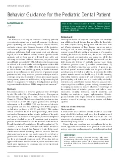
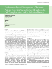
Dental intervention with certain modi64257cations must be done promptly and efficiently with attention to the patients medical history treatment protocol and health status Chemotherapy andor radiotherapy for the treatment of cancer or in preparation ID: 52395

Dental intervention with certain

Share:

Link:

Embed:

Download Presentation from below link

Download Pdf The PPT/PDF document "AMERICAN ACADEMY OF PEDIATRIC DENTISTRY ..." is the property of its rightful owner. Permission is granted to download and print the materials on this web site for personal, non-commercial use only, and to display it on your personal computer provided you do not modify the materials and that you retain all copyright notices contained in the materials. By downloading content from our website, you accept the terms of this agreement.


Presentation Transcript

\r\f  The American Academy of Pediatric Dentistry ( AAPD ) recognizes that the pediatric dental professional plays an im- portant role in the diagnosis, prevention, stabilization, and treatment of oral and dental problems that can compromise the child’s quality of life before, during, and after immuno- suppressive therapy which lowers the body’s normal immune response. This can be deliberate as in lowering the immune response to prevent the rejection of an organ or hematopoietic cell transplant * ( HCT of chemotherapy, radiation therapy, or HCT conditioning. Dental intervention with certain modications must be done promptly and eciently, with attention to the patient’s medical history, treatment protocol, and health status. Immunosuppressive therapy may cause many acute and long-term side effects in the oral cavity. Furthermore, any existing or potential sources of oral/dental infections and/or soft tissue trauma can compromise the medical treatment, leading to morbidity, mortality, and higher hospitalization costs. It is imperative that the pediatric dentist be familiar with the patient’s medical history as well as oral manifestations of the underlying condition  Developed by the Clinical Aairs Committee as Management of Pediatric Dental Patients Receiving Chemotherapy and/or Radiation and adopted in 1986, this document was last revised in 2013. e revision by the Council of Clinical Aairs included a new literature search of the PubMed ® /MEDLINE database using the terms: pediatric cancer, pediatric oncology, hematopoietic cell transplantation, bone marrow transplanta- tion, immunosuppressive therapy, mucositis, stomatitis, chemo- therapy, radiotherapy, acute eects, long-term eects, dental care, oral health, pediatric dentistry, practice guideline; eld: all; limits: within the last 10 years, humans, English, birth through age 18. Two thousand sixty-ve articles matched these criteria. Ninety-ve papers were chosen for review from this list and from the references within selected articles. When data did not appear sucient or were inconclusive, recommen- experienced researchers and clinicians.    A multidisciplinary approach involving physicians, nurses, dentists, social workers, dieticians, and other related health professionals is essential in caring for the child before, during and after any immunosuppressive therapy. 1,2 Oral and asso- ciated systemic complications that may occur as a sequelae of immunosuppressive therapy include pain, mucositis, oral ulcerations, bleeding, taste dysfunction, secondary infections (e.g., candidiasis, herpes simplex virus), dental caries, salivary gland dysfunction (e.g., xerostomia), neurotoxicity, mucosal brosis, gingival hypertrophy, post-radiation osteonecrosis, bisphosphonate-related osteonecrosis, soft tissue necrosis, tem- poromandibular dysfunction (e.g., trismus), craniofacial and dental developmental anomalies, and oral graft versus host disease ( GVHD ). 1,3,4 have an oral examination prior to initiation of treatment. 1,2 Prevention and treatment of pre-existing or concomitant oral disease is essential to minimize complications in this popu- lation. 5 e key to success in maintaining a healthy oral cavity during therapy is patient compliance. e child and the parents should be educated regarding the possible acute side eects and the long-term sequelae of immunosuppressive therapies in the oral cavity. 3,5-9 Every patient should be managed on an * \r\r\r  \f \n\t   \r\f  \n\t\b\n \n\b\t\b\n\b\t  \n\f \n\n  \t ­\b \t\b \f €\b ­ ­\n\t\n\n\n\n\t\b‚­\n  ƒ„ \n\n\f   \n\n \r\f …\t‚†\n\t­\b\n\n  \n\f \b\b\f \n\t\b\r  \b\n‚\t‡ \f „\n†\n\n\t­\n\t  \n\n\f ­\n  \b\b\b\b‚\r  \t\b†\n\t\n\b\n\t    \f  \n\t \b \b\b \n\n\t \t   ˆ‰Š \n‡\n \n‚\n\b\t\b\b\n\n\n\b†\b‡       \t\n\b†\n­\n\t  ‹ \t\b\b   ­\n\t    \r\f \n\t\b    \r\f\r\f\f\f  \f \r\fŒ  individual basis; consultations with the patient’s physicians and, when appropriate, other dental specialists should be sought before dental care is instituted. 7 \t  Dental and oral care before the initiation of immunosup- pressive therapy Objectives e objectives of a dental/oral examination before therapy starts are three-fold: 9 to identify and stabilize or eliminate existing and poten- tial sources of infection and local irritants in the oral cavity—without needlessly delaying the treatment or inducing complications. to communicate with the medical team regarding the patient’s oral health status, plan, and timing of treatment. to educate the patient and parents about the importance of optimal oral care in order to minimize oral problems/ discomfort before, during, and after treatment and about the possible acute and long-term eects of the therapy in the oral cavity and the craniofacial complex. Initial evaluation Medical history review: should include, but not be limited to, disease/condition (type, stage, prognosis), treatment proto - col (conditioning regimen, surgery, chemotherapy, radiation, transplant), medications (including bisphosphonates), allergies, surgeries, secondary medical diagnoses, hematological status (complete blood count [ CBC ]), coagulation status, immuno- suppression status, presence of an indwelling venous access line, and contact of medical team/primary care physician(s). 1 For HCT patients, include type of transplant, HCT source (i.e., bone marrow, peripheral stem cells, cord blood stem cells), matching status, donor, conditioning protocol, expected date of transplant, and GVHD prophylaxis. Patients with a compromised immune system may not be able to tolerate a transient bacteremia following invasive dental procedures. e decision regarding the need for antibiotic prophylaxis for dental procedures should be made in consultation with the child’s physician. Unless advised otherwise by the physician, the American Heart Association’s standard regimen to prevent endocarditis is an accepted option. 2,10 Dental history review: includes information such as fluo- ride exposure, habits, trauma, symptomatic teeth, previous care, preventive practices, oral hygiene, and diet assessment . Oral/dental assessment: should include thorough head, neck, and intraoral examinations, oral hygiene assessment and training, and radiographic evaluation based on history and clinical ndings. Preventive strategies Oral hygiene: Oral hygiene includes brushing of the teeth and tongue two to three times daily with regular soft nylon brush or electric toothbrush, regardless of the hematological status. 7,8,11,12 Ultrasonic brushes and dental floss should be allowed only if the patient is properly trained. 8 If capable, the patient’s teeth should be gently flossed daily. If pain or excessive bleeding occurs, the patient should avoid the aected area, but oss the other teeth. 1 Patients with poor oral hygiene and/or periodontal disease may use chlorhexidine rinses daily until the tissue health improves or mucositis develops. 13 e high alcohol content of commercially-available chlorhex- idine mouthwash may cause discomfort and dehydrate the tissues in patients with mucositis; thus, an alcohol-free chlorhexidine solution is indicated in this situation. Diet: Dental practitioners should discuss the importance of a healthy diet to maintain nutritional status with an empha - sis on foods that do not promote caries. Patients and parents should be advised about the high cariogenic potential of di- etary supplements rich in carbohydrates and oral pediatric medications rich in sucrose. 6 ey should also be instructed that sharp, crunchy, spicy, and highly acidic foods and alcohol should be avoided during chemotherapy, radiation, and HCT. 1 Fluoride: Preventive measures include the use of uoridated toothpaste, uoride supplements if indicated, neutral uoride gels/rinses, or applications of uoride varnish for patients at risk for caries and/or xerostomia. 6,8 A brush-on technique is convenient and may increase the likelihood of patient com- pliance with topical uoride therapy. 8 Lip care: Lanolin-based creams and ointments are more eective in moisturizing and protecting against damage than petrolatum-based products. 8 Trismus prevention/treatment: Patients who receive radiation therapy to the masticatory muscles may develop trismus. Thus, daily oral stretching exercises/physical therapy should start before radiation is initiated and continue throughout treatment. 7,14 Reduction of radiation to healthy oral tissues: In cases of radiation to the head and neck, the use of lead-lined stents, prostheses, and shields, as well as salivary gland sparing techniques (e.g., three-dimensional conformal or intensity modulated radiotherapy, concomitant cytoprotectants, surgical transfer of salivary glands), should be discussed with the radiation oncologist. Education: Patient and parent education includes the im- portance of optimal oral care in order to minimize oral problems and discomfort before, during, and after treatment and the possible acute and long-term effects of the therapy in the craniofacial complex. 1 \r\f Dental care  Hematological considerations: Absolute neutrophil count ( ANC ): — �2,000 per cubic millimeters ( /mm 3 ): no need for antibiotic prophylaxis; 1,14 — 1000 to 2000/mm 3 : Use clinical judgment 1 based on the patient’s health status and planned procedures. Some authors 1,7 suggest that antibiotic coverage (dosed per AHA recommendations 13 ) may be prescribed when the ANC is between 1,000 and 2,000/mm 3 . If infec- tion is present or unclear, more aggressive antibiotic therapy may be indicated and should be discussed with the medical team; and — 3 : defer elective dental care. 4 In dental emergency cases, discuss antibiotic coverage (antibiotic prophylaxis versus antibiotic coverage for a period of time) with medical team before proceeding with treatment. e patient may need hospitalization for dental management. Platelet count 7 : �— 75,000/mm 3 : no additional support needed; — 40,000 to 75,000/mm 3 : platelet transfusions may be considered pre- and 24 hours post-operatively. Local- ized procedures to manage prolonged bleeding may include sutures, hemostatic agents, pressure packs, and/ or gelatin foams; and — 3 : defer care. In dental emergency cases, contact the patient’s physician to discuss supportive measures (e.g., platelet transfusions, bleeding control, hospital admission and care) before proceeding. In addition, localized procedures (e.g., microbrillar col- lagen, topical thrombin) and additional medications as recommended by the hematologist/oncologist (e.g., aminocaproic acid, tranexamic acid) may help control bleeding. 1 Other coagulation tests may be in order for individual patients. Dental procedures: Ideally, all dental care should be completed before im- munosuppressive therapy is initiated. When that is not feasible, temporary restorations may be placed and non- acute dental treatment may be delayed until the patient’s hematological status is stable. 1,7 e patient’s blood counts normally start falling ve to seven days after the beginning of treatment cycle, staying low for approxi- mately 14 to 21 days, before rising again to normal levels for a few days until the next cycle begins. Prioritizing procedures: When all dental needs cannot be treated before therapy is initiated, priorities should be infections, extractions, periodontal care (e.g., scaling, prophylaxis), and sources of tissue irritation before the treatment of carious teeth, root canal therapy for perma- nent teeth, and replacement of faulty restorations. 4,14 Pain and the risk for pulpal infection determine which carious lesions should be treated rst. 8 Incipient to small carious lesions may be treated with uoride, silver diamine uoride, and/or sealants until denitive care can be accomplished. 7 Some patients requiring an organ transplant will be best able to tolerate dental care at least three months after transplant when overall health im- proves. 2 It is important for the practitioner to be aware that the signs and symptoms of periodontal disease may be decreased in immunosuppressed patients. 7 Pulp therapy in primary teeth: Few studies have eval- uated the safety of performing pulp therapy in primary teeth prior to the initiation of chemotherapy and/or radiotherapy. Many clinicians choose to provide a more denitive treatment in the form of extraction because pulpal/periapical/furcal infections during immunosup- pression periods can become life-threatening. 7,8 Teeth that already have been treated pulpally and are clinically and radiographically sound should be monitored periodically for signs of internal resorption or failure due to pulpal/ periapical/furcal infections. Endodontic treatment in permanent teeth: Symptomatic non-vital permanent teeth should receive root canal treatment at least one week before initiation of therapy to allow sucient time to assess treatment success before the chemotherapy. 4,7,14 If that is not possible, extraction is indicated. Extraction is also the treatment of choice for teeth that cannot be treated by denitive endodontic treatment in a single visit. In that case, the extraction should be followed by antibiotic therapy (penicillin or, for penicillin-allergic patients, clindamycin) for about one week. 7,14 Endodontic treatment of asymptomatic non-vital permanent teeth may be delayed until the hematological status of the patient is stable. 4,14 It is im- portant that the etiology of periapical lesions associated with previously endodontically treated teeth be deter- mined because they can be due to a number of factors including pulpal infections, inammatory reactions, apical scars, cysts, and malignancy. 8 If a periapical lesion is associated with an endodontically treated tooth and no signs or symptoms of infection are present, there is no need for retreatment or extraction since the radiolucency likely is due to an apical scar. 15 Orthodontic appliances and space maintainers: Poorly- tting appliances can abrade oral mucosa and increase the risk of microbial invasion into deeper tissues. 7 Appliances should be removed if the patient has poor oral hygiene and/or the treatment protocol or HCT conditioning regimen carries a risk for the development of moderate to severe mucositis. 4 Simple appliances (e.g., band and loops, xed lower lingual arches) that are not irritating to the soft tissues may be left in place in patients who present good oral hygiene. 4,8 Removable appliances and retainers that t well may be worn as long as tolerated by the patient who maintains good oral care. 7,8 Patients should be instructed to clean their appliance daily and \r\fŽ  routinely clean appliance cases with an antimicrobial solution to prevent contamination and reduce the risk of appliance-associated oral infections. 7 Consider removing orthodontic bands or adjusting prosthesis if a patient is expected to receive cyclosporine or other drugs known to cause gingival hyperplasia. If band removal is not possible, vinyl mouth guards or orthodontic wax should be used to decrease tissue trauma. 8 Periodontal considerations: Partially erupted molars can become a source of infection because of pericoronitis. e overlying gingival tissue should be excised if the dentist believes it is a potential risk and if the hematological status permits. 8,14 Patients should have a periodontal as- sessment and appropriate therapy prior to receiving bis- phosphonates as part of cancer treatment. 16-18 Extraction is the treatment of choice for teeth with a poor prognosis that cannot be treated by denitive periodontal therapy. If the patient has had bisphosphonates and an invasive periodontal procedure is indicated, risks must be discussed with the patient, parents, and physicians prior to the procedure. Extractions: ere are no clear recommendations for the use of antibiotics for extractions. 4 Recommendations generally have been empiric or based on anecdotal ex- perience. Surgical procedures must be as atraumatic as possible, with no sharp bony edges remaining and satisfactory closure of the wounds. 7,8,14 If there is documented infection associated with the tooth, anti- biotics (ideally chosen with the benet of sensitivity testing) should be administered for about one week. 7,8,14 To minimize the risk of development of osteone- crosis, osteoradionecrosis, or bisphosphonate-related osteonecrosis of the jaw ( BRONJ ), patients who will receive radiation to the jaws or bisphosphonate treatment as part of the cancer therapy must have all oral surgical procedures completed before those measures are insti- tuted. 16-18 If the patient has received bisphosphonates or radiation to the jaws and an oral surgical procedure is necessary, risks must be discussed with the patient, parents, and physician prior to the procedure. In patients undergoing long-term potent, high-dose intravenous bisphosphonates, there is an increased risk of BRONJ after a tooth extraction or with periodontal disease, 16-18 although most of the evidence has been described in the adult population. 17 Patients with a high risk of BRONJ are best managed by a dental specialist in coordination with the medical team in the hospital setting. Loose primary teeth should be allowed to exfoliate naturally. Nonrestorable teeth, root tips, teeth with perio- dontal pockets greater than six millimeters, symptomatic impacted teeth, and teeth exhibiting acute infections, signicant bone loss, involvement of the furcation, or mobility should be removed ideally two weeks (or at least seven to 10 days) before therapy is initiated to allow adequate healing. 7,8,14 Some practitioners prefer to extract all third molars that are not fully erupted, particularly prior to HCT, while others favor a more conservative approach, recom- mending extraction of third molars at risk for pulpal infection or those associated with signicant pathology, infection, periodontal disease, or pericoronitis or if the tooth is malpositioned or non-functional. 8,19,20 Communication: It is vital that the dentist communicate the comprehensive oral care plan with the medical team. Information to be shared includes the severity of dental caries (number of teeth involved and which teeth need immediate treatment), endo- dontic needs (pulpal versus periapical infection), periodontal status, number of teeth requiring extraction, soft tissue path- ology, and any other urgent care needed. Furthermore, it is important for the dentist to discuss with the medical team how much time is needed for the stabilization of oral disease as this will also affect the timing of the treatment or conditioning protocols. 1 Dental and oral care during immunosupression periods Preventive strategies Oral hygiene: Intensive oral care is of paramount importance because it reduces the risk of developing moderate/severe mu- cositis without causing an increase in septicemia and infections in the oral cavity. 1,3,5-8,11,14,21 rombocytopenia should not be the sole determinant of oral hygiene as patients are able to brush without bleeding at widely dierent levels of platelet count. 8 Patients should use a soft nylon brush two to three times daily and replace it on a regular (every two to three months) basis. 8,11 Fluoridated toothpaste may be used but, if the patient does not tolerate it during periods of mucositis due to oral burning or stinging sensations, it may be discon- tinued and the patient should switch to mild-avored non- uoridated toothpaste. If moderate to severe mucositis develops and the patient cannot tolerate a regular soft nylon toothbrush or an end-tufted brush, foam brushes or super soft brushes soaked in chlorhexidine may be used. 9 Other- wise, foam or super soft brushes should be discouraged because they do not allow for eective cleaning. e use of a regular brush should be resumed as soon as the mucositis improves. 8,11 Brushes should be air-dried between uses. 8 Electric or ultrasonic brushes are acceptable if the patient is capable of using them without causing trauma and irrita- tion. If patients are skilled at ossing without traumatizing the tissues, it is reasonable to continue ossing throughout treatment. Toothpicks and water irrigation devices should not be used when the patient is pancytopenic to avoid tissue trauma. 8,10 Dental care During immunosuppression, elective dental care should not be provided. If a dental emergency arises, the treatment plan should be discussed with the patient’s physician who will ‘\r\f make recommendations for supportive medical therapies (e.g., antibiotics, platelet transfusions, analgesia). e patient should be seen every six months (or in shorter intervals if there is a risk of xerostomia, caries, trismus, and/or chronic oral GVHD) for an oral health evaluation during treatment, in times of stable hematological status and always after re- viewing the medical history. Management of oral conditions related to immunosuppressive therapies Mucositis: e Multinational Association of Supportive Care in Cancer/International Society of Oral Oncology ( MASCC / ISOO ) has published guidelines for treatment of mucosi- tis. 11,22 The most common prescriptions for management of mucositis include good oral hygiene, analgesics, non- medicated oral rinses (e.g., 0.9 percent saline or sodium bicarbonate mouth rinses four to six times/day), and parenteral nutrition as needed. 1,11,21 Mucosal coating agents (e.g., Amphojel ® , Kaopectate ® , hydroxypropylmethylcellulose) and lm-forming agents (e.g., Zilactin ® and Gelclair ® ) also have been suggested. 1 Effective interventions for mucositis pre- vention include the use of palifermin, low-level laser therapy ( LLLT ), and cryotherapy. 22 e use of sucralfate, antimicro- bial lozenges, pentoxifylline, and granulocyte–macrophage- colony stimulating factor mouthwash for oral mucositis are not recommended. 11,22 Palifermin (keratinocyte growth factor-1) is a drug approved by the U.S. Food and Drug Administration for the prevention and treatment of oral mucositis. 23 It is recommended for mu- cositis prophylaxis for patients undergoing conditioning with high-dose chemotherapy and total body irradiation followed by HCT. 22 Palifermin is believed to stimulate epithelial cell reproduction, growth, and development so that mucosal cells damaged by chemotherapy and radiation are replaced quickly, accelerating the healing process. 23 e current MASCC/ISOO guidelines support the use of low-level laser therapy to prevent oral mucositis for patients undergoing HSC conditioning with high-dose chemotherapy with or without total body irradiation as well as patients un- dergoing radiation treatment for head and neck cancer. 22 LLLT can decrease pain and the duration and severity of chemo- therapy-induced mucositis in children. 24-26 LLLT may not be available at all cancer treatment centers due to the cost of the equipment and the need for trained personnel. Appropriate protocol must be followed when using LLLT to prevent con - tamination and occupational risks to the child and dental team. Oral cryotherapy, the cooling of intraoral tissue with ice during chemotherapy treatment, is recommended as mu- cositis prophylaxis for patients receiving bolus infusion of chemotherapy drugs with short half-lives. 22,27 This includes patients treated with uorouracil as well as patients receiving high-dose melphalan as conditioning for HCT. 22 Oral cryo- therapy reduces blood flow to the mouth by narrowing the blood vessels, limiting the amount of chemotherapy drugs delivered to the tissues. Cryotherapy is inexpensive and readily available, but further research is needed to confirm the eectiveness of oral cryotherapy in pediatric oncology. 27 Studies on the use of chlorhexidine for mucositis have given conicting results. Most studies have not demonstrated a prophylactic impact or a reduction in the severity of mucositis, although reduced colonization of candidal species has been shown. 14,28,29 Chlorhexidine is no longer recom- mended for preventing oral mucositis in patients undergoing radiotherapy. 11,22,30 Patient-controlled analgesia has been helpful in relieving pain associated with mucositis, reducing the requirement for oral analgesics. ere is no signicant evidence of the eec- tiveness of mixtures containing topical anesthetics (e.g., Philadelphia mouthwash, magic mouthwash). 22,30 e use of topical anesthetics has been suggested for pain management, 11 although there are no studies available to assess the benefit and potential for toxicity. Topical anesthetics only provide short term pain relief. 11,30 Lidocaine use may obtund or diminish taste and the gag reex 30 and/or result in a burning sensation, in addition to possible cardiovascular and central nervous system eects. Oral mucosal infections: e signs of inammation and infec- tion may be greatly diminished during neutropenic periods. us, the clinical appearance of infections may dier signi- cantly from the normal. 14 Close monitoring of the oral cavity allows for timely diagnosis and treatment of fungal, viral, and bacterial infections. Prophylactic nystatin is not eective for the prevention and/or treatment of fungal infections. 7,31 Oral cultures and/or biopsies of all suspicious lesions should be performed and prophylactic medications should be initiated until more specic therapy can be prescribed. 1,7,8,14 Oral bleeding: Oral bleeding occurs due to thrombocytopenia, disturbance of coagulation factors, and/or damaged vascular integrity. Management should consist of local approaches (e.g., pressure packs, antibrinolytic rinses or topical agents, gelatin sponges) and systemic measures (e.g., platelet trans- fusions, aminocaproic acid). 7,8,14 Dental sensitivity/pain: Tooth sensitivity could be related to decreased secretion of saliva during radiation therapy and the lowered salivary pH. 7,8,14 Patients who are using plant alkaloid chemotherapeutic agents (e.g., vincristine, vinblastine) may present with deep, constant pain (aecting the mandibular molars with greater frequency) in the absence of odontogenic pathology. e pain usually is transient and generally subsides shortly after dose reduction and/or cessation of chemo- therapy. 7,8,14 Xerostomia: Sugar-free chewing gum or candy, sucking tablets, special dentifrices for oral dryness, saliva substitutes, frequent sipping of water, alcohol-free oral rinses, and/or oral moistur- izers are recommended. 8,32 Placing a humidier by bedside at \r\f’  night may be useful. 14 Saliva stimulating drugs are not approved for use in children. Fluoride rinses and gels are recommended highly for caries prevention in these patients. Trismus: Daily oral stretching exercises/physical therapy must continue during radiation treatment. Management of trismus may include prosthetic aids to reduce the severity of brosis, trigger-point injections, analgesics, muscle relaxants, and other pain management strategies. 7,33 Hematopoietic cell transplantation Hematopoietic cell transplantation can be used in children to treat malignancies and hematologic disorders, as well as certain metabolic syndromes. Examples include: 34 malignant disorders treated with autologous HCT – brain tumors. Ewing sarcoma. – germ cell tumors. Hodgkin lymphoma. – leukemia. neuroblastoma. non-Hodgkin lymphoma. Wilms tumor. malignant disorders treated with allogenic HCT – acute lymphocytic leukemia. acute myeloid leukemia. – high-risk solid tumors. juvenile myelomonocytic leukemia. myelodysplastic syndrome. non-malignant disorders treated with allogenic HCT bone marrow failure syndromes. chronic granulomatous disease. Fanconi anemia. metabolic storage disorders. osteopetrosis. severe aplastic anemia. – sickle cell anemia. – thalessemia. Wiskott-Aldrich syndrome. Specic oral complications can be correlated with phases of HCT. 1-8 Phase I: Preconditioning e oral complications are related to the current systemic and oral health, oral manifestations of the underlying condition, and oral complications of recent medical therapy. Oral com- plications observed include oral infections, gingival leukemic inltrates, bleeding, ulceration, and temporomandibular dys- function. 1 Most of the principles of dental and oral care before the transplant are similar to those discussed for pediatric immunosuppressive therapy. 9 e two major dierences in HCT are: 1) the patient receives all the chemotherapy and/or total body irradiation in just a few days before the transplant, and 2) there will be prolonged immunosuppression following the transplant. Elective dentistry will need to be postponed until immunological recovery has occurred, at least 100 days following HCT, or longer if chronic GVHD or other complications are present. 7,8 erefore, all dental treat- ment should be completed before the patient becomes immunosuppressed. Phase II: Conditioning neutropenic phase In this phase, which encompasses the day the patient is ad- mitted to the hospital to begin the transplant conditioning to 30 days post-HCT, the oral complications are related to the conditioning regimen and supportive medical therapies. 8 Mucositis, xerostomia, oral pain, hemorrhage, opportunistic infections, taste dysfunction, neurotoxicity (including dental pain, muscle tremors), and temporomandibular dysfunction (including jaw pain, headache, joint pain) may be seen, typically with a high prevalence and severity of oral complica- tions. 1 Oral mucositis usually begins seven to 10 days after initiation of conditioning, and symptoms continue approxi- mately two weeks after the end of conditioning. 1 Among allogeneic transplant patients, hyperacute GVHD can occur, causing more severe inammation and severe mucositis symptoms, although its clinical presentation is dicult to diagnose. 1 e patient should be followed closely to monitor and manage the oral changes and to reinforce the importance of optimal oral care. Dental procedures usually are not allowed in this phase due to the patient’s severe immunosuppression. If emergency treatment is necessary, the dentist should consult and coordinate with the attending transplant team. Phase III: Engraftment to hematopoietic recovery e intensity and severity of complications begin to decrease normally three to four weeks after transplantation. Oral fungal infections and herpes simplex virus infection are most notable. 1 Acute GVHD can become a concern for allogeneic graft recipients. Xerostomia, hemorrhage, neurotoxicity, tem- poromandibular dysfunction, and granulomas/papillomas sometimes are observed. 1 A dental/oral examination should be performed and invasive dental procedures, including dental cleanings and soft tissue curettage, should be done only if authorized by the HCT team because of the patient’s conti- nued immunosuppression. 8 Patients should be encouraged to optimize oral hygiene and avoid a cariogenic diet. Attention to xerostomia and oral GVHD manifestations is crucial. HCT patients are particularly sensitive to intraoral thermal stimuli between two and four months post-transplant. 8 e mecha- nism is not well understood, but the symptoms usually resolve spontaneously within a few months. Topical application of neutral uoride or desensitizing toothpastes helps reduce the symptoms. 8 Phase IV: Immune reconstitution/recovery from systemic toxicity After day 100 post-HCT, the oral complications predomin- antly are related to the chronic toxicity associated with the conditioning regimen, including salivary dysfunction, Š\r\f craniofacial growth abnormalities, late viral infections, oral chronic GVHD, and oral squamous cell carcinoma. 1,8 Xerostomia and relapse-related oral lesions also may be observed. 1 Unless the patient is neutropenic or with severe chronic GVHD, mucosal bacterial infections are less frequently seen. Periodic dental examinations with radiographs can be performed, but invasive dental treatment should be avoided in patients with profound impairment of immune function. 8 Consultation with the patient’s physician and parents regarding the risks and benets of orthodontic care is recommended. Dental and oral care after the immunosuppressive therapy is completed Objectives e objectives of a dental/oral examination after immuno- suppressive therapy ends are three-fold: • to maintain optimal oral health. to reinforce to the patient/parents the importance of optimal oral and dental care for life. to address and/or treat any dental issues that may arise as a result of the long-term eects of immuno-suppressive therapy. Dental care Periodic evaluation: e patient should be seen at least every six months (or in shorter intervals if issues such as chronic oral GVHD, xerostomia, or trismus are present). Patients who have experienced moderate or severe mucositis and/or chronic oral GVHD should be followed closely for malignant transformation of their oral mucosa (e.g., oral squamous cell carcinoma). 5,35 Education: e importance of optimal oral and dental care for life must be reinforced. It is also important to emphasize the need for regular follow-ups with a dental professional, especially for patients who are at risk for or have developed GVHD and/or xerostomia and those who were younger than six years of age during treatment due to potential dental de- velopmental problems. Orthodontic treatment: Orthodontic care may start or resume after completion of all therapy and after at least a two-year disease-free survival when the risk of relapse is decreased and the patient is no longer using immunosuppressive drugs. 4 A thorough assessment of any dental developmental disturbances caused by the therapy must be performed before initiating orthodontic treatment. e following strategies should be considered when providing orthodontic care for patients with dental sequelae: (1) use appliances that minimize the risk of root resorption, (2) use lighter forces, (3) terminate treatment earlier than normal, (4) choose the simplest method for the treatment needs, and (5) do not treat the lower jaw. 36 How- ever, specic guidelines for orthodontic management, including optimal force and pace, remain undened. Patients who have used or will be given bisphosphonates in the future present a challenge for orthodontic care. Although bisphosphonate inhibition of tooth movement has been reported in animals, it has not been quantied for any dose or duration of therapy in humans. 36 Consultation with the patient’s parents and physician regarding the risks and benets of orthodontic care in this situation is recommended. Oral surgery: Consultation with an oral surgeon and/or periodontist and the patient’s physician is recommended for non-elective oral surgical and invasive periodontal procedures in patients who have used or are using bisphosphonates or those who received radiation therapy to the jaws in order to devise strategies to decrease the risk of osteonecrosis and osteoradionecrosis, respectively. 16-18 Elective invasive procedures should be avoided in these patients. 37 Patients with a high risk of BRONJ are best managed by in coordination with the oncology team in the hospital setting. Long-term concerns: Craniofacial, skeletal, and dental developmental issues are some of the complications faced by survivors 1,4,8 and usually develop among children who were less than six years of age at the time of their cancer therapy. 4,8 Long term eects of immunosuppressive therapy may include tooth agenesis, mi- crodontia, crown disturbances (size, shape, enamel hypoplasia, pulp chamber anomalies), root disturbances (early apical closure, blunting, changes in shape or length), reduced man- dibular length, reduced alveolar process height, and reduced vertical growth of the face. 4 e severity of the dental de- velopmental anomaly will depend on the age and stage of development during exposure to cytotoxic agents or ionizing radiation. Patients may experience permanent salivary gland hypofunction/dysfunction or xerostomia. 33 Relapse or sec- ondary malignancies can develop at this stage. 1 Routine periodic examinations are necessary to provide comprehensive oral healthcare. Careful examination of extra-oral and intra- oral tissues (including clinical, radiographic, and/or additional diagnostic examinations) are integral to diagnosing any secondary malignancies in the head and neck region. Dental treatment may require a multidisciplinary approach, involving a variety of dental specialists to address the treatment needs of each individual. Consultation with the patient’s physician is recommended if relapse or the patient’s immunologic status declines. \t­ 1.National Cancer Institute. PDQ ® Oral Complications of Chemotherapy and Head/Neck Radiation. Bethesda, Md.: National Cancer Institute; Modied December 16, 2016. Available at: “http://cancer.gov/cancertopics/pdq/ supportivecare/oralcomplications/HealthProfessional.” Accessed September 28, 2017. (Archived by WebCite ® at: “http://www.webcitation.org/70FodU3oF”) \r\f“  2. National Institute of Dental and Craniofacial Research. Dental management of the organ or stem cell trans - plant patient. Bethesda, Md.: National Institute of Dental and Craniofacial Research; Modied July, 2016. Available at: “https://www.nidcr.nih.gov/sites/default/ files/2017-09/dental-management-organ-stem-cell- transplant.pdf”. Accessed June 18, 2018. (Archived by WebCite ® at: “http://www.webcitation.org/70HPdWcYn”) 3. Hong CH, Brennan MT, Lockhart PB. Incidence of acute oral sequelae in pediatric patients undergoing chemotherapy. Pediatr Dent 2009;31(5):420-5. 4. da Fonseca M. Childhood cancer. In: Nowak AJ, Casa- massimo PS, eds. e Handbook of Pediatric Dentistry. 5th ed. Chicago, Ill.: American Academy of Pediatric Dentistry; 2018:361-9. 5. Elad S, ierer T, Bitan M, Shapira MY, Meyerowitz C. A decision analysis: e dental management of patients prior to hematology cytotoxic therapy or hematopoietic stem cell transplantation. Oral Oncol 2008;44(1):37-42. 6. Hong CH, Napeñas JJ, Hodgson BD, et al. A systematic review of dental disease in patients undergoing cancer therapy. Support Care Cancer 2010;18(8):1007-21. 7. Lalla RV, Brennan MT, Schubert MM. Oral complica- tions of cancer therapy. In: Yagiela JA, Dowd FJ, Johnson BS, Marrioti AJ, Neidle EA, eds. Pharmacology and Therapeutics for Dentistry. 6th ed. St. Louis, Mo.: Mosby-Elsevier; 2011:782-98. 8. Schubert MM, Peterson DE. Oral complications of hematopoietic cell transplantation. In: Appelbaum RF, Forman SJ, Negrin RS, Blume KG, eds. omas’ Hema- topoietic Cell Transplantation: Stem Cell Transplanta- tion. 4th ed. Oxford, UK: Wiley-Blackwell; 2009: 1589-607. Hong CH, daFonseca M. Considerations in the pediatric population with cancer. Dent Clin North Am 2008;52 (1):155-81. 10. American Academy of Pediatric Dentistry. Antibiotic prophylaxis for dental patients at risk for infection. Pediatr Dent 2018;40(6):386-91. 11. Peterson DE, Boers-Doets CB, Bensadoun RJ, Herrstedt J, Roila F, ESMO Guidelines Working Group. Manage- ment of oral and gastrointestinal mucosal injury: ESMO clinical practice guidelines for diagnosis, treatment, and follow-up. Ann Oncol 2015;26(Suppl_5): vi139-v151. 12. Wilson W, Taubert KA, Gewitz M, et al. Prevention of infective endocarditis: Guidelines from the American Heart Association: A guideline from the American Heart Association Rheumatic Fever, Endocarditis, and Kawasaki Disease Committee, Council on Cardiovascular Disease in the Young, and the Council on Clinical Cardiology, Council on Cardiovascular Surgery and Anesthesia, and the Quality of Care and Outcomes Research Interdis- ciplinary Working Group. Circulation 2007;116(15): 1736-54. Erratum in Circulation 2007;116(15):e376-7. 13. Hong CH, Allred R, Napenas JJ, Brennan MT, Baddour LM, Lockhart PB. Antibiotic prophylaxis for dental procedures to prevent indwelling venous catheter-related infections. Am J Med 2010;123(12):1128-33. 14. Little JW, Miller CS, Rhodus NL. Cancer and oral care of patients with cancer. In: Little and Falace’s Dental Management of the Medically Compromised Patient. 9th ed. St. Louis, Mo.: Elsevier; 2018:501-13. Yamagata K, Onizawa K, Yanagawa T, et al. A prospective study to evaluate a new dental management protocol before hematopoietic stem cell transplantation. Bone Marrow Transplant 2006;38(3):237-42. 16. Saad F, Brown JE, Van Poznak C, et al. Incidence, risk factors, and outcomes of osteonecrosis of the jaw: Inte- grated analysis from three blinded active-controlled phase III trials in cancer patients with bone metastases. Ann Oncol 2012;23(5):1341-7. 17. Kuhl S, Walter C, Acham S, Pfeffer R, Lambrecht JT. Bisphosphonate-related osteonecrosis of the jaws – A review. Oral Oncology 2012;48(10):938-47. 18. Dodson TB. Intravenous bisphosphonate therapy and bisphosphonate-related osteonecrosis of the jaws. J Oral Maxillofac Surg 2009;67(suppl 1):44-52. 19. American Academy of Pediatric Dentistry. Management considerations for oral surgery and oral pathology. Pediatr Dent 2018;40(6):373-82. American Association of Oral and Maxillofacial Surgeons. Management of third molar teeth. 2016. White Paper Accessed June 21, 2018. Available at: “https://www.aaoms. org/docs/govt_aairs/advocacy_white_papers/management _third_molar_white_paper.pdf”. (Archived by WebCite ® at: “http://www.webcitation.org/70KgisFtO”) 21. Stiff PJ, Emmanouilides C, Bensinger WI, et al. Palifermin reduces patient-reported mouth and throat soreness and improves patient functioning in the hemato- poietic stem-cell transplantation setting. J Clin Oncol 2006;24(33):5186-93. 22. Lalla RV, Bowen J, Barasch A, et al. MASCC/ISOO clinical practice guidelines for the management of mucositis secondary to cancer therapy. Cancer 2014;120 (10):1453-61. 23. U.S. Food and Drug Administration. Questions and an- swers on palifermin (keratinocyte growth factor). August 26, 2013. Available at: “https://www.fda.gov/Drugs/ DrugSafety/PostmarketDrugSafetyInformationforPatients andProviders/ucm110263.htm”. Accessed October 12, 2017. (Archived by WebCite ® at: “http://www.webcitation. org/70FsywwYs”) 24. He M. Zhan, B, Shen N, Wu N, Sun J. A systematic review and meta-analysis of the eect of low level laser therapy (LLLT) on chemotherapy-induced oral mucositis in pediatric and young patients. Eur J Pediatr 2018; 177(1):7-17. References continued on the next page. Œˆ\r\f 25. Amadori F, Bardellini E, Conti G, et al. Low-level laser therapy for treatment of chemotherapy-induced oral mucositis in childhood: A randomized double-blind controlled study. Lasers Med Sci 2016;31(6):1231-6. 26. Kuhn A, Porto FA, Miraglia P, Brunetto AL. Low-level infrared laser therapy in chemotherapy-induced oral mucositis: A randomized placebo-controlled trial in children. J Pediatr Hematol Oncol 2009;31(1):33-7. 27. Peterson DE, Ohrn K, Bowen J, et al. Systematic review of oral cryotherapy for management of oral mucositis caused by cancer therapy. Support Care Cancer 2013;21 (1):327-32. 28. Clarkson JE, Worthington HV, Furness S, McCabe M, Khalid T, Meyer S. Interventions for treating oral mucositis for patients with cancer receiving treatment (Review). Cochrane Database Syst Rev 2010;4(8): CD001973. 29. Cardona A, Balouch A, Abdul MM, Sedghizadeh PP, Enciso R. Efficacy of chlorhexidine for the prevention and treatment of oral mucositis in cancer patients: A systematic review with meta-analysis. J Oral Pathol Med 2017;46(9):680-8. 30. McGuire DB, Fulton JS, Park J, et al. Systematic review of basic oral care for the management of oral mucositis in cancer patients. Support Care Cancer 2013;21(11): 3165-77. 31. Gøtzche PC, Johansen HK. Nystatin prophylaxis and treatment in severely immunocompromised patients. Cochrane Database Syst Rev 2002;(2):CD002033. Up- date in Cochrane Database Syst Rev 2002;(4):CD002033. 32. Nieuw Amerongen AV, Veerman EC. Current therapies for xerostomia and salivary gland hypofunction associ- ated with cancer therapies. Support Care Cancer 2003; 11(4):226-31. 33. Jensen SB, Pedersen AM, Vissink A, et al. A systematic review of salivary gland hypofunction and xerostomia induced by cancer therapies: Prevalence, severity, and impact on quality of life. Support Care Cancer 2010;18 (8):1039-60. 34. Majhail NS, Farnia SH, Carpenter PA, et al. Indications for autologous and allogenic hematopoietic cell trans- plantation: Guidelines from the American Society for Blood and Marrow Transplantation. Biol Blood Marrow Transplant 2015;21(11):1863-96. Euvrard S, Kanitakis J, Claudy A. Skin cancers after organ transplantation. N Engl J Med 2003;348(17):1681-91. 36. Zahrowski JJ. Bisphosphonate treatment: An orthodontic concern for a proactive approach. Am J Orthod Dento- facial Orthop 2007;131(3):311-20. Dahllöf G, Jönsson A, Ulmner M, Huggare J. Orthodontic treatment in long-term survivors after bone marrow transplantation. Am J Orthod Dentofacial Orthop 2001; 120(5):459-65.