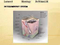
/
Chapter  The Cutaneous Senses  Cutaneous System Skin  heaviest organ in the body  Epidermis Chapter  The Cutaneous Senses  Cutaneous System Skin  heaviest organ in the body  Epidermis

Chapter The Cutaneous Senses Cutaneous System Skin heaviest organ in the body Epidermis - PDF document

conchita-marotz
conchita-marotz . @conchita-marotz
Follow
667 views
Uploaded On 2015-03-11

Chapter The Cutaneous Senses Cutaneous System Skin heaviest organ in the body Epidermis - PPT Presentation

brPage 3br Mechanoreceptors Merkel receptor diskshaped receptor located near the border betwe en the epidermis and dermis Meissner corpuscle stack of flattened disks in the dermis just below epidermis Ruffini cylinder branched fibers inside a cyl ID: 43907

brPage 3br Mechanoreceptors Merkel

Share:

Link:

Embed:

Download Presentation from below link

Download Pdf The PPT/PDF document "Chapter The Cutaneous Senses Cutaneous..." is the property of its rightful owner. Permission is granted to download and print the materials on this web site for personal, non-commercial use only, and to display it on your personal computer provided you do not modify the materials and that you retain all copyright notices contained in the materials. By downloading content from our website, you accept the terms of this agreement.


Presentation Transcript

Chapter 14: The CutaneousSenses CutaneousSystem Skin -heaviest organ in the body–Epidermis is the outer layer of the skin, which is made up of dead skin cells–Dermis is below the epidermis and contains four kinds of mechanoreceptorsthat respond to stimuli such as pressure, stretching, and vibration. MechanoreceptorsMerkel receptor-disk-shaped receptor located near the border between the epidermis and dermis Meissnercorpuscle-stack of flattened disks in the dermis just below epidermisRuffinicylinder-branched fibers inside a cylindrical capsulePaciniancorpuscle-onion-like capsule located deep in the skin Mechanoreceptors•Temporal Properties (adaptation) •Rapidly adapting fibers (RA) found in Meissnerreceptor and Paciniancorpuscle -fire at onset and offset of stimulation •Slowly adapting fibers (SA) found in Merkel and Ruffinireceptors -fire continuously as long as pressure is appliedSA1SA2 RA2RA1 Surface Deep Mechanoreceptors SA1SA2 RA2RA1 SA1SA2 RA2RA1 •Spatial Properties (detail resolution)Deep receptors: RA2 fibers (Paciniancorpuscle) and Ruffini(SA2) have large receptive fields and respond to high vibration rates.Surface receptors: Merkel receptors (SA1) and Meissnerreceptors (RA1) have small receptive fields and respond to slow vibration rates.Adapting RateSlowRapidVibration frequencyLowHigh Merkel receptors (SA1)Paciniancorpuscle (RA2)Meissnerreceptors (RA1)Ruffini(SA2) Surface receptors have smaller receptive fields than deep receptors. RA1SA1 RA2SA2 Adapting RateSlowRapidVibration FrequencyLowHigh Merkel receptors (SA1)Paciniancorpuscle (RA2)Meissnerreceptors (RA1)Ruffini(SA2) Properties of the four mechanoreceptor types. Pathways from Skin to Cortex•Nerve fibers travel in bundles (peripheral nerves) to the spinalcord•Two major pathways in the spinal cord:–Medial lemniscalpathway consists of large fibers that carry proprioceptiveand touchinformation–Spinothalamicpathway consists of smaller fibers that carry temperature and paininformation–These cross over to the opposite side of the body and synapse inthe thalamus, and then on to the Somatosensory cortex, or SA1 Maps of the Body on the Cortex•Signals travel from the thalamus to the somatosensory receiving area (S1) and the secondary receiving area (S2) in the parietal lobe•Body map (homunculus) on the cortex shows more cortical space allocated to parts of the body that are responsible for detail Discovered by Penfield in 1950 The ‘Somatosensory Homunculus’ The tongue may be used to as a substitute for sight. For more, see the ScienceNewsarticle The Seeing Tongue. http://courses.washington.edu/psy333/other/ScienceNews_Sensory_Substitution_2001.pdf Phantom Limb DisorderThe persistent sensation of an appendage, after removal by amputation or simple denervation. Ramachandranand colleagues has shown that touching the face of a phantom limb patient leads to sensations in the missing hand and arm.This lead to the hypothesis that the brain is ‘filling in’for the missing stimulation in the hand and arm representation in the somatosensory cortex. Phantom Limb DisorderTouching the chin stimulated the finger representation best, indicating that maybe Penfield got the face representation upside down.Sure enough, an fMRI experiment in 1999 showed that Ramachandranwas right and the somatosensory homunculus shown in textbooks (and Penfield) is wrong. Phantom Limb DisorderPhantom limb disorder can be painful and uncomfortable.Ramachandranused a ‘mirror box’to simulate the presence of the amputated hand which alleviated the symptoms in most of his patients. Plasticity in neural functioning leads to multiple homunculi and changes in how cortical cells are allocated to body parts Maps of the Body on the Cortex•Focal dystoniaor “musician’s cramp”-loss of skilled hand movements–Research examining the cortex has found that musicians with thisdisorder have “fused”cortical areas belonging to the affected hand–Fortunately (??) this only happens in about 1% of musicians Perceiving Details•Measuring tactile acuity–Two-point threshold -minimum separation needed between two points to perceive them as two units–Grating acuity -placing a grooved stimulus on the skin and asking the participant to indicate the orientation of the grating Tactile acuity thresholds are determined by Merkel receptors (SA1) Receptor Mechanisms for Tactile Acuity•There is a high density of Merkel receptor/SA1 fibers in the fingertips•Merkel receptors are densely packed on the fingertips -similar to cones in the fovea•Both two-point thresholds and grating acuity studies show these results Cortical Mechanisms for Tactile Acuity•Body areas with high acuity have larger areas of cortical tissuedevoted to them•This parallels the “magnification factor”seen in the visual cortex for the cones in the fovea Receptive field sizes correlate with tactile spatial acuity. SA1SA2 RA2RA1 Recall from yesterday:Deep receptors: RA2 fibers (Paciniancorpuscle) and Ruffini(SA2) have large receptive fields and respond to high vibration rates.Surface receptors: Merkel receptors (SA1) and Meissnerreceptors (RA1) have small receptive fields and respond to slow vibration rates.Adapting RateSlowRapidVibration frequencyLowHigh Merkel receptors (SA1)Paciniancorpuscle (RA2)Meissnerreceptors (RA1)Ruffini(SA2) 1 2 3 4 0 0.2 0.4 0.6 0.8 1 1.2 1.4 1.6 Finger NumberMean Threshold (mm)Acuity decreases (thresholds increase) from the index to the pinky, but the density of Merkel receptors is the same across the fingers. But there is a larger representation of the index finger in S1.S1, not the Merkel receptors, seem to be the limiting factor in tactile acuity. (Duncan and Boynton, 2007) left rightSubjects with better (lower) acuity thresholds have larger representations of the fingers in S1 -5 -4 -3 -2 -1 0 -1 -0.5 0 0.5 1 1.5 Cortical area size for finger representation (S1)Psychophysical acuity thresholdsr = -0.47, p .05 (Duncan and Boynton, 2007) Perceiving Vibration In the 60’s, Werner Lowenstein stimulated the paciniancorpuscle itself (location A), and also after dissecting it so that he could stimulate nearthe nerve fiber.Mechanical stimulation at location A caused the usual rapid adapting response.Mechanical stimulation at location B did not produce rapid adaptation; responsecontinued during the entire period of stimulation.So, the onion-like structure of the pacinancorpuscle must be responsible for the rapid adaptation. ‘Duplex Theory’of Texture Perception•Katz (1925) proposed that perception of texture depends on two cues:–Spatial cues are determined by the size, shape, and distributionof surface elements–Temporal cues are determined by the rate of vibration as skin ismoved across finely textured surfaces•Two receptors may be responsible for this process -called the duplex theory of texture perception