/
Kaan Yücel  M.D., Ph.D. Kaan Yücel  M.D., Ph.D.

Kaan Yücel M.D., Ph.D. - PowerPoint Presentation

conchita-marotz
conchita-marotz . @conchita-marotz
Follow
357 views
Uploaded On 2019-11-21

Kaan Yücel M.D., Ph.D. - PPT Presentation

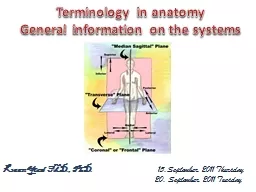
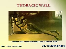
Kaan Yücel MD PhD Salvador Dali Anthropomorphic Chest of Drawers 1936 THORACIC WALL 31 102014 Friday 1 THORAX the part between the neck and the abdomen Chest Xray 11 REGIONST E ID: 766495

Share:

Link:

Embed:

Download Presentation from below link

Download Presentation The PPT/PDF document "Kaan Yücel M.D., Ph.D." is the property of its rightful owner. Permission is granted to download and print the materials on this web site for personal, non-commercial use only, and to display it on your personal computer provided you do not modify the materials and that you retain all copyright notices contained in the materials. By downloading content from our website, you accept the terms of this agreement.


Presentation Transcript

Kaan Yücel M.D., Ph.D. Salvador Dali - Anthropomorphic Chest of Drawers, 1936 THORACIC WALL 31. 10.2014 Friday

1. THORAXthe part between the neck and the abdomen Chest X-ray

1.1. REGIONS/T ERMSThoracic cavitycavity between neck and abdomen protected by the thoracic wall.Thoracic wallbounds the thoracic cavity. formed by the skin, bones, fasciae, and muscles. Thoracic cagebony portion of the thoracic wallthoracic skeleton

1.2. SURFACES OF THE THORAXPosterior surface 12 thoracic vertebræ & posterior parts of the ribs Anterior surfacesternum & costal cartilages Lateral surfaces ribs, separated by the intercostal spacesSTERNUM & COSTAL CARTILAGES anteriorly12 THORACIC VERTEBRAE & POST. RIBS posteriorlyRIBS & INTERCOSTAL SPACES laterally

1.3. BOUNDARIES OF THE THORAXSuperiorJugular notchSternoclavicular jointSuperior border of clavicleAcromionSpinous processes of C7Inferior Xiphoid processCostal arch12th and 11th ribsVertebra T12

1.4. CONTENTS OF THE THORAXOrgans of the cardiovascular, respiratory, digestive, reproductive, immune, and nervous systems

2. THORACIC WALLthoracic cage (skeleton) muscles between the ribs skin subcutaneous tissuemuscles, and fascia covering its anterolateral aspect. The mammary glands of the breasts lie within the subcutaneous tissue of the thoracic wall.

2.1. Functions of the thoracic wallProtects vital thoracic and abdominal organsResists the negative (sub-atmospheric) internal pressures generated by the elastic recoil of the lungs and inspiratory movements.Provides attachment for and support the weight of the upper limbs.Provides the origin s of many of the muscles that move and maintain the position of the upper limbs relative to the trunk.Provides attachments for muscles of the abdomen, neck, back, and respiration.

3. Skeleton of THE Thoracic Wall12 pairs of ribs and associated costal cartilages 12 thoracic vertebrae and the intervertebral (IV) discs interposed between themSternum

4. THORACIC APERTURES ‘Thoracic inlet’ ‘Thoracic outlet ’

4.1. Superior thoracic aperture“doorway” between the thoracic cavity and the neck and upper limbbounded: Posteriorly vertebra T1Laterally 1st pair of ribs and their costal cartilagesAnteriorly superior border of the manubrium TracheaEsophagus nerves, and vessels that supply and drain the head, neck, and upper limbs.

4.2. Inferior thoracic apertureBy closing the inferior thoracic aperture, the diaphragm separates the thoracic and abdominal cavities almost completely. bounded:Posteriorly 12th thoracic vertebraPosterolaterally 11th and 12th pairs of ribs Anterolaterally joined costal cartilages of ribs 7-10 costal margins Anteriorly xiphisternal joint

5. JOINTS OF THE THORACIC WALL 1. Costotransverse jointsbetween tubercle of a rib & transverse process of its own vertebra2. Sternocostal jointbetween the sternum and costal cartilages3. Costachondralis jointbetween the rib and costal cartilage4 . Intercondral joints Synovial joints between the costal cartilages of 6th and 7th, 7th and 8th , & 8th and 9th ribs . 5 . Sternal Joints between the m anubrium , body, xiphoid process of the sternum.

6. Muscles of the Thoracic Wall Serratus posterior Levator costarum Intercostal muscles(External, internal and innermost) Subcostal Transverse thoracic

6. Muscles of the Thoracic Wallexternal intercostal muscles most superficialinternal intercostal musclesbetween external & innermost muscles intercostal musclesthree flat muscles found in each intercostal space

6. Muscles of the Thoracic Wallpass obliquely anteroinferiorly external intercostal musclesextend from the inferior margins of the ribs above to the superior margins of the ribs below

6. Muscles of the Thoracic Wallin ternal intercostal musclesmost active during expirationbetween most inferior lateral edge of costal grooves of the ribs above, to the superior margins of ribs belowin the opposite direction to those of the external intercostal musclesobliquely posteroinferiorly Attachment to interosseus parts move down during expiration!

6. Muscles of the Thoracic Wallsame orientation as the internal intercostalsinnermost intercostal musclesleast distinct of the intercostal musclesneurovascular bundles (V.A.N.) in the costal grooves in a plane between innermost & internal intercostal muscles.

6. Muscles of the Thoracic Wall transversus thoracis muscleson the deep surface of the anterior thoracic wall & @ same plane as innermost intercostalslie deep to the internal thoracic vessels secure these vessels to the wall.

6. Muscles of the Thoracic WallFibers parallel the course of the internal intercostal muscles. Extend from the angle of the ribs to more medial positions on the ribs below.subcostales @ same plane as innermost intercostals

6.1. Accessory muscles of respiration upper accessory muscles assist with inspiration.upper chest, and abdominal muscles assist with expiration.

6.1. Accessory muscles of respiration Scalene muscles elevate, fix & expanding the upper chest during inspiration. S ternocleidomasteoid muscles raise the sternum expand the chest’s A-P & longitudinal dimensions.

6.1. Accessory muscles of respiration Axioappendicular muscles act primarily on the upper limbs. pectoralis major Attachment to first seven costal cartilagespectoralis minor Attachment to anterior surfaces of the 3rd-5th ribs inferior part of the serratus anteriorAttachment to lateral surfaces of upper 8-9 ribs & deep fascia overlying related intercostal spaces help elevate the ribs to expand the thoracic cavity .

6.1. Accessory muscles of respiration Trapezius raises the thoracic cage.Attachment to spinous processes of the thoracal vertebrae

6.1. Accessory muscles of respiration ExpirationAbdominal muscles pull the lower chest down depress the lower limbscompress the abdominal content & exerts pressure on chest.

7. MOVEMENTS OF THE THORACIC WALL One of the principal functions of the thoracic wall and the diaphragm is to alter the volume of the thorax and thereby move air in and out of the lungs.During breathing, the dimensions of the thorax change in vertical, lateral, and A-P directions.Diaphragm contracts DepressionDiaphragm relaxes  Elevation ( during passive expiration) Elevation &depression of the ribs

Diaphragm conracts; thoracic cavity descends, compressing the abdominal viscera expiration (back to neutral position)Lungs produces sub-atmospheric pressure As a result, the diaphragm ascends, diminishing the vertical dimension. Vertical dimension inspiration

Movement of the ribs (primarily 2nd-6th) at the costovertebral joints causes the anterior ends of the ribs to rise+ Slight elevation in A-P of the sternum.A-P dimension intercostal muscles contractpump-handle movement

Transverse dimensionintercostal muscles contract, raising the middle (lateral-most parts) of the ribs (especially the lower ones )bucket-handle movement

9. VASCULATURE OF THE THORACIC WALL Mainly posterior and anterior intercostal arteriespass between adjacent ribs in intercostal spaces.

9.1. Arteries of THE Thoracic WallArterial supply to the thoracic wall derives fromThoracic aorta [posterior intercostal & subcostal arteries] Subclavian artery [internal thoracic & supreme intercostal arteries] Axillary artery [superior & lateral thoracic arteries]

course through the thoracic wall between the ribs. Each intercostal space is supplied by a large posterior intercostal arterysmall pair of anterior intercostal arteriesException last two intercostal spacesintercostal arteries

Anterior intercostal arteries (paired)directly or indirectly from internal thoracic artery 1st-6th (from internal thoracic artery) 7th-9th (from musculophrenic-branch of internal thoracic )Posterior intercostal arteries (large, unpaired) 1st-2nd (from supreme intercostal artery- branch of costocervical trunk) 3rd-11th (from thoracic aorta) Subcostal artery (from thoracic aorta)

internal thoracic arteryVery first branch of the subclavian arteryPasses vertically through the superior thoracic aperture Lies posterior to the costal cartilages of the upper 6 ribs Lies 1 cm lateral to the sternum. @ level of 6th intercostal space, divides into 2 terminal branches superior epigastric artery & musculophrenic artery

9.2. Veins of THE Thoracic WallMost posterior intercostal veins (4-11) end @ azygos/hemiazygos venous system conveys venous blood to SVC. 1st posterior intercostal veins right & left brachiocephalic veins2nd & 3rd (sometimes 4th) form superior intercostal vein.Right superior intercostal vein final tributary of azygos vein Left superior intercostal vein empties into left brachiocephalic vein. Veins Artery V.A.N. Nerve

10. NERVES OF THE THORACIC WALL 12 pairs of intercostal nervesanterior rami of spinal nerves T1 to T11 lie in the intercostal spaces between adjacent ribs. Anterior ramus of spinal nerve T12 (subcostal nerve) inferior to rib XII.

Near the angles of the ribs, the nerves pass between internal intercostal & innermost intercostal muscles. V.A.N.Neurovascular bundles sheltered by the inferior margins of the overlying rib.

10.3. DermatomesThrough its posterior ramus and the lateral and anterior cutaneous branches of its anterior ramus, most thoracic spinal nerves (T2-T12) supply a strip-like dermatome of the trunk extending from the posterior median line to the anterior median line.Skin area supplied by a segment of the spinal cord T2- Sternal angleT4- NippleT6- Xiphoid processT8- Costal arch T10-UmbliculusT12-Midpoint between umbilicus and symphysis pubis

11. breasts Mammary glands & associated skin -connective tissues. modified sweat glands in the superficial fascia anterior to the pectoral muscles and the anterior thoracic wall. Reproduction, back painAesthetics, and breast cancer

11. breasts Mammary glands: Series of ducts and associated secretory lobules. Form 15 to 20 lactiferous ducts open nipple. Nipple is surrounded by a circular pigmented area of skin areola (L. small area).

11.1. Female BreastsNON-LACTING WOMEN – PREDOMINANT COMPONENT: FATLACTING WOMEN- PREDOMINANT COMPONENT: GLANDULAR TISSUEThe breast rests on a bed extends transversely from lateral border of the sternum mid-axillary line vertically from the 2nd through 6th ribs

Lymphatic drainage of the breast75% (lateral breast quadrants) Axillary lymph nodes Most of the remaining (medial breast quadrants) parasternal lymph nodes or to the opposite breast Lymph from inferior quadrants may pass deeply to abdominal lymph nodes .

.Axillary lymph nodes Clavicular lymph nodesSubclavian lypmhatic trunk Parasternal lymph nodes Bronchomediastinal lymph trunks Right or left venous angle