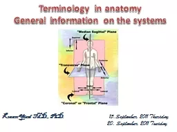
PhD 13 15September 2011 Thursday 20 September 2011 Tuesday Terminology in anatomy General information on the systems ID: 775475
Download Presentation The PPT/PDF document " Kaan Yücel M.D., " is the property of its rightful owner. Permission is granted to download and print the materials on this web site for personal, non-commercial use only, and to display it on your personal computer provided you do not modify the materials and that you retain all copyright notices contained in the materials. By downloading content from our website, you accept the terms of this agreement.
Slide1
Kaan Yücel M.D., Ph.D. 13. 15.September. 2011 Thursday 20. September. 2011 Tuesday
Terminology in anatomyGeneral information on the systems
Slide2Terminology in anatomy
It
is
important
for
medical
personnel
to
have
a
sound
knowledge
and
understanding
of
the
basic
anatomic
terms
.
The
accurate
use
of
anatomic
terms
by
medical
personnel
enables
them
to
communicate
with
their
colleagues
both
nationally
and
internationally
.
Without
anatomic
terms
,
one
cannot
accurately
discuss
or
record
the
abnormal
functions
of
joints
,
the
actions
of
muscles
,
the
alteration
of
position
of
organs
,
or
the
exact
location
of
swellings
or
tumors
.
Slide3Anatomical terms are descriptive terms standardized in an international reference guide, Terminologia Anatomica (TA).TA- International Anatomical Terminology
C
reated
by the Federative Committee on Anatomical Terminology and approved by the International Federation of Associations of Anatomists,
the
most
recent
(6th)
edition
was
published in 1998.
Slide4Many anatomical terms have both Latin and Greek
equivalents
.
Thus
the tongue is lingua (L.) and
glossa
(
Gk
), and these are the basis of such terms as lingual artery and glossopharyngeal nerve.
Slide5Various
adjectives
,
arranged
as
pairs
of
opposites
,
describe
the
relationship
of
parts
of
the
body
or
compare
the
position
of
two
structures
relative
to
each
other
.
Anatomical
directional
terms
are
based
on
the
body in
the
anatomical
position
Four
anatomical
planes
divide
the
body,
and
sections
divide
the
planes
into
visually
useful
and
descriptive
parts
.
Slide6Terms
Related
to
Position
All
descriptions
of
the
human
body
are
based
on
th
anatomic
position
.
The
various
parts
of
the
body
are
then
described
in
relation
to
certain
imaginary
planes
.
Slide7Median Sagittal PlaneThis is a vertical plane passing through the center of the body, dividing it into equal right and left halves. .
Slide8Coronal PlanesImaginary vertical planes at right angles to the median plane.Horizontal, or Transverse,or Axial PlanesAt right angles to both the median and the coronal planes.
Slide9Slide10Anatomical terms are specific for comparisons made in the anatomical position, or with reference to the anatomical planes:Superior refers to a structure that is nearer the vertex, the topmost point of the cranium (Mediev. L., skull). Inferior refers to a structure that is situated nearer the sole of the foot.
Slide11Cranial relates to the cranium and is a useful directional term, meaning toward the head or cranium. Caudal (L. cauda, tail) is a useful directional term that means toward the feet or tail region, represented in humans by the coccyx (tail bone), the small bone at the inferior (caudal) end of the vertebral column.
Slide12Posterior (dorsal) denotes the back surface of the body or nearer to the back.Anterior (ventral) denotes the front surface of the body.Rostral is often used instead of anterior when describing parts of the brain; it means toward the rostrum (L. for beak). To describe the relationship of two structures, one is said to be anterior or posterior to the other insofar as it is closer to the anterior or posterior body surface.
Slide13Slide14Medial
is
used
to
indicate
that
a
structure
is
nearer
to
the
median
plane
of
the
body.
For
example
,
the
5th
digit
of
the
hand
(
little
finger
) is
medial
to
the
other
digits
.
Lateral
stipulates
that
a
structure
is
farther
away
from
the
median
plane
.
The
1st
digit
of
the
hand
(
thumb
) is
lateral
to
the
other
digits
.
Dorsum
usually
refers
to
the
superior
aspect
of
any
part
that
protrudes
anteriorly
from
the
body,
such
as
the
dorsum
of
the
tongue
,
nose
, penis,
or
foot
Slide15.
Slide16Combined terms describe intermediate positional arrangements: inferomedial means nearer to the feet and median plane—for example, superolateral means nearer to the head and farther from the median plane.
Slide17Other terms of relationship and comparisons are independent of the anatomical position or the anatomical planes, relating primarily to the body's surface or its central core:Superficial, intermediate, and deep (Lat. Profundus, profunda) describe the position of structures relative to the surface of the body or the relationship of one structure to another underlying or overlying structure.External means outside of or farther from the center of an organ or cavity, while internal means inside or closer to the center, independent of direction.
Slide18Other terms of relationship and comparisons are independent of the anatomical position or the anatomical planes, relating primarily to the body's surface or its central core:External means outside of or farther from the center of an organ or cavity, while internal means inside or closer to the center, independent of direction.
Slide19Proximal and distal are used when contrasting positions nearer to or farther from the attachment of a limb or the central aspect of a linear structure (origin in general), respectively. For example, the arm is proximal to the forearm and the hand is distal to the forearm.
Slide20Terms
of
Laterality
Paired
structures
having
right
and
left
members
(
e.g
.,
the
kidneys
)
are
bilateral
,
whereas
those
occurring
on
one
side
only
(
e.g
.,
the
spleen
)
are
unilateral
.
Something
occurring
on
the
same
side
of
the
body as
another
structure
is
ipsilateral
.
Contralateral
means
occurring
on
the
opposite
side
of
the
body
relative
to
another
structure
.
Slide21Terms
of
Movement
Various
terms
describe
movements
of
the
limbs
and
other
parts
of
the
body.
Most
movements
are
defined
in
relationship
to
the
anatomical
position
,
with
movements
occurring
within
,
and
around
axes
aligned
with
,
specific
anatomical
planes
.
While
most
movements
occur
at
joints
where
two
or
more
bones
or
cartilages
articulate
with
one
another
,
several
non-skeletal
structures
exhibit
movement
(
e.g
.,
tongue
,
lips
,
eyelids
).
Slide22Terms of movement may also be considered in pairs of oppositing movements:Flexion and extension movements generally occur in sagittal planes around a transverse axis.
Slide23Flexion indicates bending or decreasing the angle between the bones or parts of the body. For most joints (e.g., elbow), flexion involves movement in an anterior direction, but it is occasionally posterior, as in the case of the knee joint. Lateral flexion is a movement of the trunk in the coronal plane.
Slide24Extension indicates straightening or increasing the angle between the bones or parts of the body. Extension usually occurs in a posterior direction. The knee joint, rotated 180° to other joints, is exceptional in that flexion of the knee involves posterior movement and extension involves anterior movement.
Slide25Slide26Dorsiflexion describes flexion at the ankle joint, as occurs when walking uphill or lifting the front of the foot and toes off the ground. Plantarflexion bends the foot and toes toward the ground, as when standing on your toes.
Slide27Abduction and adduction movements generally occur in a frontal plane around an anteroposterior axis. Except for the digits, abduction means moving away from the median plane (e.g., when moving an upper limb laterally away from the side of the body) and adduction means moving toward it.
Slide28Slide29Slide30Slide31Circumduction is a circular movement that involves sequential flexion, abduction, extension, and adduction in such a way that the distal end of the part moves in a circle. Circumduction can occur at any joint at which all the above-mentioned movements are possible (e.g., the shoulder and hip joints).
Slide32Slide33Rotation involves turning or revolving a part of the body around its longitudinal axis, such as turning one's head to face sideways. Medial rotation (internal rotation) brings the anterior surface of a limb closer to the median plane, whereas lateral rotation (external rotation) takes the anterior surface away from the median plane.
Slide34Pronation rotates the radius medially so that the palm of the hand faces posteriorly and its dorsum faces anteriorly. When the elbow joint is flexed, pronation moves the hand so that the palm faces inferiorly (e.g., placing the palms flat on a table). Supination is the opposite rotational movement, rotating the radius laterally and uncrossing it from the ulna, returning the pronated forearm to the anatomical position. When the elbow joint is flexed, supination moves the hand so that the palm faces superiorly.
Slide35Slide36Eversion moves the sole of the foot away from the median plane, turning the sole laterally. Inversion moves the sole of the foot toward the median plane (facing the sole medially).
Slide37Opposition is the movement by which the pad of the 1st digit (thumb) is brought to another digit pad. This movement is used to pinch, button a shirt, and lift a teacup by the handle. Reposition describes the movement of the 1st digit from the position of opposition back to its anatomical position.l
Slide38Slide39Protrusion
is a
movement
anteriorly
(
forward
) as in
protruding
the
mandible
(
chin
),
lips
,
or
tongue
.
Retrusion
is a
movement
posteriorly
(
backward
), as in
retruding
the
mandible
,
lips
,
or
tongue
.
Slide40Elevation
raises
or
moves
a
part
superiorly
, as in
elevating
the
shoulders
when
shrugging
.
Depression
lowers
or
moves
a
part
inferiorly
, as in
depressing
the
shoulders
when
standing
at
ease
.
Slide41Protraction
and
retraction
are
used
most
commonly
for
anterolateral
and
posteromedial
movements
of
the
scapula
on
the
thoracic
wall
,
causing
the
shoulder
region
to
move
anteriorly
and
posteriorly
.
Slide42Regional
terms
Slide43Slide44General
information
on
the
systems
http://sinoemedicalassociation.org/AP/bodyregions.pdf
Slide45Slide46Body Planes
Figure 1.9a
Slide47Trunk Cavities
Diaphragm: divides body cavity into thoracic and abdominopelvic cavities.Mediastinum: contains all structures of the thoracic cavity except the lungs
Slide48Ventral Body Cavity Membranes
Parietal serosa lines internal body walls
Visceral serosa covers the internal organs
Serous fluid separates the serosae
Slide49Serous Membranes
Cover the organs of trunk cavities & line the cavity
Fist represents an organInner balloon wall represents visceral serous membraneOuter balloon wall represents parietal serous membraneCavity between two membranes filled with lubricating serous fluid that is produced by the membranesInflammation of the serous membranes
Slide50Serous Membranes: Named for Their Specific Cavities and Organs
Pericardium refers to heart.
Pleura refers to lungs and thoracic
cavity
.
Peritoneum refers to
abdominopelvic
cavity
.
Slide51Other Body Cavities
Oral and digestive – mouth and cavities of the digestive organs
Nasal –located within and posterior to the nose
Orbital – house the eyes
Middle ear – contain bones (
ossicles
) that transmit sound vibrations
Synovial – joint cavities
Slide52Integumentary SystemThe skin (L. integumentum, a covering) is the body's largest organ, consists of the epidermis, a superficial cellular layer, and the dermis, a deep connective tissue layer.
Slide53Skeletal System
The skeleton is composed of cartilages and bones
.
Os
= Bone
Osteologia
The
skeletal system may be divided into two functional parts:
The
axial skeleton
consists of the bones of the head, neck, and trunk.
The
appendicular skeleton
consists of the bones of the limbs, including those forming the pectoral (shoulder) and pelvic girdles.
Slide54Slide55Joints
Joints (articulations) are unions or junctions between two or more bones or rigid parts of the skeleton. Joints exhibit a variety of forms and functions.
Some
joints have no
movement, such as the
epiphysial
plates between the epiphysis and diaphysis of a growing long bone
.
O
thers
allow only slight movement, such as teeth within their
sockets
.
S
ome
are freely movable, such as the
glenohumeral
(shoulder) joint.
Slide56Muscular System
Myologia
Mus-culus
=
Muscle
, Lat.
Little
mouse
The
muscular system consists of all the muscles of the body.
Slide57Voluntary skeletal muscles constitute the great majority of the named muscles.
All
skeletal muscles are composed of one specific type of muscle tissue.
However
, other types of muscle tissue constitute a few named muscles (e.g., the
ciliary
and detrusor muscles, and the
arrector
muscles of hairs) and form important components of the organs of other systems, including the cardiovascular, alimentary, genitourinary, integumentary, and visual systems.
Slide58There are three types of muscles:Striated muscle (skeletal muscles)-voluntarily controlled, though exceptions existNon-striated muscle (smooth muscle) - involuntaryCardiac muscle
Slide59Cardiovascular System
The heart consists of two muscular pumps that dividing the circulation into two components: the pulmonary and systemic circulations or circuits.
Slide60The
right ventricle propels low-oxygen blood returning from the systemic circulation into the lungs. Carbon dioxide is exchanged for oxygen in the capillaries of the lungs, and then the oxygen-rich blood is returned to the heart's left atrium. This circuit, from the right ventricle through the lungs to the left atrium, is the pulmonary circulation.
Slide61The
left ventricle propels the oxygen-rich blood returned to the heart from the pulmonary circulation, exchanging oxygen and nutrients for carbon dioxide in the remainder of the body's capillaries. Low-oxygen blood returns to the heart's right atrium. This circuit, from left ventricle to right atrium, is the systemic circulation.
Slide62Slide63Blood VesselsThere are three types of blood vessels: arteries, veins, and capillaries.Blood under high pressure leaves the heart and is distributed to the body by a branching system of arteries.
Slide64The final distributing vessels, arterioles, deliver oxygen-rich blood to capillaries. Capillaries form a capillary bed, where the interchange of oxygen, nutrients, waste products, and other substances with the extracellular fluid occurs. Blood from the capillary bed passes into venules, which resemble wide capillaries. Venules drain into small veins that open into larger veins. The largest veins return low-oxygen blood to the heart.
Slide65Large elastic arteries (conducting arteries) have many elastic layers (sheets of elastic fibers) in their walls. These large arteries initially receive the cardiac output. Examples of large elastic arteries are the aorta, the arteries that originate from the arch of the aorta (brachiocephalic trunk, subclavian and carotid arteries), and the pulmonary trunk and arteries.
Slide66Medium muscular arteries (distributing arteries) have walls that consist chiefly of circularly disposed smooth muscle fibers. Their ability to decrease their diameter (vasoconstrict) regulates the flow of blood to different parts of the body as required by circumstance (e.g., activity, thermoregulation). Most of the named arteries, including those observed in the body wall and limbs during dissection such as the brachial or femoral arteries, are medium muscular arteries.
Slide67Small arteries and arterioles have relatively narrow lumina and thick muscular walls. The degree of filling of the capillary beds and level of arterial pressure within the vascular system are regulated mainly by the degree of tonus (firmness) in the smooth muscle of the arteriolar walls. If the tonus is above normal, hypertension (high blood pressure) results.
Slide68Anastomoses (communications) between multiple branches of an artery provide numerous potential detours for blood flow in case the usual pathway is obstructed by compression due to the position of a joint, pathology, or surgical ligation.
If
a main channel is occluded, the smaller alternate channels can usually increase in size over a period of time, providing a collateral circulation that ensures the blood supply to structures distal to the
blockage
.
Slide69Lymphoid System
C
onstitutes
a
n
“
overflow” system that provides for the drainage of surplus tissue fluid and leaked plasma proteins to the bloodstream, as well as for the removal of debris from cellular decomposition and infection
.
Lymph nodes, small masses of lymphatic tissue located along the course of lymphatic vessels through which lymph is filtered on its way to the venous system
.
Slide70Nervous System
The nervous system enables the body to react to continuous changes in its internal and external environments.
It
also controls and integrates the various activities of the body, such as circulation and respiration.
Nervous system controls the systems in the body and regulates its functions according to the impulses received from the outer world (sensations
)
The nervous system controls and integrates the activities of the different parts of the body together with the endocrine system,
Slide71Nervous System
Nervous
system controls the systems in the body and regulates its functions according to the impulses received from the outer world (sensations
)
The nervous system controls and integrates the activities of the different parts of the body together with the endocrine
system
.
Slide72For descriptive purposes, the nervous system is divided:
•
Structurally
1. C
entral
nervous system (
CNS
) (MSS in
Turkish
)
brain
and spinal
cord
2. P
eripheral
nervous system (PNS
)
remainder
of the
nervou
s
system
outside of the CNS
.
•
Functionally
1. S
omatic
nervous system (SNS)
2. A
utonomic
nervous system (ANS
)
Slide73Slide74Slide75Nervous tissue consists of two main cell types: Neurons (nerve cells) Neuroglia (glial cells), which support the neurons.
Slide76Neurons are the structural and functional units of the nervous system specialized for rapid communication. Neuroglia (glial cells or glia), approximately five times as abundant as neurons, are non-neuronal, non-excitable cells that form a major component of nervous tissue, supporting, insulating, and nourishing the neurons.
A
smear from a mammalian spinal cord showing an isolated neuron (large arrow) and the nuclei of the surrounding
neuroglial
cells (small arrows).
Slide77The principal roles of the CNS are to integrate and coordinate incoming and outgoing neural signals and to carry out higher mental functions, such as thinking and learning.
Slide78The
peripheral nervous system (PNS) consists of nerve fibers and cell bodies outside the CNS that conduct impulses to or away from the CNS. The PNS is organized into nerves that connect the CNS with peripheral structures.
Slide79PNS
is anatomically and operationally continuous with the CNS.
Its
afferent (sensory) fibers convey neural impulses to the CNS from the sense organs (e.g., the eyes) and from sensory receptors in various parts of the body (e.g., in the skin).
Its
efferent (motor) fibers convey neural impulses from the CNS to effector organs (muscles and glands).
Slide80Nerves are either cranial nerves or spinal nerves, or derivatives of them
.
Cranial nerves
exit the cranial cavity through foramina (openings) in the cranium (G.
kranion
, skull) and are identified by a descriptive name (e.g., “trochlear nerve”) or a Roman numeral (e.g., “CN IV”).
Slide81Spinal
(segmental) nerves
exit the vertebral column (spine).
Spinal
nerves arise in bilateral pairs from a specific segment of the spinal cord.
The
31 spinal cord segments and the 31 pairs of nerves arising from them are identified by a letter and number (e.g., “T4”) designating the region of the spinal cord and their superior-to-inferior order (C, cervical; T, thoracic; L, lumbar; S, sacral; Co, coccygeal).
Slide82Cranial NervesSome cranial nerves convey only sensory fibers, some only motor fibers, and some carry a mixture of both types of fibers.
Cranial
nerve
i -
olfactory
(
sensory
)
Cranial
nerve
ii -
optic
(
sensory
)
3.
cranial
nerve
iii -
Oculomotor
(
sensory
and
motor)
4.
Cranial
nerve
iv -
trochlear
(motor)
5.
Cranial
nerve
v -
trigeminal
(
sensory
and
motor)
6.
Cranial
nerve
vi -
abducent
(motor)
7.
Cranial
nerve
vii -
facial
(
sensory
and
motor)
8.
Cranial
nerve
viii -
vestibule
cochlear
(
sensory
)
9.
Cranial
nerve
ix -
glosspharyngeal
(
sensory
and
motor)
10.
Cranial
nerve
x -
Vagus
(
sensory
and
motor)
11.
Cranial
nerve
xi
accessory
(motor)
12.
Cranial
nerve
xii -
hypoglossal
(motor)
Slide83Somatic
and Visceral
Fibres
The types of fibers conveyed by cranial or spinal
nerves
:
Somatic
fibers
General
sensory fibers
(general somatic afferent [GSA] fibers) transmit sensations from the body to the CNS; they may be
exteroceptive
sensations from the skin (pain, temperature, touch, and pressure) or pain and proprioceptive sensations from muscles, tendons, and joints
.
Somatic motor fibers
(general somatic efferent [GSE] fibers) transmit impulses to skeletal (voluntary) muscles.
Slide84Visceral fibers
Visceral sensory fibers
(general visceral afferent [GVA] fibers) transmit pain or subconscious visceral reflex sensations (information concerning distension, blood gas, and blood pressure levels, for example) from hollow organs and blood vessels to the CNS
.
Visceral motor fibers
(general visceral efferent [GVE] fibers) transmit impulses to smooth (involuntary) muscle and glandular tissues.
Slide85Somatic Nervous
System
The somatic nervous system (SNS), composed of somatic parts of the CNS and PNS, provides
sensory
and
motor
innervation to all parts of the body (G. soma), except the viscera in the body cavities, smooth muscle, and glands.
Slide86The
somatic sensory system
transmits sensations of touch, pain, temperature, and position from sensory receptors
.
Most of these sensations reach conscious levels (i.e., we are aware of them).
The
somatic motor system innervates only skeletal muscle, stimulating voluntary and reflexive movement by causing the muscle to contract, as occurs in response to touching a hot iron.
Slide87Autonomic Nervous
System
(
Visceral
Nervous
System
)
The autonomic nervous system (ANS), classically described as the visceral nervous system or visceral motor system, consists of motor fibers that stimulate smooth (involuntary) muscle, modified cardiac muscle (the intrinsic stimulating and conducting tissue of the heart), and glandular (secretory) cells.
Slide88The efferent nerve fibers and ganglia of the ANS are organized into two systems or divisions: Sympathetic divisionParasympathetic division(craniosacral) division
Slide89Respiratory System
The respiratory apparatus consists of the nose, nasopharynx, paranasal sinuses, larynx, trachea, bronchi, lungs, and pleuræ.
Slide90NoseConsists of the external nose and the nasal cavity, both of which are divided by a septum into right and left halves. Opens into the nasopharynx. An amazing humidifier and warmer of air.
Slide91The paranasal sinuses are cavities found in the interior of the maxilla, frontal, sphenoid, and ethmoid bones. They are filled with air; they communicate with the nasal cavity through relatively small apertures. Infection of the paranasal sinuses is a common complication of nasal infections.We still are unsure as to all the functions of these air-filled spaces. Multiple theories of function exist.
Slide92NasopharynxIt is located posterior to the nose and superior to the soft palate. Being one of the three parts of the pharynx, a structure belonging to the digestive system, nasopharynx has a respiratory function. It is the posterior extension of the nasal cavities. The nose opens into the nasopharynx .
Slide93Larynx
O
rgan
of voice
voice
box
Located
between
the trachea and the root of the tongue, at the upper and forepart of the neck, where it presents a considerable projection in the middle line.
P
laced
at the upper part of the air
passage
Although
most commonly known for its role as the phonating mechanism for voice production, its most vital function is to guard the air passages, especially during swallowing when it serves as the “sphincter” or “valve” of the lower respiratory tract, thus maintaining a patent airway.
C
omposed
of nine cartilages connected by membranes and ligaments and containing the vocal folds.
Slide94Slide95TracheaExtending from the larynx into the thorax, terminates inferiorly as it divides into right and left main bronchi. Transports air to and from the lungs.A fibrocartilaginous tube, supported by incomplete cartilaginous tracheal cartilages (rings), that occupies a median position in the neck. The tracheal cartilages keep the trachea patent.
Slide96PleuraEach pulmonary cavity (right and left) is lined by a pleural membrane (pleura) that also reflects onto and covers the external surface of the lungs occupying the cavities. Each lung is invested by and enclosed in a serous pleural sac.
Slide97LungsVital organs of respiration. Main function is to oxygenate the blood Although cadaveric lungs may be shrunken, firm or hard, and discolored, healthy lungs in living people are normally light, soft, and spongy, and fully occupy the pulmonary cavities.
Slide98Tracheobronchial Tree Beginning at the larynx, the walls of the airway are supported by horseshoe- or C-shaped rings of hyaline cartilage. The trachea constitutes the trunk of the tree. It bifurcates into main bronchi (right and left main bronchi), one to each lung. Within the lungs, the bronchi branch in a constant fashion to form the branches of the tracheobronchial tree.
Slide99Slide100Digestive System
The apparatus for the digestion of the food consists of the digestive tube and of certain accessory organs.
The
Digestive Tube (alimentary canal) is a
musculomembranous
tube, about 9
metres
long, extending from the mouth to the anus, and lined throughout its entire extent by mucous membrane.
Slide101It
has received different names in the various parts of its course
:
A
t
its commencement is the mouth, where provision is made for the mechanical division of the food (mastication), and for its admixture with a fluid secreted by the salivary glands (insalivation
)
B
eyond
this are the organs of deglutition, the pharynx and the esophagus, which convey the food into the stomach, in which it is stored for a time and in which also the first stages of the digestive process take
place
.
Slide102Peristalsis, a series of ring-like contraction waves, begins around the middle of the stomach and moves slowly toward the pylorus. It is responsible for mixing the masticated (chewed) food mass with gastric juices and for emptying the contents of the stomach into the duodenum.
Slide103T
he
stomach is followed by the small intestine, which is divided for purposes of description into three parts, the duodenum, the jejunum, and ileum.
In
the small intestine the process of digestion is completed and the resulting products are absorbed into the blood and lacteal vessels.
Finally
the small intestine ends in the large intestine, which is made up of cecum, colon, rectum, and anal canal, the last terminating on the surface of the body at the anus.
Slide104The accessory organs are the teeth, for purposes of mastication; the three pairs of salivary glands—the parotid, submaxillary, and sublingual—the secretion from which mixes with the food in the mouth and converts it into a bolus and acts chemically on one of its constituents.
Slide105The liver and pancreas, two large glands in the abdomen, the secretions of which assist in the process of digestion.
Slide106Absorption of chemical compounds occurs principally in the small intestine, a coiled 5- to 6-m-long tube (shorter in life, when tonus is present, than in the cadaver) consisting of the duodenum, jejunum, and ileum.
Slide107Slide108Most reabsorption of water occurs in the ascending colon.Feces form in the descending and sigmoid colon and accumulate in the rectum before defecation.
Slide109Peritoneum and Periotenal CavityThe peritoneum is a continuous, serous membrane which lines the abdominopelvic cavity and invests the viscera. The peritoneal cavity is within the abdominal cavity and continues inferiorly into the pelvic cavity.
Slide110Oral region includes: Oral cavityTeethGingivaeTonguePalateRegion of the palatine tonsils
Slide111The oral cavity is where food is ingested and prepared for digestion in the stomach and small intestine
.
Food
is chewed by the teeth, and saliva from the salivary glands facilitates the formation of a manageable food bolus (L. lump).
Slide112PharynxThe pharynx is the superior expanded part of the alimentary system posterior to the nasal and oral cavities, extending inferiorly past the larynx.EsophagusThe esophagus is a muscular tube that conveys food from the pharynx to the stomach.
Slide113StomachThe stomach is the expanded part of the digestive tract between the esophagus and small intestine. It is specialized for the accumulation of ingested food, which it chemically and mechanically prepares for digestion and passage into the duodenum. The stomach acts as a food blender and reservoir; its chief function is enzymatic digestion.
Slide114Small IntestineThe small intestine, consisting of the duodenum, jejunum, and ileum, is the primary site for absorption of nutrients from ingested materials.
Slide115The duodenum (L. breadth of 12 fingers), the first and shortest part of the small intestine, is also the widest and most fixed part. The second part of the small intestine is the jejunum, whereas third part is, the ileum. Together, the jejunum and ileum are 6-7 m long.
Slide116Large IntestineThe large intestine consists of the cecum; appendix; ascending, transverse, descending, and sigmoid colon; rectum; and anal canal.
Slide117The large intestine is where water is absorbed from the indigestible residues of the liquid chyme, converting it into semi-solid stool or feces that is stored temporarily and allowed to accumulate until defecation occurs.
Slide118Excretory System
Functions
Collect
water and filter body fluids.
Remove
and concentrate waste products from body fluids and return other substances to body fluids as necessary for homeostasis.
Eliminate
excretory products from the body.
Slide119Kidneys
The
ovoid kidneys remove excess water, salts, and wastes of protein metabolism from the blood while returning nutrients and chemicals to the blood.
The
kidneys produce urine that is conveyed by the ureters to the urinary bladder in the pelvis.
Slide120The superomedial aspect of each kidney normally contacts a suprarenal gland. A weak fascial septum separates the glands from the kidneys so that they are not actually attached to each other. The suprarenal glands function as part of the endocrine system, completely separate in function from the kidneys.
Slide121UretersThe ureters are muscular ducts with narrow lumina that carry urine from the kidneys to the urinary bladder.
Slide122Urinary bladderThe urinary bladder, a hollow viscus with strong muscular walls, is characterized by its distensibility. The urinary bladder is a temporary reservoir for urine and varies in size, shape, position, and relationships according to its content and the state of neighboring viscera.
Slide123UrethraThe male urethra is a muscular tube that conveys urine from the internal urethral orifice of the urinary bladder to the external urethral orifice, located at the tip of the glans penis in males. The urethra also provides an exit for semen (sperms and glandular secretions). The female urethra passes anteroinferiorly from the internal urethral orifice of the urinary bladder.
Slide124Endocrine System
The endocrine system is made up of glands that produce and secrete hormones.
These
hormones regulate the body's growth, metabolism (the physical and chemical processes of the body), and sexual development and function.
Slide125Endocrine System
The hormones are released into the bloodstream and may affect one or several organs throughout the body
.
Hormones are chemical messengers created by the body
.
They transfer information from one set of cells to another to coordinate the functions of different parts of the body.
Slide126The
major glands of the endocrine system
Hypothalamu
s
Pituitary
gland
T
hyroid
P
arathyroids
S
uprarenal
glands
P
ineal
body
T
he
reproductive organs (ovaries and
testes
)
The
pancreas
is also a part of this system; it has a role in hormone production as well as in digestion.
Slide127The endocrine system is regulated by feedback in much the same way that a thermostat regulates the temperature in a room.
For
the hormones that are regulated by the pituitary gland, a signal is sent from the hypothalamus to the pituitary gland in the form of a "releasing hormone," which stimulates the pituitary to secrete a "stimulating hormone" into the circulation.
Slide128The
stimulating hormone then signals the target gland to secrete its hormone.
As
the level of this hormone rises in the circulation, the hypothalamus and the pituitary gland shut down secretion of the releasing hormone and the stimulating hormone, which in turn slows the secretion by the target gland.
This
system results in stable blood concentrations of the hormones that are regulated by the pituitary gland
.
Slide129Slide130Slide131Slide132Reproductive
System
Slide133Slide134Slide135Slide136M
ale
internal genital organs
Testes
E
pididymides
(singular = epididymis
)
D
uctus
deferentes
(singular =
ductus
deferens
S
eminal
glands
E
jaculatory
ducts
Prostate
B
ulbourethral
glands
Slide137Slide138F
emale
internal genital organs
Ovaries
U
terine
tubes
Uterus
V
agina
Slide139