/
What Causes Wilson Disease? What Causes Wilson Disease?

What Causes Wilson Disease? - PowerPoint Presentation

conchita-marotz
conchita-marotz . @conchita-marotz
Follow
355 views
Uploaded On 2018-10-13

What Causes Wilson Disease? - PPT Presentation

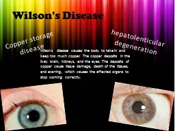
Wilson disease is caused by mutations in the ATP7B gene This gene makes an enzyme that is involved in copper transport When the enzyme is mutated not working properly copper accumulates in the liver and brain and becomes toxic ID: 689388

wilson disease tests liver disease wilson liver tests serum rings hepatitis urine hospitalization caeruloplasmin negative mutations failure copper acute

Share:

Link:

Embed:

Download Presentation from below link

Download Presentation The PPT/PDF document "What Causes Wilson Disease?" is the property of its rightful owner. Permission is granted to download and print the materials on this web site for personal, non-commercial use only, and to display it on your personal computer provided you do not modify the materials and that you retain all copyright notices contained in the materials. By downloading content from our website, you accept the terms of this agreement.


Presentation Transcript

Slide1
Slide2
Slide3
Slide4

What Causes Wilson Disease?

Wilson disease is caused by mutations in the

ATP7B

gene.

This gene makes an enzyme that is involved in copper transport.

When the enzyme is mutated (not working properly) copper accumulates in the liver and brain and becomes toxic

.

MUTATIONS ON Chromosome 13Slide5

Wilson Disease

PathogenesisSlide6

How Does it Run in Families?

Wilson disease is inherited in an autosomal recessive pattern.

Affected individuals have mutations in both copies of

ATP7B

Carriers (mutation in only one copy) do not have symptoms

Slide7
Slide8

Wilson Disease

Clinical spectrum

Hepatic

Neuropsychiatric

Other

Diagnostic tests

Blood tests

Urine tests

Liver biopsy

Genetic testing

Treatment options

General chelators

Metallothionein

inducers

Liver Transplantation

Follow-up

Blood tests

Urine testsSlide9

Wilson

Disease

Hepatic

Manifestations

Asymptomatic hepatomegaly

Persistently abnormal AST and ALT

Fatty liver

Cirrhosis

Acute hepatitis

Similar to viral or autoimmune etiologies

Acute liver failure

Coomb

s negative hemolytic anemia

Acute renal failureSlide10
Slide11

Kayser

-Fleischer RingsSlide12

Wilson Disease

Coombs-negative hemolytic anemia

Rapid progression to renal failure

Modest rise in AST/ALT (< 2000 IU/L)

Normal or markedly subnormal alkaline

phosphatase

(< 40 IU/L

)

Serum

ceruloplasmin

usually decreased

Serum and urine copper increased

Slide13
Slide14

Wilson Disease

Histopathology

Chronic hepatitis

Fatty infiltration

Apoptosis

CirrhosisSlide15
Slide16
Slide17
Slide18

Case presentation

An eight-year-old African boy, previously well, was referred to our unit on account of

nephrotic

syndrome. He had presented to our emergency department a day before with as cites, facial swelling and reduced urinary output in the preceding two weeks. After two days of hospitalization, he became deeply jaundiced with worsening of the generalized edema. Slide19

In the second week of hospitalization, our patient developed tremors of his hands while at rest and when reaching for objects. He became clumsy when performing chores involving the use of his hands. His gait was noticed to be shuffling with a tendency to fall forward when trying to walk. At the same time, his face retained a wry smile and his speech became slurred and

dysarthric

. Slide20

He was also noticed to be emotionally labile; he cried inconsolably when asking for food. He was reviewed by a pediatric neurologist who pointed out the possible presence of

Kayser

-Fleischer (KF) rings on both eyes. A slit-lamp examination by an ophthalmologist promptly revealed the presence of both KF rings and sunflower cataracts.Slide21
Slide22

Initial investigations showed

proteinuria

of grade 1+ Table 

1

 shows the results for his liver function test during hospitalization. His serum electrolytes, urea and

creatinine

were within the normal reference

Serology for human immunodeficiency virus, hepatitis B and hepatitis C viruses were negative. A serum sample for caeruloplasmin level was returned as having 5mg/

dL

of caeruloplasmin, using an

immunoturbidimetric

method (reference range: 25 to 45mg/dL). range. Slide23

0.1-1.2,<0.3mg/dl

64-83 g/l

35-52 g/l

13-40 U/L

10-59U/L

53-128U/L

1-30U/LSlide24

Using the scoring system proposed by the 8

th

 International Meeting of Wilson Disease and

Menkes

Disease, our patient achieved a score of 6 (compatible neuropsychiatric features = 2; K-F rings = 2 and caeruloplasmin level of 5mg/

dL

=2) and a diagnosis of WD was made.Slide25

إعداد :

أحمد رفيق الدلو

حسين محمد دكة

Thank you