/
Shortly before his life was cut short by the guillotine during the Fre Shortly before his life was cut short by the guillotine during the Fre

Shortly before his life was cut short by the guillotine during the Fre - PDF document

daisy
daisy . @daisy
Follow
342 views
Uploaded On 2022-09-01

Shortly before his life was cut short by the guillotine during the Fre - PPT Presentation

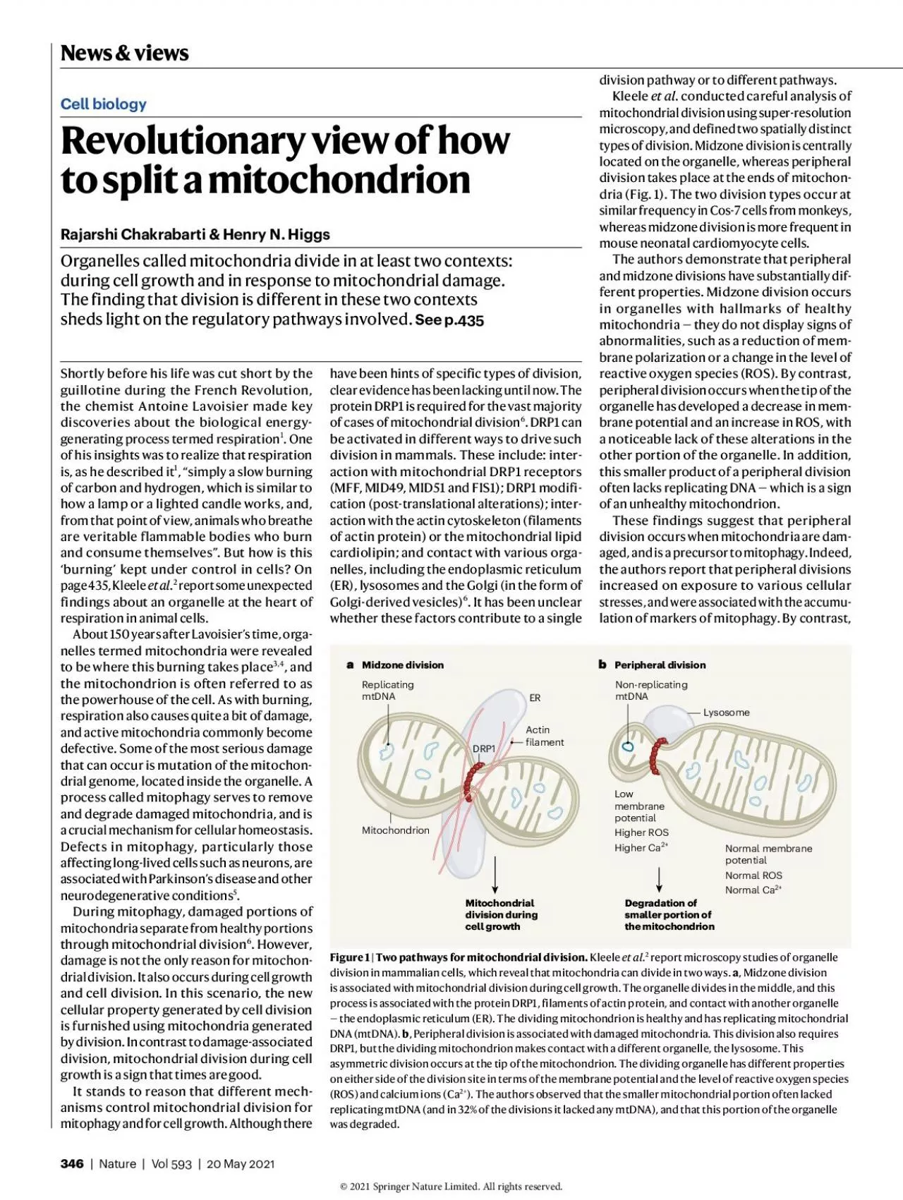
Revolutionary view of how to split a mitochondrion Rajarshi Chakrabarti Henry N HiggsOrganelles called mitochondria divide in at least two contexts during cell growth and in response to mitochondr ID: 945688

mitochondrial division cell peripheral division mitochondrial peripheral cell midzone organelle mitochondria membrane fis1 mitochondrion 146 drp1 150 calcium cells

Share:

Link:

Embed:

Download Presentation from below link

Download Pdf The PPT/PDF document "Shortly before his life was cut short by..." is the property of its rightful owner. Permission is granted to download and print the materials on this web site for personal, non-commercial use only, and to display it on your personal computer provided you do not modify the materials and that you retain all copyright notices contained in the materials. By downloading content from our website, you accept the terms of this agreement.


Presentation Transcript

Shortly before his life was cut short by the guillotine during the French Revolution, the chemist Antoine Lavoisier made key discoveries about the biological energy-generating process termed respirationof his insights was to realize that respiration is, as he described it, “simply a slow burning of carbon and hydrogen, which is similar to how a lamp or a lighted candle works, and, from that point of view, animals who breathe are veritable flammable bodies who burn and consume themselves”. But how is this ‘burning’ kept under control in cells? On page435, Kleele etal report some unexpected findings about an organelle at the heart of respiration in animal cells.About 150 years after Lavoisier’s time, organelles termed mitochondria were revealed to be where this burning takes place, and the mitochondrion is often referred to as the powerhouse of the cell. As with burning, respiration also causes quite a bit of damage, and active mitochondria commonly become defective. Some of the most serious damage that can occur is mutation of the mitochondrial genome, located inside the organelle. A process called mitophagy serves to remove and degrade damaged mitochondria, and is a crucial mechanism for cellular homeostasis. Defects in mitophagy, particularly those affecting long-lived cells such as neurons, are associated with Parkinson’s disease and other neurodegenerative conditionsDuring mitophagy, damaged portions of mitochondria separate from healthy portions through mitochondrial division. However, damage is not the only reason for mitochondrial division. It also occurs during cell growth and cell division. In this scenario, the new cellular property generated by cell division is furnished using mitochondria generated by division. In contrast to damage-associated division, mitochondrial division during cell growth is a sign that times are good.It stands to reason that different mechanisms control mitochondrial division for mitophagy and for cell growth. Although there have been hints of specific types of division, clear evidence has been lacking until now. The protein DRP1 is required for the vast majority of cases of mitochondrial divisionbe activated in different ways to drive such division in mammals. These include: interaction with mitochondrial DRP1 receptors (MFF, MID49, MID51 and FIS1); DRP1 modification (post-translational alterations); interaction with the actin cytoskeleton (filaments of actin protein) or the mitochondrial lipid cardiolipin; and contact with various organelles, including the endoplasmic reticulum (ER), lysosomes and the Golgi (in the form of Golgi-derived vesicles). It has been unclear whether these factors contribute to a single division pathway or to different pathways. . conducted careful analysis of mitochondrial division using super-resolution microscopy, and defined two spatially distinct types of division. Midzone division is centrally located on the organelle, whereas peripheral division takes place at the ends of mitochondria (Fig.1). The two division types occur at similar frequency in Cos-7 cells from monkeys, whereas midzone division is more frequent in mouse neonatal cardiomyocyte cells.The authors demonstrate that peripheral and midzone divisions have substantially different properties. Midzone division occurs in organelles with hallmarks of healthy mitochondria—they do not display signs of abnormalities, such as a reduction of membrane polarization or a change in the level of reactive oxygen species (ROS). By contrast, peripheral division occurs when the tip of the organelle has developed a decrease in membrane potential and an increase in ROS, with a noticeable lack of these alterations in the other portion of the organelle. In addition, this smaller product of a peripheral division often lacks replicating DNA— which is a sign of an unhealthy mitochondrion.These findings suggest that peripheral division occurs when mitochondria are damaged, and is a precursor to mitophagy. Indeed, the authors report that peripheral divisions increased on exposure to various cellular stresses, and were associated with the accumulation of markers of mitophagy. By contrast, Revolutionary view of how to split a mitochondrion Rajarshi Chakrabarti & Henry N. HiggsOrganelles called mitochondria divide in at least two contexts: during cell growth and in response to mitochondrial damage. The finding that division is different in these two contexts sheds light on the regulatory pathways involved. See p. \r\f \n\r\f \n\f \n\f\t\b\t Figure 1 | Two pathways for mitochondrial division. report microscopy studies of organelle division in mammalian cells, which reveal that mitochondria can divide in two ways. , Midzone division is associated with mitochondrial division during cell growth. The organelle divides in the middle, and this process is associated with the protein DRP1, filaments of actin protein, and contact with another organelle — the endoplasmic reticulum (ER). The dividing mitochondrion is healthy and has replicating mitochondrial DNA (mtDNA). , Peripheral division is associated with damaged mitochondria. This division also requires DRP1, but the dividing mitochondrion makes contact with a different organelle, the lysosome. This asymmetric division occurs at the tip of the mitochondrion. The dividing organelle has different properties on either side of the division site in terms of the membrane potential and the level of reactive oxygen species (ROS) and calcium ions (Ca). The authors observed that the smaller mitochondrial portion often lacked replicating mtDNA (and in 32% of the divisions it lacked any mtDNA), and that this portion of the organelle was degraded. 346NatureVo  May  midzone division increased after stimulation of cell proliferation.Both types of division are associated with DRP1 accumulation. However, there are differences in other molecular players involved. Midzone division is associated with contact with the ER and with the polymerization of actin filaments through the ER-bound actin-polymerization protein INF2. In addition, the data suggest that MFF has a role in midzone, but not in peripheral, division. Peripheral division is associated with lysosomal contact and with FIS1. Kleele and colleagues’ careful work is valuable, because it c

learly demonstrates that there is more than one type of mitochondrial division, thus enabling a more nuanced analysis of division factors based on the reason for division. Moreover, this work is a reminder that we need to walk before we can run when trying to map complicated biological processes such as mitophagy. Otherwise, our understanding of them might be hampered by an incomplete grasp of the earlier processes that lead up to them. This work also raises exciting questions. Do other factors participate specifically in peripheral or midzone division? In this respect, MID51 and MID49 are particularly interesting because the current work does not provide conclusive results about their role. Other factors worth examining include cardiolipin, Golgi-derived vesicles and post-translational modifications of DRP1. Another issue to explore is whether cell-type-specific differences make a major contribution, a feature hinted at by the authors’ investigation of different cell types.A fascinating aspect to consider further is the complete compartmentalization of a different profile of calcium, ROS and membrane potential to the smaller portion of a mitochondrion undergoing peripheral division. Different characteristics on either side of the division site have been demonstrated previously for mitochondrial divisionOne possible mechanism for this compartmentalization is that the inner mitochondrial membrane (the inner of the two membranes surrounding the organelle) undergoes division before the outer membrane, as has been suggested previously. However, compartmentalization in the absence of an independent division of the inner mitochondrial membrane might be possible. This idea is supported by the observation that infoldings of the inner membrane, termed cristae, can maintain membrane potentials that are different from each other, even when in close proximity in a mitochondrion. Another matter to consider is the source of the rising calcium levels in the smaller portion of a peripherally dividing mitochondrion. Calcium transfer from lysosomes is a possibilityThere are some other puzzles. The role of FIS1 in mammalian mitochondrial division has been controversial. Kleele and colleagues’ work suggests that FIS1 is the DRP1 receptor for peripheral divison, and another study also suggests that FIS1 is a DRP1 receptorHowever, other studies indicate that FIS1 depletion has a minimal effect on division, and alternative functions for FIS1 have been described. Two explanations for this apparent contradiction are that the other studies on FIS1 were in contexts that did not favour peripheral division, or that the role of FIS1 in peripheral division might be indirect. Something else to consider is the absence of an increase in mitochondrial calcium levels during midzone division. Previous studies8,14have shown that an increase in mitochondrial calcium precedes division events resembling the midzone division described by Kleele and colleagues.It would be interesting to examine the effect of suppressing the mitochondrial calcium uniporter (a protein that pumps calcium across the membrane) on midzone and peripheral division.A final question is whether there are only two types of mitochondrial division in mammalian cells. Given the large number of regulatory mechanisms, it is possible that variations on these two pathways, or completely independent pathways, remain to be found.Rajarshi Chakrabarti and Henry N. Higgs are at the Geisel School of Medicine at Dartmouth, Hanover, New Hampshire 03755, USA.e-mails: henry.higgs@dartmouth.edu;rajarshi.chakrabarti@dartmouth.edu.Lavoisier, A. L. in Œuvres de Lavoisier Vol. II, – (Imprimerie impériale, )..Kleele, T. et al. Nature, – ().Kennedy, E. P. & Lehninger, A. L. J. Biol. Chem. – ().Ernster, L. & Schatz, G. J. Cell Biol., s–s ()..Martinez-Vicente, M. Front. Mol. Neurosci. ,  ()..Kraus, F., Roy, K., Pucadyil, T. J. & Ryan, M. T. Nature– ()..Abrisch, R. G., Gumbin, S. C., Wisniewski, B. T., Lackner,L.L. & Voeltz, G. K. J. Cell Biol. , e ()..Chakrabarti, R. et al. J. Cell Biol., – ()..Wolf, D. M. et al.EMBO J. , e ()..Peng, W., Wong, Y. C. & Krainc, D. Proc. Natl Acad. Sci. USA, – ()..Simpson, C. L. et al.Cell Rep.  ()..Yamano, K., Fogel, A. I., Wang, C., van der Bliek, A. M. & Youle, R. J. eLife, e ().Yu, R., Jin, S. B., Lendahl, U., Nistér, M. & Zhao, J. EMBO J., e ().Cho, B. et al.Nature Commun.,  ().This article was published online on  May . Conversation has been described as “the primordial site of human sociality”. We all have a lifetime’s experience to draw on if asked how it works, or when we reflect on the conversations we have participated in. But because conversation is something that we know tacitly how to do, scientific attempts to understand it are often relegated to the ‘soggy’ end of social psychology. Conversation certainly differs from other subjects of scientific scrutiny. For instance, black holes do not exist to be understood by people, whereas conversation exists only to be understood by people and to help us understand each other. Writing in Proceedings of the National Academy of Sciences, Mastroianni etal. report how they have taken up the challenge of researching conversation scientifically. The authors focused on the question of whether conversations end when people want them to, and gathered data from two studies. In the first one, individuals (806 in total) taking part in an online survey were asked to recall the most recent conversation they had in person, report its duration and indicate whether it ended when they wanted it to. If they indicated that the conversation didn’t end when they wanted, they were asked to estimate how much longer or shorter they would have liked it to have been. Participants were also asked how they thought the person they were speaking to might have answered the same questions. These conversations were mostly between people who were familiar to each other; 88% were between those who had known each other for at least a year, and 84% PsychologyThe sense of a conversational endingElizabeth StokoeHow we feel about the duration of our conversations has rarely been studied. New research has asked people about the lengths of their conversations, and whether they end when they want them to. NatureVo  May 347 News & view