/
Histology Dr.  Mayssaa Histology Dr.  Mayssaa

Histology Dr. Mayssaa - PowerPoint Presentation

danya
danya . @danya
Follow
342 views
Uploaded On 2022-06-11

Histology Dr. Mayssaa - PPT Presentation

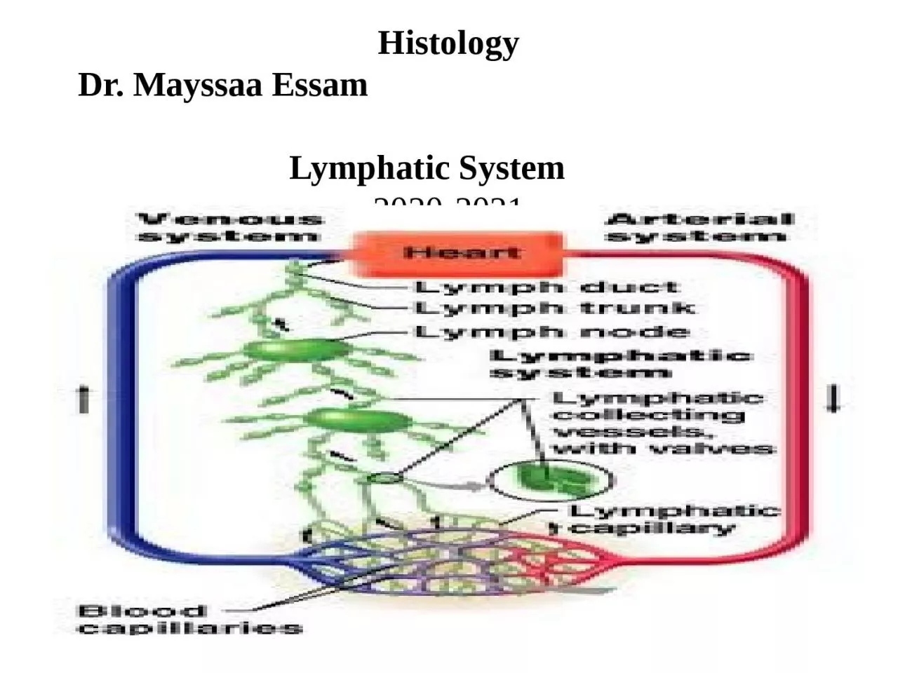
Essam Lymphatic System 20202021 Lymph vascular system ID: 916971

system lymph vessels cells lymph system cells vessels tissue lymphatic blood nodes lymphocytes composed called cortex immune tonsils thymus

Share:

Link:

Embed:

Download Presentation from below link

Download Presentation The PPT/PDF document "Histology Dr. Mayssaa" is the property of its rightful owner. Permission is granted to download and print the materials on this web site for personal, non-commercial use only, and to display it on your personal computer provided you do not modify the materials and that you retain all copyright notices contained in the materials. By downloading content from our website, you accept the terms of this agreement.


Presentation Transcript

Slide1

HistologyDr. Mayssaa Essam Lymphatic System 2020-2021

Slide2

Lymph vascular system The interconnected system of spaces and vessels between body tissues and organs by which lymph circulates throughout the body.Functions of the Lymph Vascular System :-The lymph vessels return to the blood extracellular fluid from connective tissue spaces. This system ensures the return of water, electrolytes and plasma proteins to the blood. The lymph vascular system plays as role in homeostasis of the volume of extracellular fluid. The lymph vascular system also returns lymphocytes from the lymph nodes to the blood. The system also transports immunoglobulins (antibodies) from the lymph nodes to the blood.

Slide3

The lymphatic System consist of:-1)Lymphatic vessels 2) Lymphoid organsLymphatic vessels:These vessels covey fluids from the tissues to the blood stream . L.V. Found in most tissue and organs but absent from central nervous system, cartilage, bone and epidermis , this L.V. Including of:1)lymph capillaries vessels which they are small thin walled and building ended tube . its similarly blood capillaries vessels but the endothelial cells overlap each other but there is gap between them and no peri-cytes and continuous basal membrane is absent.Lymph vessels are devoted to propulsion of the lymph from the lymph capillaries, which are mainly concerned with absorption of interstitial fluid from the tissues.

Slide4

Lymph capillaries are slightly larger than their counterpart capillaries of the vascular system. Lymph vessels that carry lymph to a lymph node are called the afferent lymph vessel, and one that carries it from a lymph node is called the efferent lymph vessel, from where the lymph may travel to another lymph node, may be returned to a vein, or may travel to a larger lymph duct. Lymph ducts drain the lymph into one of the subclavian veins and thus return it to general circulation. Generally, lymph flows away from the tissues to lymph nodes and eventually to either the right lymphatic duct or the largest lymph vessel in the body, the thoracic duct. These vessels drain into the right and left subclavian veins respectively.

Slide5

2(. Lymphatic collecting vessels or larger lymphatic vessels:- They larger vessels have thicker walls in which the typical three layers can sometimes be distinguished the tunica intimae consists of endothelium and a thin layer of longitudinal elastic fibers, the tunica media composed of arranged circularly smooth muscle fibers and the tunica adventitia is the thickest and composed of longitudinal collagen elastic fiber and few smooth muscle fibers. These vessels possess valves which are more numerous than those in vein.

Slide6

3(- lymphatic trunks or lymphatic ducts:-there are only two lymph ductA-Thoracic lymphatic ducts.B-Right lymphatic duct.The walls of these duct resemble those of large veins , but are thinner and posses valves .The ducts drain into the venous system by joining the subclavian vein at the root of the neck . the right lymph ducts collected lymph from right parts of head and right parts of thorax , whereas the thoracic ducts collected lymph from the parts remnants of body . Lymph : This is a clear fluid that travels via the lymphatic channels. This contains fluid, debris, chemicals, toxins, bacteria, viruses and lymphocytes on its way back from the tissues.

Slide7

Lymphoid organs: A structure composed principally of lymphatic tissue. It includes lymph nodes, spleen, tonsils, and thymus.Lymph nodes.Any of the small bodies located along the vessels of the lymphatic system (in humans notably in the neck, armpits, and groin) Some lymph nodes are located deeper within the body at the chest (between the two lobes of the lungs), around the coils of the intestines, in the pelvis etc., that filter bacteria and foreign particles from lymph fluid. During infection, lymph nodes may become swollen with activated lymphocytes. Also called lymph gland. The lymph nodes are small bean shaped glands or bulbs that tend to occur in clusters much like grapes.

Slide8

The structure of lymph nodes consists of:-Cortex Darker region composed of lymphocyte aggregate is called (lymph nodules )the lymph nodules contain the germinal center, and covered by sub capsular sinus.MedullaIt is lighter region and composed modularly cords (consists of lymphatic tissue ) this region contain more lymph sinuses and blood capillaries and macrophages than the cortex.Lymph node is surround by thick connective tissue called (capsule). Lymph nodes have a hilum where blood vessels and nerve enter and leave and the lymphatic vessels leave the node, the capsule sends septa into the node divided the parenchyma into incomplete compartment .

Slide9

Function of lymph nodesProduct of lymphocytes and plasma cells.Immunology function the lymphocytes in lymph nodes are able to recognized and reaction with foreign antigen. Flattened epithelial.

Slide10

The thymus is composed of two identical lobes and is located anatomically in the anterior superior mediastinum, in front of the heart and behind the sternum. Thymus is surrounded by connective tissue called (capsule) from which trabeculae (septa ) extend into organ , particularly subdivided into lobules composed into . 1)cortex the dark color.2)medulla. The lighter color.

Slide11

Both cortex and medulla have the same cells type ad which including:-Reticular epithelial cells.These cells more in medulla than cortex ,lymphocytes or thymocytes. Thymocytes are hematopoietic progenitor cells present in the thymus. Thymopoies is the process in the thymus by which thymocytes differentiate into mature T lymphocytes. The primary function of thymocytes is the generation of T lymphocytes (T cells).These more in cortex than the medulla.macrophages.Present in medulla more than the cortex .

Slide12

Hassals corpuscles are special and specific bodies present in the thymic medulla. The medulla of thymus is pale staining and composed of scattered thymocytes, reticular cells and characteristic bodies called Hassals Corpuscles. The Hassal’s Corpuscles are large, acidophilic, rounded bodies consisting of central degenerated Hyaline mass surrounded by concentrically arranged reticular cells . They are specific to thymus .The article deals with different clinical conditions associated with Hassals Corpuscle. Increase or decrease in number of Hassals Corpuscle with different diseases and their histopathological importance .

Slide13

Function of thymus:-*Reduction of T-lymphocytes which are responsible for cellular immunity and help hormonal immune response .Lymphocyte production take place in cortex.*Filtration of blood , product the thyroxin hormone,this hormone produced by the reticule epithelial cells and influence the maturation of T- lymphocytes and stimulate their activity in the blood.

Slide14

Spleen is the largest haem lymph organ in the body . its situated in the abdomen under the left half of diaphragm.Its surrounded by connective tissue called (capsule ) contain few smooth muscle . The capsule sends trabecula extend into the organ , particularly subdivided into lobules composed into:-

Slide15

1.Red pulp This occupies the centrical masses soft dark -red mass (histologically) consist of large irregular thin walled of blood vessels called (splenic sinusoids).these sinusoids separated by cord of reticular tissue called (splenic cords or billroths). The red pulp contains monocytes , plasma cells , red blood cells and less number from lymphocytes .2. White pulp or splenic pulp. This pulp distributed in roughly spherical masses consist of lymphoid tissue .this pulp structure resemble the cortex of lymphoid nodules .its filled with lymphocytes and some macrophage and plasma cells .

Slide16

The space between the trabeculae is filled with lymphatic tissue .Spleen is covered by capsule except where it is attached by the peritoneum.Function of spleen:-1) Immune defense (forming the antibody).2) Blood forming such as lymphocyte +monocytes. 3(Blood storage .4(Filtration of blood .

Slide17

Slide18

4-Tonsils pair of soft tissue masses located at the rear of the throat (pharynx). Each tonsil is composed of tissue similar to lymph nodes, covered by pink mucosa, aggregation of lymphoid tissue, they form a discontinuous ring .There are three types of tonsils:1. palatine tonsils.: Located in the lateral walls of the pharynx ,these tonsils are lined by a non keratinized stratified squamous epithelium tissue . 2. lingual tonsils :Located in the posterior third of the tongue, this tonsils lined by non keratinized stratified squamous epithelium tissue.3. pharyngeal tonsils.: Located in the root of the nasopharynx ,this lined by a with ciliated pseudo stratified epithelium tissue.Function of tonsil.1-Formation of lymphocyte.2- Defense of organism toward involving microorganism.

Slide19

Immune System The immune system is a system of biological structures and processes within an organism that protects against disease. To function properly, an immune system must detect a wide variety of agents, known as pathogens, from viruses to parasitic worms, and distinguish them from the organism's own healthy tissue.

Slide20

Basic types of immune reactions The immune system protects the body from possibly harmful substances by recognizing and responding to antigens. Antigens are substances (usually proteins) on the surface of cells, viruses, fungi, or bacteria. Nonliving substances such as toxins, chemicals, drugs, and foreign particles (such as a splinter) can also be antigens. The immune system recognizes and destroys substances that contain antigens.Antibodies can come in different varieties known as isotopes or classes. In placental mammals there are five antibody isotopes known as IgA, IgD, IgE, IgG, and IgM. They are each named with an "Ig" prefix that stands for immunoglobulin, another name for antibody, and differ in their biological properties, functional locations and ability to deal with different antigens.

Slide21

IsotypesIgA: Found in mucosal areas, such as the gut, respiratory tract and urogenital tract, and prevents colonization by pathogens.. Also found in saliva, tears, and breast milk.IgD:Functions mainly as an antigen receptor on B cells that have not been exposed to antigens. It has been shown to activate basophils and mast cells to produce antimicrobial factors. IgE:Binds to allergens and triggers histamine release from mast cells and basophils, and is involved in allergy. Also protects against parasitic worms.

Slide22

IgG: In its four forms, provides the majority of antibody-based immunity against invading pathogens. The only antibody capable of crossing the placenta to give passive immunity to the fetus.IgM: Expressed on the surface of B cells (monomer) and in a secreted form (pentamer) with very high avidity. Eliminates pathogens in the early stages of B cell-mediated (humoral) immunity before there is sufficient .