PPT-In the name of GOD Dysphagia
Author : dardtang | Published Date : 2020-06-15
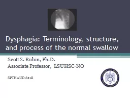
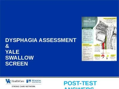
Dysphagia is an alarm symptom that warrants prompt evaluation to define the exact cause and initiate appropriate therapy It may be due to a structural
Presentation Embed Code
Download Presentation
Download Presentation The PPT/PDF document "In the name of GOD Dysphagia" is the property of its rightful owner. Permission is granted to download and print the materials on this website for personal, non-commercial use only, and to display it on your personal computer provided you do not modify the materials and that you retain all copyright notices contained in the materials. By downloading content from our website, you accept the terms of this agreement.
In the name of GOD Dysphagia: Transcript
Download Rules Of Document
"In the name of GOD Dysphagia"The content belongs to its owner. You may download and print it for personal use, without modification, and keep all copyright notices. By downloading, you agree to these terms.
Related Documents