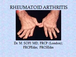
and Pathogenesis Rheumatoid arthritis RA is one of the most common inflammatory arthritides Affected patients suffer from chronic articular pain disability and excess mortality It primarily affects the small ID: 920822
Download Presentation The PPT/PDF document "Rheumatoid Arthritis Epidemiology, Path..." is the property of its rightful owner. Permission is granted to download and print the materials on this web site for personal, non-commercial use only, and to display it on your personal computer provided you do not modify the materials and that you retain all copyright notices contained in the materials. By downloading content from our website, you accept the terms of this agreement.
Slide1
Rheumatoid Arthritis Epidemiology, Pathology,and Pathogenesis
Slide2Rheumatoid arthritis (RA) is one of the most common inflammatory arthritides. Affected patients suffer from chronic
articular
pain, disability, and excess mortality.
It primarily affects the small
diarthrodial
joints of the hands and feet, although larger weight-bearing and
appendicular
joints can also be involved.
Extra-
articular
manifestations and systemic symptoms also occur, but in a minority of patients.
Slide3RA is a heterogeneous disease variable severity and unpredictable response to therapy. Genetic and environmental factors are clearly implicated in its etiology
and pathogenesis.
Translational research efforts have led to novel targeted therapies, although the treatment of RA remains a significant unmet medical need.
Slide4The Role of HLA-DR and the SharedEpitope Hypothesis
The most potent genetic risk for RA is conveyed by certain major
histocompatibility
complex alleles
(
MHC )
or HLA for human leukocyte antigen Increased prevalence of RA was reported to be associated with a subset of DR4 alleles in most Western European populations or a subset of DR1 alleles in other populations such as Spanish, Basque, and Israeli cohorts.
Current HLA typing can discriminate allelic variants at the nucleotide level and reveals that a conserved amino acid sequence is over-represented in patients with RA.
Slide5Different models have been proposed to explain the role of the shared epitope
in RA. Susceptibility alleles could bind efficiently to
arthritogenic
peptides, such as those either derived from a self-antigen or a microbial pathogen, lead to the positive or negative selection of autoimmune T cells in the thymus, lead to inadequate numbers of regulatory T cells, become the target of T cells themselves due to molecular mimicry between QKRAA and pathogens implicated in RA, such as Escherichia coli ,or Epstein–Barr virus (EBV) peptides.
Slide6Nongenetic Risk FactorsInfluence of Sex
Women are two to three times more likely to develop RA than men. Hormonal factors like
estrogen
and progesterone could potentially explain some of the gender effect.
Estrogen
might have detrimental effects through its ability to decrease apoptosis of B cells, potentially permitting the selection of auto reactive clones.
Hormones also have a complex influence on the balance of T-cell subsets with distinct cytokine profiles. For instance, administration of
estrogen
in animal models can enhance or suppress T-helper (
Th
) 1-mediated immunity,
Slide7The situation during pregnancy exemplifies complex influence that sex has on RA. Seventy-five percent of pregnant women with RA experience spontaneous remission, although the disease typically flares within weeks after delivery.
Soluble mediators released by the placenta like transforming growth factor (TGF) beta, IL-10, or alpha-fetoprotein might contribute to this effect. Alternatively, the immune system in pregnant women displays a shift towards a Th2 bias, which could suppress the characteristic Th1 profile of RA.
Slide8Tobacco Exposure to various environmental factors increase the risk for RA, and cigarette smoke is one of the best characterized. Of interest, smoking also enhances the risk of developing anti-CCP positive RA in patients with the SE .
The mechanism of anti-CCP antibody generation from inhaled smoke probably relates to inflammation and activation of innate immunity in the airway, which then induces peptide
citrullination
. In a susceptible host, such as someone carrying the SE and with genetically determined immune
hyperreactivity
, these repeated insults followed by chronic exposure to
citrullinated
peptides could lead to the production of anti-CCP antibodies and other antibodies like rheumatoid factors.
Slide9Bacteria and Their Products: Infectious agents have long been considered prime candidates as initiating factors for RA, although the search for a specific etiologic agent has been unrewarding.
Bacterial stimulate synovial innate immune responses. Even nonspecific bacterial products could thus play a role in
synovitis
by activating cytokine networks or acting enhancing adaptive autoimmune responses.
Viruses :Several viruses have been implicated as possible etiologic factors in RA. A relationship between RA and EBV was suggested by several observations.
Slide10SYNOVIAL PATHOLOGYThe complex histological architecture of the synovial tissue in RA is the result of a dynamic process involving coordinated molecular signals (
chemokines
, adhesion molecules, cytokines, and growth factors) and cellular events (apoptosis, proliferation, cell migration, and survival).
Increased numbers of both type A and B
synoviocytes
augment the depth of the lining layer, sometimes to 10 cell layers, and mononuclear cells infiltrate the
sublining
The lining is the primary source of inflammatory cytokines and proteases, thus participating in joint destruction in concert with activated
chondrocytes
and
osteoclasts
.
Slide11Villous projections protrude into the joint cavity, invading the underlying cartilage and bone where the proliferating tissue is called
pannus
. In the synovial
sublining
region,
edema
, blood vessel proliferation, and increased
cellularity
lead to a marked increase in tissue volume.
T and B lymphocytes, plasma cells,
interdigitating
and follicular
dendritic
cells (IDC and FDC), and natural killer cells (NK cells) accumulate in rheumatoid
synovium
and can be distributed diffusely throughout the
sublining
or organized into lymphoid aggregates. The dominant cells, CD4+ T cells, CD4+ T cells are especially enriched in aggregates, whereas CD8+ T cells are present in the periphery of the aggregates or scattered throughout the
sublining
Slide12B-Cell Autoimmunity and Autoantibodies
Antibodies directed against joint-specific and systemic
autoantigens
are commonly detected in the blood of RA patients.
Rheumatoid Factors
IgG
and
IgM
RFs are found in up to 90% of RA patients. Testing for
IgM
RF is about 70% sensitive and 80% specific for RA. However, these
autoantibodies
can also be produced during chronic infections, malignancy, and in a variety of inflammatory and autoimmune syndromes.
RFs are also detectable in 1% to 4% of healthy individuals, and up to 25% of healthy individuals over the age of 60 years. They can be detected in the blood up to 10 years before the onset of RA, with an increasing incidence in the period immediately before clinical symptoms develop . Therefore, the mere presence of RF is not sufficient to cause arthritic symptoms. The presence of RF in RA, however, has prognostic significance.
Sero
positive patients have more aggressive disease while
sero
negative patients tend to experience less severe arthritis with fewer bone erosions.
Slide13Anti-Cyclic Citrullinated PeptideAntibodies
Anti-CCP testing has a sensitivity of up to 80% to 90% and a specificity of 90% for RA, which increases to >95% specificity if combined with the presence of
IgM
RF .
Anti-CCP antibodies are occasionally produced in other inflammatory diseases, such as psoriatic arthritis, autoimmune hepatitis, and pulmonary tuberculosis (TB). Similar to RF, anti-CCP antibodies are a risk factor for more aggressive disease and are produced early in disease.
Slide14T-Cell Autoimmunity
Naive CD4+ T cells can be differentiated into multiple
effector
types, including Th1 and Th2 phenotypes. Experimental systems have shown that precursor cells can be polarized towards one of these phenotypes depending on the nature of the antigen, characteristics of the antigen-presenting cells, and the cytokine milieu.
Th1 cells are involved in the
defense
against intracellular pathogens and have been implicated in many autoimmune diseases. Th2 cells participate in host
defense
against parasitic worms but can also contribute to allergy and asthma.
Each subtype is induced by cytokines present in the milieu (mainly IL-12 for Th1 cells, IL-4 for Th2 cells) and secretes characteristic
effector
cytokines (IFN-gamma and IL-2 by Th1 cells, IL-4 and IL- 10 by Th2 cells). IL-4 and IL-10 inhibit Th1 cells, while IFN-gamma suppresses Th2 function.
Slide15MACROPHAGE AND FIBROBLAST CYTOKINES
Macrophages and fibroblasts are the primary sources of cytokines in the rheumatoid
synovium
. Synovial macrophages and fibroblasts produce a plethora of
proinflammatory
factors in the joint involved in the cytokine network (Figure 1), including IL-1, IL-6, IL-8, IL-12, IL-15, IL-16, IL-18, IL-32, TNF-alpha, granulocyte macrophage colony-stimulating factor (GM-CSF), and multiple
chemokines
.
These cytokines can participate in
paracrine
and
autocrine
networks that enhance and perpetuate synovial inflammation. For instance, macrophages and fibroblasts in the
intimal
lining can activate adjacent cells that, in turn, can produce mediators that can stimulate their
neighbors
. The concept of cytokine networks dominated by synovial lining cells played
amajor
role in the advent of
anticytokine
therapy in RA.
Slide16Slide17Although proinflammatory cytokines can be counterbalanced by the suppressive cytokines (IL-10,
TGFbeta
), soluble receptors (TNF-alpha), binding proteins (IL-18), and naturally occurring receptor antagonists (IL-1Ra), all of which are produced by macrophages and fibroblasts in the synovial
intima
, the concentrations are below those required to suppress inflammation.
Although the cytokine network can be highly redundant, disease control can be achieved in many patients by inhibiting a single cytokine. TNF-alpha antagonists are the most salient example, in which one third to one half of patients have dramatic clinical responses to cytokine blockade
Slide18In RA, TNF-alpha is mainly produced by synovial macrophages. The stimulating signals have not been defined but could involve a family of receptors that recognize specific molecular patterns and activate the innate immune system, and other cytokines like IL-15.
TNF-alpha can then bind to two ubiquitously expressed receptors (TNF-RI and TNF-RII) to induce the release of other cytokines and
metalloproteases
by fibroblasts, decrease the synthesis of
proteoglycans
by
chondrocytes
, and promote the differentiation of
monocytes
to
osteoclasts
in the presence of RANKL.
Slide19Interleukin 1 exhibits many properties that can contribute to inflammation in RA, including increased synthesis of IL-6, chemokines
, GM-CSF, prostaglandin and
collagenase
.
Of the two forms of IL-1, IL-1 beta is secreted, whereas IL-1 alpha is expressed within cells and associated with cell membranes. Interleukin 18 is another
proinflammatory
member of the IL-1 family and induces the production of IFN-gamma, IL-8, GM-CSF, and TNF-alpha by synovial macrophages.
Slide20Interluekin 6 has pleiotropic
effects and influences systemic inflammation through its actions on
hematopoiesis
and many cell types of the immune system.
IL-6 is perhaps the major factor that induces acute phase proteins like CRP by the liver. Very high levels of IL-6 are present in the synovial fluid of RA patients and type B
synoviocytes
are the major source.
IL-6 is also implicated in the activation of the endothelium and contributes to bone erosion by stimulating the maturation of
osteoclasts
. In RA, IL-6 levels decrease dramatically after treatment with TNF inhibitors.
Slide21MECHANISM OF JOINT DESTRUCTION
The generation of new blood vessels is required to provide nutrients to the expanding synovial membrane and is an early event in the development of
synovitis
. The expanding tissue can ultimately outstrip angiogenesis in RA; synovial fluid oxygen tension is quite low and is associated with low pH and high lactate levels.
Hypoxia is a potent stimulus for angiogenesis in the
synovium
, and factors that promote blood vessel growth, such as vascular endothelial growth factor (VEGF), IL-8, angiopoietin-1, and many others, are expressed in RA. Several anti-angiogenesis approaches can markedly attenuate arthritis in animal models. For instance, targeting the
integrin
alpha-v beta-3 expressed by proliferating blood vessels in the
synovium
or treating with antibodies to the type 1 VEGF receptor (VEGF-R1) suppress clinical and
histologic
evidence of disease.
Slide22Activated type-B synoviocytes are a major source of inflammatory mediators and
metalloproteinases
in RA.
Synoviocytes
can be grown in vitro to study signal transduction systems that relay information from the environment to the nucleus and activate gene expression. Fibroblast like cells derived from the
synovium
of RA patients exhibit some unique aggressive properties.
Insufficient
synoviocyte
apoptosis in RA probably contributes to
intimal
lining hyperplasia of the
synovium
due to several mechanisms, including low expression of anti-apoptotic genes and abnormal function of
tumor
suppressor genes .
Slide23Cartilage Destruction
Aggressive
synoviocytes
at sites of
pannus
overgrowth, cytokine-activated
chondrocytes
, and PMNs are major cell types responsible for destruction of the cartilage in RA.
They release destructive enzymes in response to IL-1, TNF-alpha, IL-17, and immune complexes. Once the cartilage is compromised, mechanical stress works as an accelerating factor to enhance destruction.
A variety of enzymes participate in extracellular matrix degradation of the joint, such as matrix
metalloproteinases
(MMPs;
collagenases
,
gelatinases
, and
stromelysin
), serine proteases (
trypsin
,
chymotrypsin
), and
cathepsins
. Reversible loss of
proteoglycans
occurs early, most likely due to the catabolic effect of cytokines and the production of
stromelysins
and
aggrecanases
.
Slide24Bone Destruction
Focal bone erosions are a hallmark of RA that can occur early in the disease and cause significant morbidity due to
subchondral
and the cortical bone damage.
RA is also associated with
periarticular
bone loss adjacent to inflamed joints and generalized
osteopenia
, leading to increased risk of fracture in both the
appendicular
and axial skeleton. The cellular and molecular mechanisms underlying cartilage destruction and focal bone erosions are distinct.
Synoviocytes
,
chondrocytes
, and
neutrophils
are probably the major effectors of the former. Bone erosions are mainly caused by
osteoclasts
, which are derived from macrophage precursors . They accumulate at the
pannus
–bone interface and the
subchondral
marrow space.
Slide25Receptor activator of NF-
κB
(RANK) and its
ligand
RANKL form the most important receptor–
ligand
pair that modulates bone
resorption
in RA.
RANK is expressed by
osteoclasts
and modulates their maturation and activation. Expression of the RANKL on T cells and fibroblast like
synoviocytes
is promoted by cytokines such as TNF-alpha, IL-1, and IL-17.
The RANK–RANKL system is antagonized by a soluble decoy receptor,
osteoprotegerin
(OPG), that binds to RANKL. Injection of OPG or deletion of the RANKL gene in animal models inhibits bone destruction but does not suppress inflammation. Of interest, anti–TNF-alpha agents can slow the progression rate of bone erosions in RA, even in patients without clinical improvement. Therefore, the inflammatory and destructive mechanisms in RA can be distinct.
Slide26Innate and adaptive immunity both contribute to the pathogenesis of RA.
B cell help
Autoantibody production