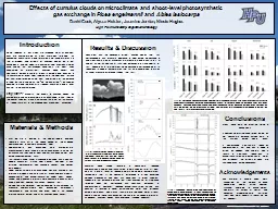
/
Effect of cumulus cells and Effect of cumulus cells and

Effect of cumulus cells and - PowerPoint Presentation

debby-jeon
debby-jeon . @debby-jeon
Follow
396 views
Uploaded On 2017-07-03

Effect of cumulus cells and - PPT Presentation

vitrification protocol on survival and subsequent development Golestan jahromi PhD student Introduction Several lines of evidence indicate that surrounding cumulus cells play a fundamental role in the maturation process and full development competence ID: 566289

cumulus oocytes ice vitrification oocytes cumulus vitrification ice mii discussion 1000 vitrified cells bovine blastocyst rate enclosed stage cleavage

Share:

Link:

Embed:

Download Presentation from below link

Download Presentation The PPT/PDF document "Effect of cumulus cells and" is the property of its rightful owner. Permission is granted to download and print the materials on this web site for personal, non-commercial use only, and to display it on your personal computer provided you do not modify the materials and that you retain all copyright notices contained in the materials. By downloading content from our website, you accept the terms of this agreement.


Presentation Transcript

Slide1
Slide2

Effect of cumulus cells and

vitrification

protocol on survival and subsequent development

Golestan

jahromiPhD studentSlide3

Introduction Several lines of evidence indicate that surrounding

cumulus cells

play a fundamental role in the maturation process and full development competence Slide4

Introduction cumulus cells are beneficial to oocyte survival after cryopreservation may minimize the release of cortical granules prevent premature zona hardening

maintaining fertilization capacity of cryopreserved oocytes Slide5

Introduction Chian et al. reported that bovine oocytes matured without cumulus cells had a higher survival rate after vitrification.

Moreover, the rate of embryo development to the 8-cell stage in cumulus-cells free oocytes was significantly higher than that of cumulus cell-intact oocytes. Slide6

IntroductionThe need to maintain cumulus cells during cryopreservation of oocytes is still a matter of debate. Considering that the functions of cumulus cells are different in immature (GV) and mature (MII) oocytes, it is necessary to determine the effects of cumulus cells on bovine oocyte vitrification at both the GV and the MII stage, respectivelySlide7

IntroductionWater permeability (Lp), solute permeability (Ps), and activation energies for Lp and Ps (Ea

) were determined using a two parameter model. Osmotic tolerance limits and toxic tolerance limits were determined based on oocyte developmental competence following exposure to different CPAs.Slide8

IntroductionIce growth and recrystallization are considered to be important factors in determining vitrification outcomes. Synthetic ice blockers, which specifically inhibit the formation/emergence of ice nuclei and ice crystal growth, have recently been used to supplement

vitrification

solutionsSlide9

IntroductionUnlike conventional cryoprotectants that inhibit freezing by interacting with water, ice blockers are believed to bind to the surface of growing ice crystals and inhibit the addition of any further water molecules in specific planes of growth

This selective attraction to surfaces of ice growth permits ice blockers to exert significant effects even while present at

very low concentrations. Slide10

IntroductionSmall quantities of ice blocker can therefore modify the number and size of ice crystals and thereby change the vitrification tendency of a solution without adding additional toxicity Slide11

IntroductionThe commercially available ice blockers are SuperCool X-1000 and SuperCool Z-1000. Slide12

The SuperCool X-1000 is a copolymer of polyvinyl alcohol (PVA) of mean molecular mass 2000 Da, with 20% of the hydroxyls replaced by acetate groups. The SuperCool Z-1000 is a copolymer of polyglycerol (PGL) of mean molecular mass 750 Da Slide13

To evaluate the effect of the presence of cumulus cells on the outcome of

vitrification

of GV or MII oocytes The effect of adding ice blockers SuperCool

X-1000 and SuperCool Z-1000 to vitrification media on oocyte survival and subsequent embryonic developmentSlide14

Materials & methodsSlide15
Slide16

In vitro maturationGroups of 50 COCs were placed in 500 μl maturation medium in four-well plates and cultured for 24 h at 39 °C in a 5% CO2 humidified air atmosphere. The maturation medium was Tissue Culture Medium 199 (TCM 199) supplemented with 10% Fetal Bovine Serum (FBS) and 10

ng

/ml epidermal growth factor (EGF).Slide17

Vitrification and warmingThe holding medium (HM) used for handling oocytes during vitrification and warming was HEPES-buffered TCM 199 supplemented with 20% (v/v) FBS. All manipulations were performed on a 39 °C heated stage in a warm room (25–27 °C).

All the media were used at room temperature, except for the warming solution which was used at 37 °C.Slide18

According to the manufacturer, the cooling and warming rates of the Cryotop are 23,000 and 42,000 °C/min, respectivelySlide19

Control and vitrified GV and MII oocytes were fertilized using the same conditions. All oocytes were washed four times in fertilization medium, and then transferred in groups of 50 into four-well dishes containing 250 μl of fertilization medium (Tyrode’s

medium with 25

mM bicarbonate, 22 mM sodium lactate, 1 mM sodium pyruvate and 6 mg/ml fatty acid-free BSA). In addition, 10

μg/ml heparin–sodium salt was addedSlide20

Motile spermatozoa were obtained by centrifugation of frozen–thawed semenSlide21

Zygotes were transferred in groups of 25 to 25 μl culture droplets of synthetic oviductal fluid medium (SOF) under mineral oilSlide22

Day 2

after insemination

Day 8 after

insemination

Cleavage rates

proportion of

oocytes

developing to the

blastocyst

stageSlide23

Experiment 1Slide24

Experiment 2: Effect of ice blocker X-1000 and Z-1000

VS +1% (v/v) X-1000

VS +1% (v/v)

Z-1000

VS +1% (v/v) Z-1000 and 1% (v/v) X-1000Slide25

Statistical analysis The data for survival, cleavage and blastocyst rates were expressed as mean ± SD and analyzed using one-way ANOVA. Differences were considered significant at a level of P < 0.05.Slide26

ResultsSlide27

development of bovine GV stage

Oocytes treated

N

Survived, n (%)Cleaved, n (%)Blastocyst, n (%)Blastocyst/cleavage (%) Cumulus-enclosed control

141141 (100 ± 0.0)121 (86.3 ± 1.9)47 (33.8 ± 1.8)(39.5 ± 2.2)Partially-denuded control

118

117

(99.3 ±0.8)

89

(75.8 ± 3.9)

14

(11.5 ± 4.2)

(14.8 ± 5.2)

Cumulus-enclosed vitrified

177

166

(93.8 ± 2.5)

108

(65.8 ± 5.6)

19

(11.3 ± 1.7)

(18.0 ± 3.5)b

Partially-denuded vitrified

143

117

(81.3 ± 3.6)c

56

(47.3 ± 4.0)c

4

(4.0 ± 2.3)c

(7.8 ± 4.5)b Slide28

embryo development of bovine MII stage

Oocytes treated

N

Survived, n (%)Cleaved, n (%)Blastocyst, n (%)Blastocyst/cleavage (%) Cumulus-enclosed control

130126 (96.8 ± 1.5)a 115 (91.6 ± 2.5)a 45 (35.6 ± 2.8)a (39.0 ± 4.1)a Partially-denuded control

122

119

(97.4 ± 1.1)a

92

(78.4 ± 4.7)b

32 (27.2 ± 2.3)b

(34.6 ± 2.7)

a,b

Cumulus-enclosed vitrified

158

147

(93.0 ± 2.3)a

51

(35.2 ± 4.6)c

7 (5.0 ± 4.3)c

(12.6 ± 9.7)

b,c

Partially-denuded vitrified

167

153

(91.8 ± 2.4)a

57

(36.8 ± 3.2)c

7 (4.4 ± 1.4)c

(10.8 ± 3.5)c Slide29

development of bovine GV stage cumulus-enclosed oocytes vitrified with different ice blocker media

Oocytes treated

N

Survived, n (%)

Cleaved, n (%)Blastocyst, n (%)Blastocyst/cleavage (%) Control 110

104

(94.4 ± 1.9)a

77

(74.6 ± 3.2)

24

(23.0 ± 10.4)

(31.2 ± 1.9)

Basic media

114

98

(86.0 ± 2.7)b

38

(38.0 ± 3.2)

2 (2.0 ± 1.3)

(6.2 ± 4.1)

Basic media + X-1000

111

97

(88.4 ±2.9)

36

(37.2 ± 2.4)

3 (2.8 ± 1.2)

(7.8 ± 3.2)

Basic media

+ Z-1000

110

93

(83.6 ± 4.8)

37

(40.0 ± 3.0)

2(2.2 ± 1.4)

(4.8 ± 3.0)

Basic media

+ X-1000

+ Z-1000

114

101

(88.0 ± 3.6)

43

(41.4 ± 6.7)

0 (0)

(0)Slide30

Cumulus-enclosed oocytes vitrified at the GV stage exhibited a significantly higher cleavage rate and blastocyst rate than those vitrified at MII stage (P < 0.05). Slide31

Why?

DiscussionSlide32

DiscussionThe role of the cumulus cells during vitrification of MII oocytes remains controversial. Some investigators reported that cumulus presence would

protect MII oocytes against

vitrification-induced damage. Slide33

DiscussionZhang et al. found no difference in the development of vitrified ovine MII oocytes with or without cumulus cells. Gasparrini et al. reported that the presence of cumulus cells severely reduced the cleavage rate of MII buffalo oocytes following

vitrificationSlide34

Discussion It is generally accepted that cumulus-oocyte communication via an intact corona radiata is necessary for oocytes to attain full cytoplasmic maturation during

IVM

and improve fertilization rates during IVF Slide35

Discussion we established that the cleavage rate of denuded (GV and MII) bovine oocytes was significantly

reduced

compared to cumulus-enclosed oocytes, and almost

no denuded bovine oocytes developed up to blastocyst stage after in vitro fertilization.Slide36

In other species, the effect of cumulus cells on immature oocytes following vitrification was evaluated by the maturation rate, spindle and chromatin quality. Slide37

DiscussionTharasanit et al. [33] reported that cumulus removal from equine oocytes prior to IVM or vitrification resulted in reduced meiotic competence, MII spindle and chromatin quality

.

while the cumulus does protect immature equine oocytes during vitrification it does so by mechanisms other than support during maturationSlide38

Discussion In contrast, Bogliolo et al. reported that immature ovine oocytes vitrified without cumulus cells showed a significantly

higher

survival and meiotic maturation rate than those with cumulus cells, and no differences in spindle and chromatin organization between two groups were observed.Slide39

In this study:The survival, cleavage and blastocyst rate of cumulus-enclosed vitrified oocytes are significantly higher than that of partially-denuded vitrified oocytes.Slide40

Kuwayama et al. reported higher survival and blastocyst rate for cumulus-enclosed than cumulus-free human MII oocytes after vitrificationSlide41

Tharasanit et al. found that cumulus-enclosed equine MII oocytes preserved their meiotic spindle and chromatin quality better during vitrification than denuded oocytes. Conversely, other investigators believed cumulus cells are not necessary or detrimental to MII oocytes during vitrification

. Slide42

DiscussionIn the present study:No significant differences were detected between vitrified cumulus-enclosed and partially-denuded oocytes in the survival, cleavage and blastocyst rate. Slide43

DiscussionThe possible explanation is the cumulus was detrimental to vitrification, which comprises the benefits of cumulus in IVF procedure. From another point of view, the

intracytoplasmic

sperm injection technique rather than conventional IVF has been used to achieve fertilization, which can circumvent the detrimental effects of removing the cumulus on subsequent zona penetrability.Slide44

DiscussionMoreover, the oocytes were easier to handle for vitrification using a Cryotop when the oocytes were denuded completely from their cumulus cells. Therefore, cumulus removal prior to

vitrification

is at present a standard practice during cryopreservation of MII human oocytes.Slide45

DiscussionThe cell cycle stage during meiosis appears to affect the results of bovine oocyte vitrification due to varying sensitivity to cooling procedures. Chilling injury is reported to be

higher

in vitrified immature oocytes, owing to

low membrane stability and susceptibility of the cytoskeletonSlide46

Discussion However, an increase in chromosomal abnormality has been observed in vitrified mature oocytes, owing to alterations in the meiotic spindle. Slide47

DiscussionThe results indicate that cumulus-enclosed oocytes vitrified at the GV stage exhibited a significantly higher cleavage and blastocyst rate than those vitrified at MII. Slide48

Discussion This may be due to the increase in volume associated with cumulus expansion during maturation. It may also be due to the

higher water permeability (

Lp) and solute permeability (Ps) of MII than GV bovine oocytes.

That means the changes of cell volume and intracellular CPA concentrations are more severe in MII than GV bovine oocytes during CPA addition and dilution process, which make it more sensitive.Slide49

DiscussionThe osmotic tolerance limits tests indicated that the safe volume changes of GV bovine oocytes were from 150% to 46% of their volume. Toxicity tolerance limits tests suggested that Me2SO are more toxic than EG, and the exposure time to 30% and 40% EG should be less than 1 min.Slide50

DiscussionIn the present study, we report for the first time the effect of ice blockers on the bovine oocytes, however, the results indicate that the survival rate and development competence of bovine oocytes vitrified in solutions supplemented with or without X-1000 and/or Z-1000 by

Cryotop

method are not significantly different. Slide51

DiscussionIce blockers did not affect the survival rate and developmental competence of vitrified bovine oocytes. Slide52

Why?Slide53

DiscussionWhen the vitrification systems are large volume, such as organs, in which a large quantity of nucleators

exit, ice blockers can suppress nucleation and recrystallization by binding to

nucleators in solutions during vitrification and warming. Therefore, the ice growth was inhibited and damage to the systems was reduced. Slide54

DiscussionIn this study, the combination of the two ice blocker agents inhibit blastocyst development maybe because 1% X-1000 and 1% Z-1000 is not the ideal balance, which shows less effective than either agent alone.Slide55

55

cumulus-enclosed GV bovine oocytes survived

vitrification

and subsequently developed at higher rates than MII oocytes .

Ice blockers had no effect on bovine oocyte vitrification.Slide56