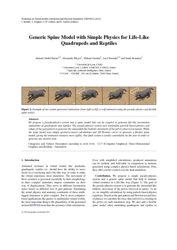
Ultrasound Evaluation of the Adnexa Ovary and Fallopian Tube Parts A amp B 23 week lecture Holdorf Contents Physiologic Cysts Follicular Cysts Corpus Luteum Cysts Corpus Luteum of Pregnancy ID: 742721
Download Presentation The PPT/PDF document "Ultrasound of the Female Pelvis" is the property of its rightful owner. Permission is granted to download and print the materials on this web site for personal, non-commercial use only, and to display it on your personal computer provided you do not modify the materials and that you retain all copyright notices contained in the materials. By downloading content from our website, you accept the terms of this agreement.
Slide1
Ultrasound of the Female Pelvis
Ultrasound Evaluation of the Adnexa- Ovary and Fallopian Tube
Parts A & B
2-3 week lecture
HoldorfSlide2
Contents
Physiologic Cysts
Follicular Cysts
Corpus Luteum Cysts
Corpus Luteum of Pregnancy
Theca Lutein Cysts
Hemorrhagic Cysts
Ovarian Torsion
Polycystic Ovarian Syndrome (PCOD)Slide3
Surface Epithelial Stromal Tumors
Serous Tumors
Mucinous Tumors
Endometrioid Tumors
Clear cell tumor
Transition cell (Brenner) Tumor
Germ Cell Tumor
Dysgerminoma
Yolk Sac (Endodermal Tumor)Slide4
Sex Cord-Stromal Tumors
Fibromas
MEIG’S syndrome
Thecoma
Granulosa
Sertoli-Leydig) (Androblastoma) Tumor
Metastatic Tumors (to the ovary)
Krukenberg Tumor
Ovarian Cancer ScreeningSlide5
CA125
Adnexal Pathology
Pelvic Inflammatory Disease (PID)
Hydrosalpinx
EndometriosisSlide6
Physiologic Cysts
Ovarian cysts can be visualized sonographically in women of all ages. The presence of a simple (unilocular, anechoic, thin-walled) cystic mass related to either ovary measuring less than 3 cm is considered within normal limits. Sonographic and clinical follow-up however, is recommended when the dimensions of a cyst exceed 3cm. approximately 60% of ovarian cysts resolve spontaneously. Slide7
Several types of benign cysts exist:
Functional cysts
Follicular cysts
Corpus luteum cysts
Corpus luteum cyst of pregnancy
Theca lutein cystsSlide8
Functional cysts
Generic hormonally active cysts that usually result from the stimulation of released pituitary gonadotropins.
They are the most common cause of ovarian enlargement in young women.
Functional cysts range in size from 0.5 mm to 3cm, and are further categorized as the following;Slide9
Follicular cystsCorpus luteum cysts
Corpus luteum cysts of pregnancy
Theca lutein cystsSlide10
Follicular cysts
Occur when a dominant follicle fails to extrude the ovum or fails to regress following ovulation. Serous fluid distends the lumen of the follicle creating cysts. Most follicular cysts are unilocular and measure 3 to 8cm. The maximum measurement of a normal dominant follicle is 3cm.
Sonographic findings
Anechoic
Thin-walled
UnilocularSlide11
Follicular CystSlide12
Corpus Luteum Cysts
Occur when the dominant follicle extrudes the ovum. In the absence of a pregnancy, the corpus luteum cyst may continue to grow or hemorrhage into the lumen. They rarely exceed 4cm in diameter, may contain internal echoes, and cause symptoms that simulate an ectopic pregnancy.
Sonographic findings
Thick, hyperechoic
Usually echogenic content
Possible solid in appearanceSlide13
Corpus Luteum CystSlide14
Corpus luteum cyst of pregnancy
Is persistent in the corpus luteum in the presence of hCG.
They can become enlarged, even grater than 6cm, and usually regress spontaneously by 14 weeks of gestation.Slide15
Corpus Luteum of PregnancySlide16
Theca Lutein cysts
Are frequently multi-locular, and are the largest of the functional cysts. They result from overstimulation by the high levels of hCG associated with Trophoblastic disease or hCG administration during infertility treatment. The ovaries are bilaterally enlarged and the cysts may persist for days or weeks.Slide17
Theca Lutein CystSlide18
Hemorrhagic cyst
Occasionally, an ovarian cyst may hemorrhage into the lumen, because of its large size, spontaneous rupture, or even torsion, Hemorrhage is most common in corpus luteum cysts. Clinically, patients present with a sudden onset of pelvic pain.Slide19
Sonographic findings
Typical cystic appearance
Acute cyst=hyperechoic, mimicking a solid mass, by with posterior acoustic enhancement
Subacute hemorrhagic cyst=complex appearance, with internal echoes, strands, rarely a fluid-fluid levelSlide20
Hemorrhagic Ovarian CystSlide21
OVARIAN TORSION
Torsion of the ovary is caused by partial or complete rotation of the ovarian pedicle on its axis.
Lymphatic and venous drainage is compromised, causing congestion and edema of the ovary, eventually leading to loss of artery perfusion and resultant infarction.
Right sided torsion can clinically mimic acute appendicitis. Slide22
Risk factors for torsion include:
Preexisting ovarian cyst or mass
Children and young females with mobile adnexa (ovary is usually normal)
PregnancySlide23
Sonographic findings
Enlarged ovary, commonly with multiple cortical follicles
Absent color and spectral Doppler flow (but this is varied depending on the degree of torsion
Possible arterial Doppler flow but absent venous flow
Possible adnexal massSlide24
Ultrasound of ovarian torsionSlide25
POLYCYSTIC OVARIAN SYNDROME (PCOS)/Disease
OR Stein-Leventhal Syndrome.
Is an endocrinology disorder associated with chronic anovulation.
It is most commonly found in adolescent girls and young women (teens to twenties). Diagnosis of PCOS is actually a clinical diagnosis, and not necessarily a sonographic diagnosis.Slide26
Clinical signs
Obesity
Oligomenorrhea or amenorrhea
Hirsutism
InfertilitySlide27
Sonographic findings
Bilateral multiple cysts of varying size throughout the subcapsular and stromal ovarian tissue
Enlarged ovaries with tiny follicles in the subcapsular periphery
Endovaginal Sonography may reveal multiple cysts present ranging from 2mm to 10mm
ALWAYS bilateral Slide28
Cartoon of PCOS/DiseaseSlide29
Ultrasound of PCOSSlide30Slide31
Surface Epithelial Stromal Tumors
Tumors arising form the surface epithelium that covers the ovary and the underlying ovarian stroma account for 65-75% of all ovarian neoplasms, and 89-90% of all ovarian malignancies.
The tumor types are divided into five categories, based on epithelial differentiation:Slide32
SerousMucinous
Endometrioid
Clear Cell
Transitional Cell (Brenner)Slide33
Proliferative changes of these tumors are further divided into three categories:
Benign
Atypically proliferating (borderline, or low potential for malignancy)
Malignant
10 to 15% of serious and mucinous tumors are borderline malignant.
Although they have cytologic features of malignancy, they do not invade the stroma and have a good prognosis.Slide34
SEROUS TUMORS
Common, accounting for 25-30% of al ovarian neoplasms. 50-70% of serous tumors are benign, but serous cystadenocarcinomas account for 40-50% of all malignant ovarian neoplasms.
Benign Serous tumors are bilateral 12-20% of the time, and occur most commonly in women 40-50 years of age.
Malignant serous tumors are bilateral 50% of the time, and occur most commonly in per-and post menopausal women.Slide35
Cystadenofibromas
A type of serous tumor with a solid component, more likely to mimic a malignant lesion.
Tumor size varies, but typically serous are smaller than mucinous tumors.
Sonographic findings of benign serous tumors
Sharply marginated
Anechoic
Large, but usually unilocular
Possibly internal thin-walled septationsSlide36
Sonographic findings of malignant serous tumors
Multi-locular
Multiple papillary projections and septations
Occasionally echogenic material within
Possibly multiple echogenic foci
AscitesSlide37
Benign serous tumor: Serous Cystadenoma: A: Unilocular Serous cystadenoma B: Multi-locular CystadenomaSlide38
MUCINOUS TUMORS
Benign mucinous tumors comprise 20-25% of all benign ovarian neoplasms,
are more common in women 30-50 years of age,
and very rarely are bilateral.Slide39
Malignant mucinous tumors account for only 5-10% of all malignant primary ovarian neoplasms,
occur most commonly in women 40-70 years of age. 15-20% are bilateral.
Penetration of the tumor capsule or rupture may spread mucin-secreting cells into the peritoneal cavity, filling it with gelatinous material known as Pseudomyxoma Peritonei.
It occurs with either benign or malignant mucinous tumors,
and may have a sonographic appearance similar to ascites, possibly with multiple septations.Slide40
Sonographic findings of benign mucinous tumors
Multi-loculated with thicker and more numerous septations
Fine, gravity-dependent echoes
Up to 50cm in diameterSlide41
Sonographic findings of malignant mucinous tumors
Multi-loculated cystic lesions measuring 15-30cm in diameter
Contain echogenic material and papillary projectionsSlide42
Benign Mucinous TumorSlide43
Mucinous CystadenomaSlide44
Malignant Mucinous Tumor: Mucinous Cystadenoma of low malignant potentialSlide45Slide46
ENDOMETRIOID TUMORS
80% of ovarian Endometrioid tumors are malignant, but have a better prognosis than either serous or mucinous carcinomas. Endometrioid tumors account for 20-25% of all ovarian carcinomas.
Histologically, Endometrioid tumors are identical to endometrial adenocarcinoma, and 30% of patients with this tumor have associated endometrial adenocarcinoma.Slide47
Sonographic findings
Cystic mass with papillary projections
In some cases, there may be a predominantly solid mass, possibly with areas of hemorrhage or necrosis
Slide48
Endometrioid TumorSlide49
CLEAR CELL TUMOR
Nearly always malignant, clear cell tumors constitute 5-10% of all malignant ovarian epithelial-stromal tumors. Most typically, they occur in women 50-70 years of age. Clear cell tumors are bilateral up to 20% of the time, and range in size up to 30cm.
Sonographic findings
Nonspecific
Complex, but predominantly cystic in appearanceSlide50
Confirmed Clear Cell TumorSlide51
Transition cell (Brenner) tumor
Only 1-2% of all primary ovarian tumors are Brenner tumors, and they are almost always benign.
Occurring in women from 40-80 years of age, the mean age is 50 years. 6-7% are bilateral, and most are smaller than 2cm in diameter (few will exceed 10cm)
Slide52
Sonographic findings
Hypoechoic solid mass
Calcifications may be present
Usually cystic areas are seen
May mimic an ovarian fibromasSlide53
Brenner TumorSlide54
GERM CELL TUMORS
Are derived from the primitive germ cells of the embryonic gonad, and account for approximately 20% of all ovarian neoplasms. In adults, the vast majority of germ cell tumors are benign, 95% being the cystic Teratomas.
In children and adolescents, more than 60% of ovarian neoplasms are of germ cell origin, and one-third of them are malignant.
Three germ cell tumors are important to sonographers:
Cystic teratoma
Dysgerminoma
Yolk sac (endodermal sinus) tumorSlide55
CYSTIC TERATOMA
Most common benign germ cell tumor of the ovary, accounting for 15-25% of ovarian neoplasms
Usually occurs in women of active reproductive years
Also know as dermoid cyst, the tumors are composed of derivatives of all three germ layers (endoderm, mesoderm, ectoderm)
In its purest form it is always benignSlide56
In approximately 2% of cases, malignant transformation can occur
Up to 15% of tumors are bilateral
Usually an incidental finding, but symptoms may include abdominal pain, abdominal mass or swelling, and abnormal bleeding
Most common complication is ovarian torsion, less commonly ruptureSlide57
Sonographic findings
A wide range of sonographic appearances exist
“DERMOID PLUG” – predominantly cystic mass with an echogenic mural nodule, which typically casts an acoustic shadow
Tip of the iceberg sign – highly echogenic mass that shadows and obscures the posterior wall of the lesion
Dermoid mesh-multiple echogenic linear interfaces floating within a cystic mass (hair fibers)
Fat/fluid or hair-fluid level parent in an adnexal mass
Calcifications present within an adnexal mass
A complex, cystic adnexal massSlide58
Predominantly Solid DermoidSlide59
Dermoid PlugSlide60
Dermoid “Tip of the Iceberg”Slide61
DYSGERMINOMA
A malignant germ cell tumor, comprising 3-5% of ovarian malignancies, Dysgerminoma occurs primarily in women under 30 years of age and are bilateral in 15% of cases. Highly radiosensitive, the 5-year survival rate is up to 90%
Sonographic findings
Muli-loculated, solid, echogenic ovarian massSlide62
A known DysgerminomaSlide63
YOLK SAC (ENDODERMAL SINUS) TUMOR
The second most common malignant ovarian germ cell neoplasm after Dysgerminoma, the yolk sac tumor occurs in 20-30 year old women. Tumors are almost always unilateral, ranging in size from 3cm to 30cm in diameter.
They are highly malignant, and metastasize via the lymphatic system and direct invasion of surrounding structures. Patients have increased levels of serum alpha-fetoprotein (AFP)
Sonographic findings
Similar in appearance to Dysgerminoma
Slide64
SEX CORD-STROMAL TUMORS
Approximately 8% of all ovarian tumors are sex chord-stromal tumors, which arise from the sex cords of the embryonic gonad, or from ovarian stroma.
One-half of these tumors are fibromas while most of the others are granulose cell tumors. Neoplasms of low-grade malignancy.
Also included in this category are the Thecoma and Sertoli-Leydig cell (Androblastoma) tumors.Slide65Slide66
Fibroma
Accounting for 4% of all ovarian neoplasms. Fibromas are benign. They occur at all ages, but most frequently during middle age.
Fibromas range in size from microscopic to very large; ascites is associated with 10-15% of fibromas over 10cm in diameter.Slide67
MEIG’S SNYDROME
Refers to ascites and pleural effusion associated with fibrous ovarian tumor (most commonly the fibromas), which disappears after excision of the tumor.Slide68
Sonographic findings
Hypoechoic with posterior acoustic attenuation, a similar sonographic appearance to uterine leiomyomas
Rarely with focal or diffuse calcifications
Rarely bilateral
Associated with ascites 10-15% of the time when the tumor is over 10cm
Similar to Brenner tumor or Pedunculated uterine fibroidSlide69
Meig’s Syndrome: PESlide70
Chest and abdominal CT, showing pleural effusion (white arrow) and ovarian teratoma (black arrow)Slide71Slide72
THECOMA
Accounting for 1% of all ovarian tumors, thecomas occur most commonly in post-menopausal women who present with clinical sings of estrogen or androgen activity. 97% of cases are unilateral, and are rarely malignant. Varying in size, thecomas range form small to fairly large (5-10cm) in diameterSlide73
Sonographic findings
Hypoechoic with posterior acoustic attenuation-similar to fibromas
Possibly an abnormally thick endometrium secondary to hormonal stimulationSlide74
GRANULOSA
95% of granulose cell tumors are of the adult type, most often occurring in postmenopausal women of 50-55 years of age.
These tumors commonly produce estrogen. Juvenile granulose cell tumors result in precocious puberty.Slide75
Sonographic findings
Small tumors are predominantly solid, similar to uterine fibroids
Large tumors are multi-loculated and cystic, similar to cystadenomasSlide76
SERTOLI-LEYDIG (ANDROBLASTOMA) TUMOR
A rare tumor, accounting for less than 0.5% of ovarian neoplasms, 75% of these tumors occur in women under 30 years of age, and are almost all unilateral.
Up to 20% are malignant. Approximately half of patients will present with symptoms of masculinization, or occasionally there is associated estrogen production.
Most tumors are between 5 to 15cm in diameterSlide77
Sonographic Findings
Appearance is similar to that of granulose cell tumorsSlide78
METASTATIC TUMORS (to the ovary)
It is estimated that approximately 5-10% of ovarian neoplasms are metastatic in nature. These neoplasms are usually bilateral sold masses.
The most common primary sites of ovarian metastases are tumors of the breast and gastrointestinal tract.Slide79
Tumors spread to the ovary by several routes:
Direct invasion
Occurs usually from carcinomas of the uterus and fallopian tubes and occasionally from colonic and retroperitoneal malignancies.
Peritoneal fluid.
Carries malignant cells from anywhere within the abdominoplevic cavity
Blood vessels and lymphatics
Bring malignant cells from more distant sites.Slide80
KRUKENBERG TUMOR
This specific type of metastatic ovarian cancer most commonly arises from a gastric carcinoma, but also from carcinomas of the large intestine, appendix and breast.
It is characterized by the presence of mucin-filled signet-ring cells, and cannot be distinguished sonographically or by MRI from Primary carcinomaSlide81
Sonographic findings of metastatic ovarian carcinoma
Bilaterally enlarged, solid ovarian masses
Possible necrotic changes resulting in a complex, predominantly cystic appearance similar to cystadenocarcinoma
Possible ascitesSlide82
Krukenberg Ovarian TumorSlide83
OVARIAN CANCER SCREENING
Ovarian cancer is the fourth leading cause of cancer death and the fifth most frequent cancer in women. Ovarian cancer causes more deaths in American women than all other forms of primary gynecologic cancers. Because of its silence during its early stages, 60-70% of women have stage III or IV at the time of diagnosis.
The overall five-year survival rate is 20-30%, but early detection in stage I increases the five-year survival rate to 80%. Protocols have recently been developed to screen for ovarian cancer and involve several components:Slide84
Risk assessment
Average age = 50-59 years
History of unsuccessful pregnancies, or nulliparity
Family history criteria:
‘5% risk with one affected first-degree relative
7% risk with two or more affected-first degree relative
Women who have used oral contraceptives are at REDUCED risk for ovarian cancer.
Slide85
CA125
A biological tumor marker that is elevated in the blood of most (80%) women with epithelial ovarian cancer, CA 125 has been found to detect less than 50% of stage 1 disease, and is insensitive to mucinous and germ cell tumors.
Elevation is suggestive of the presence of carcinoma, but serum levels may also be elevated in women with other malignancies as well as benign GYN pathology, such as endometriosis and fibroids. Use of CA 125 with Sonography for screening has been more encouraging than use of CA 125 alone.Slide86
Sonography
The presence of an ovarian mass in a post-menopausal woman with an elevated CA 125 is highly suggestive for carcinoma. However, Sonography cannot accurately distinguish benign form malignant masses; well-defined, anechoic lesions are more likely to be benign, whereas lesions with thick irregular septations, irregular walls, and solid components favor malignancy.
Scoring systems based on sonographic characteristics have been proposed. One system, using two dimensional real-time Sonography alone, claims results of 97% sensitivity and 77% specificity.Slide87
ADNEXAL PATHOLOGY
Pelvic inflammatory disease PID
PID is a generic term refereeing to inflammation of pelvic and adnexal structures. The cause is most frequently sexually transmitted diseases, which ascend through the cervix and endometrial cavity into the pelvis. Common causative organisms include Chlamydia, gonorrhea, and E. coli, to name a few.
Less commonly, infection can occur from Appendicular, diverticular, or postsurgical abscesses, or even post-abortion complications. Slide88
The inflammation may be localized or it may diffusely involve all pelvic organs.
PID predisposes women to infertility, tubal scarring, and ectopic pregnancy.
Slide89
Hydrosalpinx
HYDORSLAPINX is defined as the collection of fluid within a scarred or obstructed fallopian tube.
The most frequent cause is the replacement of pus (pyosalpinx) by serous fluid in cases of documented PID.
PID occurs in stages:
Stage I early PID or endometritis
Stage II salpingitis with or without pyosalpinx
Stage III Severe PID with tubo-ovarian abscess (TOA) or pelvic peritonitis
CHROINC long standing, Subacute condition which follows acute PIDSlide90
Clinical signs
A broad spectrum of non-specific complaints which include:
Fever
Leukocytosis
Lower abdominal pain
Pelvic tenderness, usually bilateral and diffuse
Constant dull pain worsened by sexual activity (dyspareunia)
Vaginal discharge - pus
Vaginal bleedingSlide91
HydrosalpinxSlide92
Tubo-Ovarian AbscessSlide93
ENDOMETRIOSIS
Endometriosis is defined as the presence of functional endometrial tissue outside the endometrium and myometrium.
Implants may occur anywhere within the pelvis, but most commonly occur in:
Ovaries
Fallopian tubes
Uterine ligaments
Posterior cul-de-sac
Pelvic peritoneumSlide94
Implants may be small and sonographically undetectable, or large and palpable.
Endometriosis is a benign proliferative disease that may cause extreme pain, or conversely be asymptomatic.
Endometriosis is more common in Caucasians, women of reproductive age, and women of higher socioeconomic status who postpone having children until later in life.
There are two forms of endometriosis: diffuse (scattered minute implants), or localized (endometrioma) which is a discrete mass sometimes called a chocolate cyst.Slide95
Clinical signs
Chronic pain
Infertility
4 Ds of endometriosis
Dysmenorrhea-painful menses
Dyspareunia-painful intercourse
Dysuria-difficult urination
Dyschezia-difficult defecationSlide96
Sonographic findings
Well defined, unilocular or multi-locular cystic mass, often diffusely homogenous with low-level echoes
Occasionally echo patterns may be solid, cystic, or complex
Obliteration of pelvic tissue planes
Diffuse form is rarely detected sonographically
Slide97
Cartoon of EndometriosisSlide98
Ultrasound of EndometriosisSlide99